Miraculous Recovery of Hypoxemic COVID-19 Patients with Ivermectin
Unhooking Spike Hemagglutinated Red Blood Cells
By Peter A. McCullough, MD, MPH
The past three years have generated millions of case vignettes of patients with COVID-19 respiratory illness. The most dramatic cases include critically ill inpatients with severe hypoxemia despite maximum respiratory support. By far, the most notable cases of survival have occurred with the administration of ivermectin. Former NIH researcher David Scheim, PhD, early in the pandemic proposed that SARS-CoV-2 Spike protein was acting like a grappling hook pulling together circulating red blood cells into long chains and clumps in a process called hemagglutination. This explained why the red blood cells could not carry oxygen normally and was congruent with the finding of micro blood clots in the lungs. Recently, Boschi et al have provided additional support for this mechanism.[i] In a spectacular publication, Stone et al, describes the prompt improvement of oxygenation in patients with ivermectin.[ii](See link for article)
All but three of these 34 patients had significantly increased SpO2 values within 24 h after the first IVM dose.
All patients in both of these critical series recovered.
These rapid increases in SpO2 values after IVM treatment stand in sharp contrast to declines in SpO2 and associated pulmonary function through the second week following the onset of moderate or severe COVID-19 symptoms under standard care.
is an antiviral that works against a host of RNA viruses
is powerfully anti-inflammatory
stimulates autophagy – the healing mechanism that helps the body get rid of the spike protein
improves the microbiome
“If You Had to Design a Drug for COVID, It Would Look Exactly Like Ivermectin” ~ Dr. Paul Marik
___________________
**Comment**
Hypoxemic simply means abnormally low blood oxygen level. Some COVID patients have struggled with breathlessness, which has been treated in hospitals by using ventilators that have failed to work. A front-line NY doctor pointed this out in early 2020 but nobody cares. Our corrupt government through the CARES ACT actually paid hospitals to use ineffective treatments which have caused a staggering death toll. A National Library of Medicine January 2021 report of 69 studies involving more than 57,000 patients concluded that fatality rates were 45% in COVID-19 patients receiving invasive mechanical ventilation, increasing to 84% in older patients.
Attorney Renz announced at a Truth for Health Foundation Press Conference that CMS data showed that in Texas hospitals, 84.9% percent of all patients died after more than 96 hours on a ventilator.
And this isn’t even counting the death toll from toxic remdesivir, the horrific neglect of patients due to malnourishment, and dehydration, and with the government’s “get sicker” policy.
Africa uses ivermectin regularly for parasites, and in 2021 I posted how African countries with community directed ivermectin treatment programs had much lower COVID morbidity and mortality and were the strongest predictor of improved survival and recovery rates of COVID. Yet, mainstream medicine and media simply couldn’t or wouldn’t understand this.
For a deep dive into the timeline in the War on Ivermectin:
A Timeline of Major Battles In the Global War on Ivermectin – Part 1
My chronology of the Disinformation tactics deployed to paint ivermectin as an ineffective horse dewormer against Covid. Largely taken from the ever-evolving keynote lecture I give at conferences
Pierre Kory, MD, MPA
In this three-parter, I am going to present, in approximate chronological order, the most important events regarding both the emergence of evidence of the massive efficacy of ivermectin and the countering, neutering, and destroying tactics deployed by the Disinformationists paid for by Big Pharma and/or The Bill and Melinda Gates Foundation (BMGF). Although many of these events will not be news to my long-time subscribers, there is some new stuff, and it reads (hits) different when presented chronologically and in somewhat rapid-fire format. Let’s go.
Lets start with some foreshadowing by taking a look as to where this is all heading. As of today, December 5, 2022, the evidence base for ivermectin in Covid is below, thanks to the tireless work of the c19early.com group.
93 controlled trials. 73 of them are peer-reviewed trials. 43 of them randomized controlled trials. Aside from the evidence base for hydroxychloroquine in Covid (which is larger), I know of no other medicine in any disease model in history with an evidence base this large, yet still considered “unproven” or “ineffective” by the health systems of advanced health economies around the world.
Similarly, it is unprecedented that, despite an evidence base this large and positive, these same health systems systematically persecute and punish physicians who use the medicine despite an unparalleled safety profile. How did we get to this dystopian nightmare? Slowly and deliberately, using relentless propaganda and censorship of the truth. Take a walk with me down memory lane of the Disinformation war on ivermectin. (See link for article, relevant research, & powerful video)
The Timeline of Major Battles In the Global War on Ivermectin – Part 2
In the wake of the FLCCC press conference, Senate Testimony and review paper retraction, suddenly Merck fires the first public salvo in the Disinformation war by posting brazen lies on their website.
Pierre Kory, MD, MPA
Following from all the events in December 2020 and January 2021, we continue:
FEBRUARY 4, 2021 – MERCK’S PR DEPARTMENT POSTS BRAZEN LIES ON THE COMPANY WEBSITE
The anti-ivermectin PR campaign was kicked off by Merck’s PR department when they quietly posted three brazen lies on the night of February 3rd. I already covered this action in a recent post. This ignited a media amplification of Merck’s statement, most notably by.. Reuters, posted within 6 hours of Merck’s.
The media literally started blaring unfiltered and un-fact checked Pharma lies. My confusion as to what was wrong in the world further deepened.
DISSIDENT RESPONSE
We didn’t know what to do besides attacking this action on Twitter and in interviews and podcasts (which were all on the periphery/small audiences of the independant media of the internet or on right wing-leaning outlets). Not one critical take of Merck by major media as they all assumed Merck was just trying to be helpful in their guidance. I first begin to use the phrase clown world.
The Timeline of Major Battles In the Global War on Ivermectin – Part 3
The final phases of the Disinformation war on ivermectin kicks into high gear with the launch of the “Horse Dewormer” public relations campaign followed by the publication of Pharma corrupted trials.
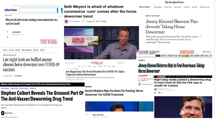
AUGUST 2021 – THE LAUNCH OF THE “HORSE DE-WORMER” PUBLIC RELATIONS CAMPAIGN AGAINST IVERMECTIN BY THE FEDERAL HEALTH AGENCIES
This was, after the manipulation of the Pharma funded trials, the biggest offensive in the war. I maintain that Weber Shandwick, the PR firm working simultaneously for Moderna, Pfizer and the CDC had constructed it well before, and were just waiting for the best time to launch it.
This is what prompted them to launch the campaign:
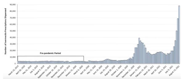
As you can see from the graph above.. ivermectin prescriptions in the U.S were rapidly increasing to a level never before seen in history. August 13, 2021 was the middle of the Delta wave.. and Delta was wicked. Much harder to treat than prior variants. Late Delta was insanely difficult to treat (October-December 2021), so much so that ivermectin alone was no longer enough, and during that time period of late Delta, I was routinely using between 3-6 different medicines to keep patients out of the hospital. But none died and nearly all avoided hospital (the one exception was a cousin who contacted me on Day 10 of her illness, already breathless, I treated her for a day and a half before she had to be admitted, however she was only in for 4 days and never ventilated).
It was carried by every major news organization around the world, like our friends at the Associated Press. No-one notices the unprecedented nature of such an action (it has been FDA approved for years) nor that they have no authority to do this. At the risk of repeating myself, just take a moment and ponder the fact that you have three major U.S medical societies calling for an immediate end to the use of a medicine supported by a meta-analysis of 60 controlled trials showing it leads to major mortality (and other) benefits. Now you know why I call our country the United States of Pharma.
Also:
September 1, 2021 – What happened next is that the horse dewormer meme explodes throughout mass media – every late night talk show host does a bit, every broadcaster and journalist. They pull a fierce “2 by 4” PR campaign (remember a “2 by 4” defines a propaganda campaign as any story or message that appears for 2 weeks on 4 different channels or major media sources. Rachel Maddow actually “led the way” on August 21, the same day as the FDA tweet that kicked off the entire campaign. Nice coordination there Weber Shandwick. CNN then followed up on August 23, blaming “right-wing” media for “pushing” a “deworming drug.” These narratives start to build as you can see:
(See link for article)
___________________
**Comment**
The entire sordid account is here for historical review and record. Dr. Kory deserves serious street cred for this poignant, humble, beyond belief account he continues to live through.
He states:
I went to bed on February 6th, 2022 as a physician (albeit clinging on to his license). On Feb. 7, 2022, I woke up to discover the U.S. Department of Homeland Security had come to the conclusion that my deeply studied scientific opinions made me a domestic terrorist.
He ends by stating he’s probably done writing about ivermectin on his Substack as he needs to move on, but that he never wants us to forget that it all started with his first patient who had a “profound and robust clinical response within 12 hours of her first dose after being ill and feverish for the prior two weeks.” He states that result kept on happening until the variants changed which required higher doses and synergistic therapies.Actually, this too has similarly been experienced in Lymeland.
Speaking of similarities, Kory points out that the atrocities that happened to Dr. Burzynski, who successfully treated patients with cancer, have happened to him. In Burzynski’s case, concerted actions by health system entities began 30 years ago and predicted exactly what has happened to Kory and other doctors now considered “dissidents.”
Kory states we are predictably looking at a nasty RSV/FLU season due to “the lunatic mass vaccination campaign against a coronavirus.” He is also busy treating the “vaccine” injured and those with long haul syndromes. Ironically, ivermectin is one of his primary therapies to treat these syndromes which has transformed the lives of many, but similarly to Lyme/MSIDS the fly in the ointment is “insufficient evidence” for this claim, despite what he sees playing out with his own eyes.
The fraudulent trials “debunking” ivermectin continue….
For more excellent reading on the chronology of the take-down of ivermectin:
The Alongshan virus was discovered in China only five years ago. Now researchers have found the novel virus for the first time in Swiss ticks. It appears to be at least as widespread as the tickborne encephalitis virus and causes similar symptoms. The team is working on a diagnostic test to assess the epidemiological situation.
________________
**Comment**
There is no test for this virus.
This too is a problem. We don’t have accurate tests for the pathogens infecting people. Sick people continue to go to doctors for help, are given inaccurate, faulty tests, are told they are “fine,” or are gaslit, and are sent packing.
Climate-change researchers overlook and do not take into accountestablished populations of I. scapularis found in the late 1960’s in the upper Midwest, as well as at Manitoba in 1991, in their climate change model maps. Even game hunters remember ticks on the heads and necks of deer in the 1950’s and 1960’s in northwestern Ontario and southern Manitoba. The faulty climate change maps are devoid of any ticks in those areas – yet experience shows otherwise. This has been the experience of patients across the globe as well. Source
The bacteria that causes Lyme in humans doesn’t hurt ticks. In fact, it might help them survive.
Dec. 7, 2022
University of Rhode Island entomologist Jannelle Couret is tipping the way we understand the bacteria that causes Lyme disease.
Instead of looking at it from the human perspective, she and an interdisciplinary team of researchers are taking the view of the tick.
While the bacteria – Borrelia burgdorferi – is the pathogen that causes Lyme disease in humans, its presence is quite different in blacklegged ticks that pick up the bacteria from feeding on white-footed mice.
For the ticks, the bacteria doesn’t cause disease. It might even be beneficial.
For the next four years, Couret’s team will research the ecological factors driving the evolution of Borrelia burgdorferi in blacklegged ticks thanks to a $2.6 million grant from the National Institutes of Health.
The grant is part of the prestigious Ecology and Evolution of Infectious Disease (EEID) program, run by the NIH, National Science Foundation, and U.S. Department of Agriculture.
“I am really interested in the factors that are driving the tick populations,” said Couret, an assistant professor of biological studies and the principal investigator on the grant.
“Their populations vary year to year. Our preliminary data suggests that the survival of the ticks during some of their early life stages is improved based on whether they host these bacteria.”
All-female research team
For the four-year study, Couret is collaborating with Associate Professor Sukanya Narasimhan of Yale Medical School, Associate Professor Jean Tsao of Michigan State University, and Associate Professor Cynthia Lord of the University of Florida – along with postdoctoral, graduate and undergraduate trainees at each institution.
“One of my favorite aspects of this work is the research team. We are all women and three of us are women of color,” said Couret, who is part Indigenous, Afro-Cuban, and American. “I think that is – unfortunately – somewhat rare in science.”
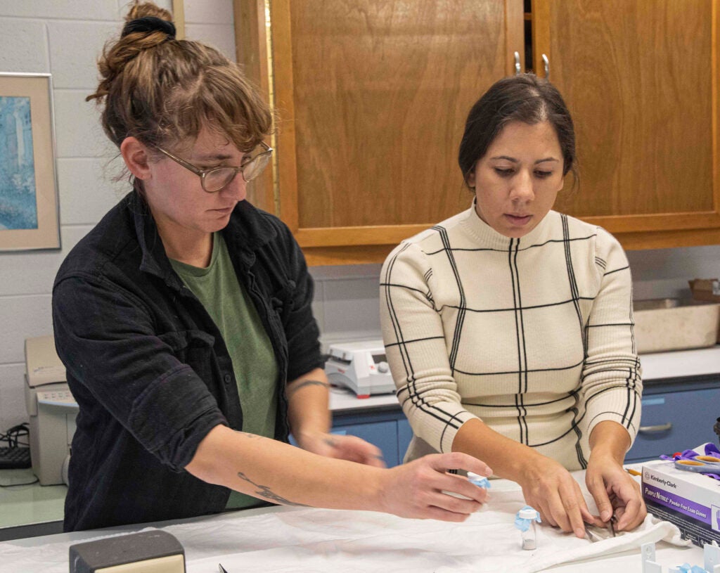
Couret (on the right) works with graduate student Samantha Schofield as they sort out tick samples. (URI photo/Nora Lewis)
Blacklegged ticks, also called deer ticks, carry seven known pathogens and are responsible for about 95% of the tick-borne diseases in the U.S., including about 30,000 cases of Lyme disease reported each year.
Deer ticks can acquire the bacteria that causes Lyme disease during any of its life stages – larvae, nymph or adult – during a blood meal from white-footed mice, the primary carriers of the Lyme disease bacterium.
(While the abundance of deer ticks is casually associated with deer, these hosts do not transmit Borrelia burgdorferi to ticks, and deer are not considered an important host for the maintenance of the bacteria in wildlife populations.)
Do ticks benefit from carrying infections?
But the bacteria doesn’t lead to Lyme disease in either the mice or the ticks. In pilot studies, Couret has seen changes in the ticks that acquire the bacteria – including behavior, metabolism, respiration, and survival. So there appears to be an advantage for those ticks, she said.
“That’s a shift in mindset,” said Couret, who joined URI in 2015 after earning her Ph.D. in the ecology of infectious diseases at Emory University.
“We mainly think of Borrelia burgdorferi as a pathogen because it causes Lyme disease in humans. We are studying the transmission cycle of the bacteria in nature between ticks and white-footed mice. It’s possible that it’s not acting as a pathogen, but rather as a beneficial symbiont of the tick, a partner. The bigger picture question is, if we view Borrelia burgdorferi with this lens, can we better understand its transmission dynamics?”
Environmental factors
In understanding the transmission cycle of Lyme disease, the researchers will explore the relationships of many influences on the bacteria in the tick, including environmental factors, such as temperature and humidity; ecological facets, such as the tick’s microbiome; and the bacteria’s interactions with other organisms in the tick.
“We’re studying the effects of the bacteria on ticks at different levels, from gene expression to behavior,’’ she said. “We’ll combine that information to look at the evolutionary fitness of ticks, and model the impacts of bacteria on annual tick populations. We also are considering the microbiome. We want a really comprehensive view of the ensemble of ecological interactions that influence ticks, Borrelia burgdorferi, and their partnership.”
For the study, Narasimhan, a molecular biologist, will look at gene expression to learn what is changing in the ticks that acquire the bacteria, along with what is changing in the bacteria.
Lord, a vector-borne disease modeler, will incorporate the experiment results in a model that can predict tick populations and rates of transmission of Borrelia burgdorferi.
Tsao, a tick ecologist, will study deer ticks in the Midwest, another hot spot of Lyme disease. Paralleling Couret’s work in Rhode Island, Tsao will study tick behavior and development in a semi-natural environment.
Tsao and Couret will also look at traits that may be affected by the presence of Borrelia burgdorferi, effects of environmental conditions, survival rates, and gene expression.
Learning ways to improve prevention
When it’s completed, the study will greatly expand our understanding of the factors driving the maintenance of Lyme disease in wildlife. Findings could eventually lead to ways to control the deer tick population or inform disease prevention measures, Couret said.
Also, by characterizing the role of the microbiome as it relates to tick-Borrelia interactions, the research could lead to novel methods of biological controls, such as finding competing bacteria within the tick that, when present, negatively impact Borrelia burgdorferi transmission.
A unique aspect of the grant is the heavy focus on providing comprehensive mentorship for trainees, centering the experiences of those who have been marginalized in science and supporting the team through professional development across all four institutions involved.
Called the Microbiome Integrated Tick Ecology Network – or MITEY Network, as in mites – the mentoring will send trainees to each partner university to sharpen science skills, promote sustainable and productive writing practices and science communication, support a growth mindset, and reduce imposter syndrome.
“We want to make sure it’s an inclusive research culture and environment for our trainees,” Couret said.
Unfortunately the wrong things are emphasized in this article. I don’t give a jot what gender, color, or beliefs researchers have and nobody else should either. What we desperately need are good, unbiased, well designed research studies that help patients by giving real answers to real problems. And true to form, this study, once again, while lining the pockets of researchers, most probably won’t help patients. I realize I’ve grown quite skeptical, but we need accurate tests, treatments, and transmission studies. Period.
We need mainstream medicine and public health to accept the fact this is a relapsing illness that persists and sequesters inside the body.
Until these foundational, fundamental issues are addressed and resolved, everything else is moot.
Quite a bit of research has already been done on this subject with regard to Ixodes ricinus – the European vector of Lyme and a species that is very closely related to the east coast U.S. tick Ixodes scapularis – the black-legged tick.
Important excerpts, showing this is not a new idea:
Survival rate of nymphal and adult I. ricinus was significantly enhanced by infection by B. burgdorferi s.l. (χ2: nymph, P = 0.008; adult, P = 0.021).
Moreover, ticks infected by B. afzelii survived better than other ticks (infected by other genospecies or not). The results here indicate that infection by B. burgdorferi s.l., and more specifically infection by B. afzelii, confers survival advantages to I. ricinus under challenging thermohygrometric conditions.
Important excerpt showing that Bb also relies upon the tick for survival:
The spirochaete relies heavily on its arthropod host for basic metabolic functions and has developed complex interactions with ticks to successfully colonize, persist and, at the optimal time, exit the tick. For example, proteins shield spirochaetes from immune factors in the bloodmeal and facilitate the transition between vertebrate and arthropod environments.
So we already know that the relationship between ticks and pathogens is beneficially symbiotic.
For the love of God, can we please move on to accurate testing, effective treatments, and transmission studies?
Molecular Detection of Anaplasma phagocytophilum, Babesia odocoilei, Babesia species and Borrelia burgdorferi Sensu Lato in Songbirds
John D Scott1 *, Elena McGoey2, Ana Morales3 and Risa R Pesapane2,4 1 Upper Grand Tick Centre, 365 St. David Street South, Fergus, Ontario, N1M 2L7, Canada 2 School of Environmental and Natural Resources, College of Food, Agricultural, and Environmental Sciences, The Ohio State University, Columbus, OH 43210, USA 3 McGill Bird Observatory, Ste Anne de Bellevue, QC, Canada H9X 0A6 4 Department of Veterinary Preventive Medicine, College of Veterinary Medicine, The Ohio State University, 1920 Coffey Rd., Columbus, OH 43210, USA
Abstract
The blacklegged tick, Ixodes scapularis, is known to carry various tick-borne zoonotic pathogens with the potential to cause debilitating human and animal diseases. Juvenile I. scapularis parasitize songbirds and, perhaps, these avifauna are competent hosts of common microbial pathogens. We extracted brachial venous blood from 18 groundforaging passerine birds that were parasitized by I. scapularis larvae and nymphs. Using molecular identification, namely PCR, DNA sequencing, and Basic Local Alignment Search Tool (BLAST), we targeted Anaplasma phagocytophilum, Babesia spp. and Borrelia burgdorferi sensu lato. Overall,
15 (83%) of 18 passerine birds were positive for 3 microbial zoonotic pathogens that comprised of A. phagocytophilum (n = 8), Babesia odocoilei (n = 6), Babesia spp. 20-5A74 (n = 1), and B. burgdorferi sensu lato (n = 9).
The pathogen load consisted of 8 singles, 5 doubles, and 2 triples.
One novel Babesia sp. (Babesia spp. 20-5A74) was found, and the remaining Babesia infections were B. odocoilei.
Our findings reveal that ground-foraging, passerine birds are avian hosts of zoonotic pathogens. We provide the first-ever documentation that songbirds are hosts of B. odocoilei. Based on our data, B. odocoilei outnumbered other Babesia spp., and elucidated the authentic fact that B. odocoilei is the predominant Babesia sp. in North America. As avian hosts, passerine birds play a significant role in the enzootic transmission cycle of B. burgdorferi sensu lato, A. phagocytophilum, and Babesia species.
Important excerpts:
In the USA, tick researchers have reported B. odocoilei in Indiana [41-43], Michigan [44] Maine [42,43], Massachusetts [41-43], New York [45], Oklahoma [46,47], Pennsylvania [48,49] Texas [50,51], Virginia [52], and Wisconsin [42,43]. As well, B. odocoilei has been detected in I. pacificus in California [53]. In Canada, B. odocoilei has been detected in Saskatchewan [54], Ontario [7,15,55-59], and Quebec [55,57,58]. And yet, acarologists and ecologists have not reported B. microti in these three provinces [7,15,21,55-59]. Babesia odocoilei, which is a sequestering Babesia sp., can be recalcitrant to treat in human patients [7].
Not only do groundfrequenting songbirds transport ticks, they may also be hosts for tick-borne, zoonotic pathogens. Migratory songbirds widely disperse zoonotic pathogens across North America and, therefore, one does not have to frequent or live in an endemic area to contract human babesiosis caused by B. odocoilei.
https://madisonarealymesupportgroup.com/2021/05/28/study-shows-babesia-odocoilei-is-pathogenic-to-humans/ Study found B. odocoilei in two of 19 participants. DNA amplicons from these two patients are almost identical matches with the type strains of B. odocoilei in GenBank. In addition, the same two human subjects had the hallmark symptoms of human babesiosis, including night sweats, chills, fevers, and profound fatigue. Based on symptoms and molecular identification, we provide substantive evidence that B. odocoilei is pathogenic to humans. Dataset reveals that B. odocoilei serologically cross-reacts with Babesia duncani.
https://www.ncbi.nlm.nih.gov/pmc/articles/PMC3998201/InAustria and Italy patients experienced a severe illness caused by EU1, a species closely related to B. odocoilei. InTaiwan it was (TW1) and in Korea (KO1). Human babesiosis is now reported from around the world. The study in this link states that reported human cases of babesiosis have been attributed, without strong molecular evidence to B. divergans: https://www.ncbi.nlm.nih.gov/pmc/articles/PMC3020600/
Pollutants in Human Plasma Found via Double-Filtration Plasmapheresis Plasma Exchange
Studies in toxicology usually study urine, feces, and other secretions and measure indirectly. Dr. Gatti, whose lab was raided for reporting detection of nanoparticles in vaccines, has a new study.
James Lyons-Weiler
Does everyone remember Drs. Gatti and Montanari? We flew them in from Italy in 2017 to the IPAK Vaccine Safety Conference in Pittsburgh, PA? Probably not.
“aflatoxin B1, chromium, lead, cadmium, arsenic, lindane, cobalt, polycyclic-aromatic-hydrocarbons, disulfoton and aluminium (listed in descending concentration).”
The aluminum was found in various forms and types, bound with and free from silicon.
They also found unknown thread-like objects.
Makes me wonder if we should all detox this way once a year?
Dr. Gatti, congratulations on your new study and on surviving the attack on your lab by Italian authorities.
It should be possible to estimate the blood and body levels of compounds to which we are exposed, say, before and after vaccination.
Citation:
Scholkmann, Felix, and Antonietta M. Gatti. 2022. “Particles in the Eluate from Double Filtration Plasmapheresis—A Case Study Using Field Emission Scanning Electron Microscopy/Energy-Dispersive X-ray Spectroscopy (FE-SEM/EDX)” Compounds 2, no. 4: 367-377. https://doi.org/10.3390/compounds2040030
_______________
**Comment**
Plasmapheresis or plasma exchange, around since the 50’s, has been used to treat autoimmune conditions, blood disorders, viral infections, chronic inflammation, pulmonary fibrosis, MS, Graves’ disease, Myasthenia gravis, transverse myelitis, HIV-related neuropathy, cancer, and even Lyme/MSIDS. Plasma is extracted from your blood, treated, and then put back into the body.
The limitation of this study is it was on a singular patient who had been treated for the following chronic infections:
Borrelia afzelii
Borrelia burgdorferi (CH)
Borrelia burgdorferi (USA)
Borrelia garinii
Chlamydia pneumoniae
Babesia divergens
Bartonella henselae
Rickettsia Helvetica
Rickettsia conorii
Rickettsia helvetica
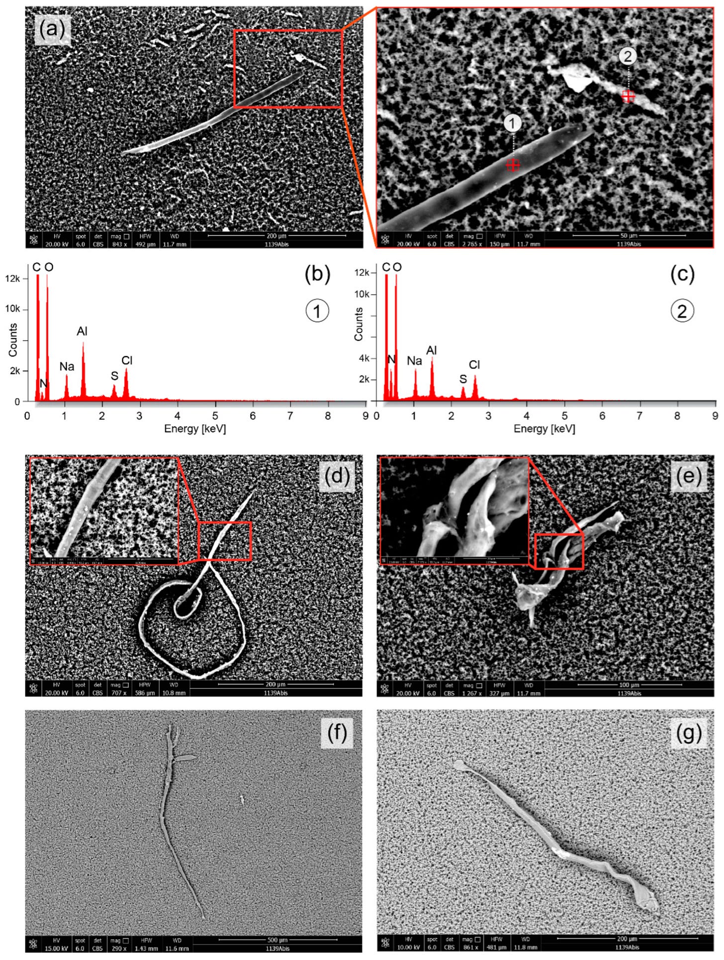
The thread-like object in Figure 4d, however, has a similar morphology and size as a thread-like parasitic nematode (roundworm) of the superfamiliy Filarioridea. Ticks can be also infected with these filarial nematodes [59,60].
Excerpt:
The pollution of nano- and microparticles is an emerging health concern [32,61] and novel ways of quantifying the individual exposure as well as methods to remove these particles from the body are of imminent interest for preventing and treating human diseases. DFPP, possibly in combination with the application of chelating agents, might be a powerful way to remove these nano- and microparticles from the body. The analysis of the eluate with FE-SEM/EDX seems be a useful approach to proof this possibility.
In summary, our analysis of the eluate obtained from a DFPP application revealed particles and objects in the nm and µm range of different shape and chemical composition. Our study is the first to date to investigate the composition of an eluate obtained by DFPP with FE-SEM/EDX.
IMO, while plasmapheresis might certainly help Lyme/MSIDS, the organism(s) often don’t remain in the blood for long but migrate to immunopriviledged sites like the brain, synovial fluid, spine, and organs. This is the problem with all treatments, and testing which rely on delivery via blood, and perfectly illustrates why the current CDC monotherapy is an absolute joke. Further, it doesn’t take into account the relapsing nature of these pathogens which change forms in the body.Savvy treatmentpurposelycycles antimicrobials which helps address these complex issues, which mainstream medicine/research is completely oblivious about.
https://madisonarealymesupportgroup.com/2021/07/07/what-is-in-the-pcr-tests/ Gatti has also done groundbreaking work showing graphene in the PCR COVID test as well as silver, aluminum, titanium, glass fibres, etc – many of which are undeclared in the package leaflet but can cause hardened mucous membranes in people who are tested often for COVID.
https://madisonarealymesupportgroup.com/2018/04/28/italian-lab-shut-down-about-to-testify-about-vaccine-contamination-damage/ Back in the 90’s, Dr. Antonietta Gatti discovered the relationship between micro- and nanoparticles as well as a great number of pathologies: cardiovascular diseases, many forms of cancer, multiple neurological diseases, and autoimmune diseases. Then she found nanoparticles pollute nearly all vaccines. Her home was raided by the Italian police and all digital assets with years of her work and research were because she was about to testify in parliament about vaccine damage.