Archive for the ‘research’ Category

Harvard Student, Queen’s Prof, Collaborating to Rapidly Identify Lyme-Infected Ticks

https://www.coastreporter.net/national-news/harvard-student-queens-prof-collaborating-to-rapidly-identify-lyme-infected-ticks-

Harvard student, Queen’s prof, collaborating to rapidly identify Lyme-infected ticks

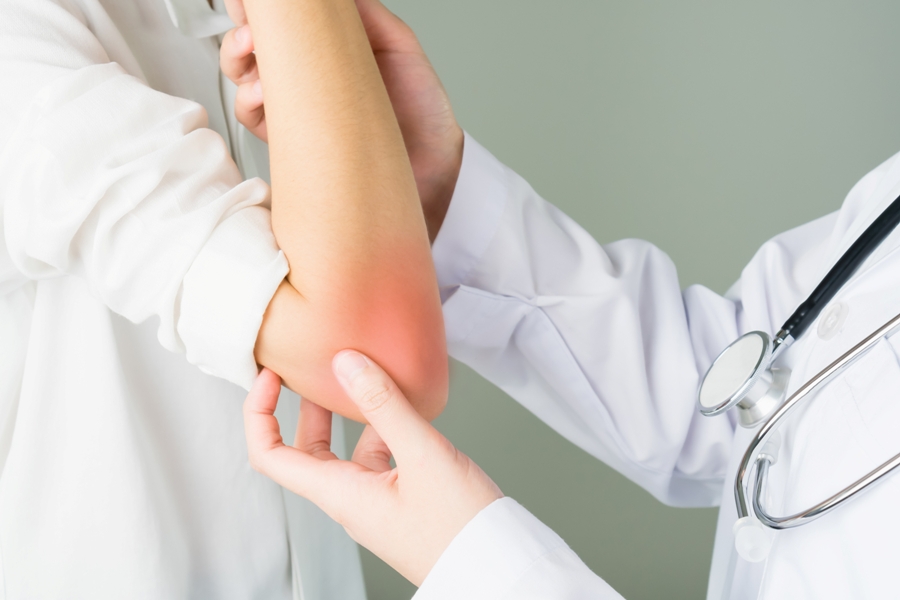
OTTAWA — Harvard University microbiology student Indumathi Prakash was just seven years old when a tick bite near her home in Massachusetts gave her Lyme disease and left her to spend most of her childhood battling rheumatoid arthritis.
2021071616070-60f1e62aa5ca5b9124ece6c1jpeg

OTTAWA — Harvard University microbiology student Indumathi Prakash was just seven years old when a tick bite near her home in Massachusetts gave her Lyme disease and left her to spend most of her childhood battling rheumatoid arthritis.

Now Prakash, 21, is working with a Queen’s University professor, although virtually, on a rapid test that will diagnose the bacterium that causes Lyme disease in ticks in just a few hours.

If successful, the test will open the door to better and faster treatment for the rapidly expanding disease.

“I really wanted to do something to prevent the same thing from happening to someone else so that’s kind of why I got interested in tick-borne disease in general,” said Prakash, in a phone interview from Cambridge, Mass.

Lyme disease has been around in Canada since the 1980s. Warmer winters over the last decade have allowed ticks, and the pathogens they often carry, to flourish in ways they never did before. (See link for article)

_________________

**Comment**

Interesting that this author doesn’t utilize her own countryman’s independent research showing ticks are impervious to the climate and hide under leaf litter, snow, mulch, etc. when conditions are harsh – but doing so would be defying the accepted narrative that “climate change” is behind tick and disease proliferation, and all the woes on planet earth today, despite climatologists who state otherwise, and we certainly can’t have that.

Important quote: 

Since Lyme disease can often be prevented if antibiotics are started within 24 hours of the bite, doctors routinely give people with tick bites antibiotics before it’s clear the bacteria is present.

No, I’m afraid doctors don’t routinely give people with tick bites antibiotics.  I wish they did – and they certainly should.  Doctors continue to take a “wait and see” approach, which is dooming patients to sometimes a life-time of suffering. In my opinion, it appears doctors would rather diagnose patients with anything but Lyme/MSIDS.

The article points out that ticks can be tested by results often take days to obtain results.  This is another problem with testing – not only for ticks but for humans.

Evidently the new test will also detect other coinfections that often come with Lyme disease.


Lyme Disease Skin Rash Puzzles Doctors, Leads to Misdiagnoses

https://danielcameronmd.com/lyme-disease-skin-rash-misdiagnoses/

LYME DISEASE SKIN RASH PUZZLES DOCTORS, LEADS TO MISDIAGNOSES

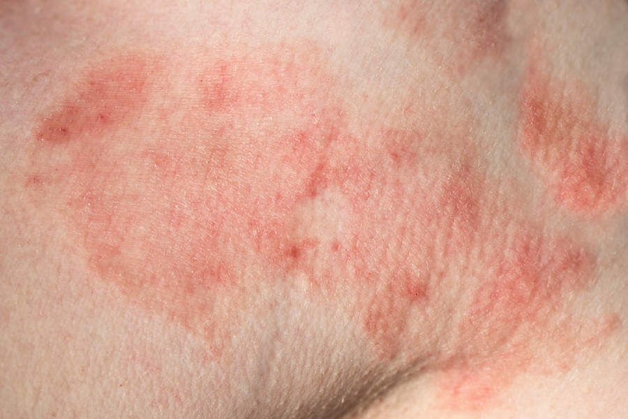
Patient with Lyme disease skin rash

Lyme disease can cause an atypical skin rash that may be mistakenly attributed to another illness, as this case report demonstrates. In their article, Suzuki and colleagues describe a 43-year-old woman who developed a rash which puzzled doctors and led to several misdiagnoses before Lyme disease was correctly identified.


Lyme disease can cause an atypical skin rash, which may be overlooked by clinicians. In this case report, a 43-year-old woman, who was an avid gardener and lived in Wisconsin, developed pink papules behind her right knee.1  Over the next several days, the rash “evolved into painless vesicles with surrounding faint erythema.” She also developed fever, chills, neck pain and malaise.

Diagnoses: shingles, cellulitis, necrotizing fasciitis

Doctors initially suspected shingles (Herpes Zoster) but treatment with valacyclovir, an antiviral medication, did not improve the skin rash or symptoms. “The blisters increased in size and showed purple discoloration,” the authors wrote.

The doctors then diagnosed her with cellulitis, a bacterial skin infection which also causes a skin rash. She was prescribed ceftriaxone, followed by cephalexin and trimethoprim-sulfamethoxazole. But the woman remained ill.

She was then referred for evaluation for suspected necrotizing fasciitis, an inflammation of the vessel walls.  At this time, she had a fever of 101.3°F.

“The atypical appearance of skin rash might confuse physicians with many differential diagnoses, such as spider bite, herpes zoster, bullous cellulitis, necrotizing fasciitis, and so on.”

Additionally:

“Physical examination showed blister and a surrounding round erythematous patch at the right popliteal fossa and right inguinal lymphadenopathy,” the authors wrote.

Lyme disease skin rash

The woman was diagnosed clinically with “early Lyme disease with bullous erythema migrans.”

“Given the season, geographic location, outdoor activity, and progression despite treatment for cellulitis and shingles, she was clinically diagnosed with early Lyme disease,” the authors report.

The woman was treated for Lyme disease with a 10-day course of doxycycline and her skin rash resolved.

Laboratory tests for Lyme disease were negative – a common occurrence with early Lyme disease.

“The atypical appearance of skin rash might confuse physicians with many differential diagnoses, such as spider bite, herpes zoster, bullous cellulitis, necrotizing fasciitis, and so on,” the authors point out.

Editor’s comments:  I have seen rashes described in this article in my Lyme disease patients. I am concerned when a patient is diagnosed with shingles, cellulitis or necrotizing vasculitis before Lyme disease is considered as a possible cause of an atypical skin rash.

If uncertain about the root cause of the rash, I will typically treat patients with a combination of medications including an antiviral agent and an antibiotic that is effective in treating cellulitis, a spider bite and Lyme disease.
References:
  1. Suzuki H, Carlson JR, Matsumoto E. 43-Year-Old Female With Fever and Bullous Skin Lesion. Clin Infect Dis. Dec 17 2020;71(10):2763-2764. doi:10.1093/cid/ciaa206

_____________________

**Comment**

Everyone knows that delayed treatment for Lyme/MSIDS is causing chronic/persistent symptoms, yet it continually appears doctors would rather diagnose patients with anything other than Lyme/MSIDS.  It’s almost like they want to run through a litany of other causes first, treat for those, and finally if nothing works throw up the white flat and admit it’s tick-borne illness.

For more:

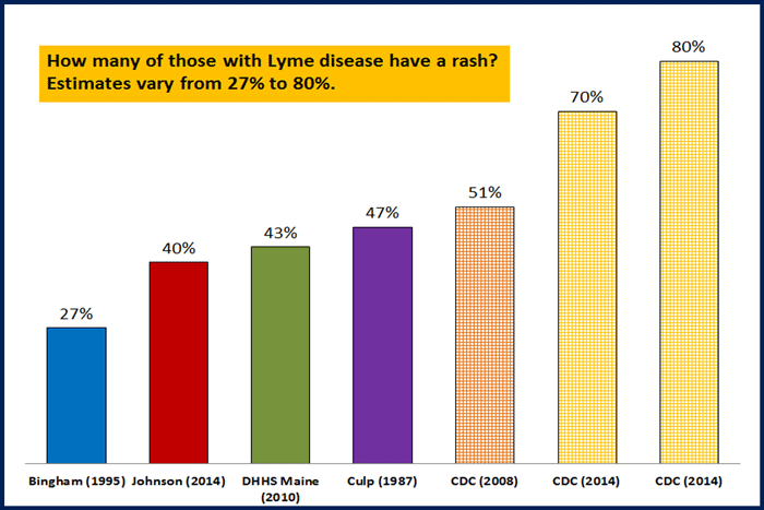
Please remember that those getting the rash is highly variable and certainly not a sure thing.  If you get it, it is diagnostic for Lyme – no testing needed; however, if you don’t get it, you can still be infected:

Rashes-larger-blog-4

Babesia in a MS Patient

https://danielcameronmd.com/babesia-microti-multiple-sclerosis-patient/  Go Here for Podcast

BABESIA MICROTI IN A MULTIPLE SCLEROSIS PATIENT

babesia microti in MS patient

Welcome to another Inside Lyme Podcast. I am your host Dr. Daniel Cameron. In this episode, I will be discussing the case of a 54-year-old male with a 12-year history of multiple sclerosis who was diagnosed with Babesia microti.

Haberli and colleagues describe the case in the journal Multiple Sclerosis and Related Disorders.1

Babesia microti, a tick-borne pathogen that infects red blood cells, can cause severe disease in immunocompromised individuals. This patient was immunocompromised due to multiple sclerosis (MS) and the immunosuppressive drug he was receiving, ocrelizumab.

Disease Modifying Therapy

Ocrelizumab (sold under the brand name Ocrevus™ in the US) was approved in 2017 for the treatment of multiple sclerosis (MS). It is a humanized anti-CD20 monoclonal antibody, which targets mature B-cells that contribute to demyelination and damage to nerve cells.

Ocrelizumab is a disease modifying therapy (DMT) approved to treat both primary progressive MS and relapse-remitting MS. Ocrevus™ can cost just over $65,000 per year if not covered by insurance.

The patient was prescribed Ocrevus™. 

“The use of ocrelizumab has been associated with increased risk of infections involving upper respiratory, gastrointestinal, and urinary tracts, in addition to herpes simplex reactivation,” wrote the authors. 

“Data from one randomized clinical trial has shown a statistically significant increase in the incidence of malignancy among ocrelizumab users compared to placebo and interferon β1,” they added.

Rituximab, a medication with a similar mechanism of action, has been associated with rare infections in non-MS patients including severe or relapsing Babesia microti infections, the authors wrote.

MS patient fails treatment

This is the first published case of Babesia microti in a patient treated with Ocrevus™.

The 54-year-old man had been diagnosed with major depressive disorder, hypertension and had a 12-year history of relapse-remitting multiple sclerosis.

He was hospitalized with malaise, fever, fatigue for one week and episodes of syncope. He failed treatment with 4 different disease modifying drugs ─ glatiramer acetate, natalizumab, dimethyl fumarate, and fingolimod.

The man refused further therapy and was lost to follow-up.

6 years later, MS symptoms worsen

Six years later, the man’s MS symptoms worsened and he was prescribed Ocrevus™.

Four months later, he was hospitalized and treated for possible sepsis. He had unexplained thrombocytopenia (low blood platelet count).

Tests positive for Babesia microti

As part of his sepsis work-up, the patient was tested for tick-borne infections with PCR test results for Babesia microti returning positive.

The man was treated with 500mg daily azithromycin and 750mg twice daily atovaquone. 

“At week 8, his therapy was discontinued due to complete resolution of pancytopenia, negative B. microti PCR, and negative Giemsa-stained blood smear,” wrote the authors.

There have been other cases of Babesia microti patients treated with Ocrevus™.  

“There were five babesiosis cases associated with ocrelizumab reported between June 2018 and August 2020 on the FAERS database,” the authors wrote.²

“There were two females and two males with an age range of 45 to 51 years old, and one female 65 to 85 years old, all of whom had serious events without reported fatalities,” wrote the authors based on a review of the FDA Adverse Event Reporting System (FAERS) public dashboard.

The following questions are addressed in this podcast episode:  

  1. What is Babesia microti?
  2. How do you diagnose Babesia?
  3. Is testing accurate?
  4. How severe is Babesia?
  5. Are there asymptomatic cases of Babesia?
  6. Why do immunosuppressive drugs pose a risk to Babesia patients?
  7. What are the risk factors for symptomatic Babesia?
  8. What is a disease modifying agent?
  9. What is Ocrevus™?
  10. What was the treatment for Babesia?
  11. What is the significance of 8 weeks of treatment with Babesia?

    Thanks for listening to another Inside Lyme Podcast. You can read more about these cases in my show notes and on my website @DanielCameronMD.com. As always, it is your likes, comments, reviews, and shares that help spread the word about Lyme disease. Until next time on Inside Lyme.

Please remember that the advice given is general and not intended as specific advice to any particular patient. If you require specific advice, then please seek that advice from an experienced professional.

Inside Lyme Podcast Series

This Inside Lyme case series will be discussed on my Facebook page and made available on podcast and YouTube.  As always, it is your likes, comments, and shares that help spread the word about this series and our work. If you can, please leave a review on iTunes or wherever else you get your podcasts.

References:
  1. Haberli N, Coban H, Padam C, Montezuma-Rusca JM, Creed MA, Imitola J. Babesia microti infection in a patient with multiple sclerosis treated with ocrelizumab. Mult Scler Relat Disord. Feb 2021;48:102731. doi:10.1016/j.msard.2020.102731
  2. Ezequiel M, Teixeira AT, Brito MJ, Luis C. Pseudotumor cerebri as the presentation of Lyme disease in a non-endemic area. BMJ Case Rep. 2018;2018.

________________________

**Comment**

This case brings up a very important point: MS can be caused or exacerbated by tick-borne infections. Immune-suppressing drugs are contraindicated for people with TBI’s. The only caveat to that is if they are used in conjunction with antibiotics.

I can’t help but wonder how many other patients are out there with this exact same scenario.

For more:

Patient With Facial Palsy From Lyme Disease Fails Treatment

https://danielcameronmd.com/patient-facial-palsy-from-lyme-disease-fails-treatment/

PATIENT WITH FACIAL PALSY FROM LYME DISEASE FAILS TREATMENT

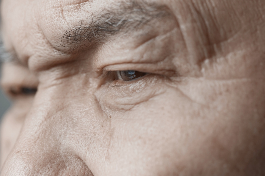
facial palsy from lyme disease in elderly man

Facial palsy, sometimes referred to as Bell’s palsy, is considered an early sign of Lyme disease. When the infection is diagnosed in the early stage it is typically easier to treat. However, this case report features an elderly patient with facial palsy from Lyme disease who remained ill following treatment.

In this case report, Hareem and colleagues describe a 71-year-old man with facial palsy from Lyme disease who failed treatment. The authors offer an inside look at what led up to the failed treatment.¹

Steroids associated with treatment failure?

The man was initially treated in August 2017 for an upper respiratory tract infection and prescribed the steroid dexamethasone. The previous year, he had a tick bite but no rash. One week later he was seen for a sore throat, right-sided ear pain, headache, dizziness, nausea, neck pain, and tinnitus. He had slight asymmetry of his smile.

Lyme disease Western blot test results were positive and the patient was prescribed doxycycline. However, several days later, the man returned with a complete facial palsy from Lyme disease. His condition had worsened.

“On his next visit, he complained of continuing right-sided headache and neck pain along with right-sided hearing loss, right-sided otalgia, dizziness, and nausea,” wrote the authors. His treatment was changed to cefuroxime.

Gait problems, hearing loss

Two weeks later his condition was even worse. “He continued to complain of gait instability and otalgia with hearing loss and neck pain,” wrote the authors. His treatment was changed to 30 days of doxycycline. He had an 80% hearing loss in the right ear and 25% in the left ear on audiology testing.

Authors’ conclusion

The man was diagnosed with “chronic Lyme disease from failed antibiotic therapy with simultaneous unilateral involvement of the seventh and eighth cranial nerves.”  He was prescribed an additional 4 weeks of doxycycline but remained ill. 

Wormer and colleagues previously discussed treatment failures in Lyme disease patients treated with steroids.2

Editor’s Note:  The 71-year-man never received a course of intravenous ceftriaxone. The authors did not discuss other diagnostic or treatment options.

References:
  1. Hareem A, Dabiri I, Zaheer N, Burakgazi AZ. Medically Refractory Neuroborreliosis Case Presented with Coexistance Involvements of Cranial 7 and 8 Nerves. Neurol Int. Mar 18 2021;13(1):125-129. doi:10.3390/neurolint13010012
  2. Wormser GP, Brady KC, Cho MS, Scavarda CA, McKenna D. Efficacy of a 14-day course of amoxicillin for patients with erythema migrans. Diagn Microbiol Infect Dis. Jun 2019;94(2):192-194. doi:10.1016/j.diagmicrobio.2019.01.003

____________________

For more:

What Does a Lyme Rash Look Like?

https://danielcameronmd.com/what-does-a-lyme-disease-rash-look-like/  Podcast Here

WHAT DOES A LYME DISEASE RASH LOOK LIKE?

what does a lyme disease rash look like

Welcome to another Inside Lyme Podcast. I am your host Dr. Daniel Cameron. In this episode, I will be discussing a question I’m asked frequently: What does a Lyme disease rash look like? As a new study has found, the erythma migrans rash can appear differently based on several factors.

The study by Rebman and colleagues, entitled “The presenting characteristics of erythema migrans vary by age, sex, duration, and body location,” was published in Infection in March 2021.¹ It addressed a frequently asked question: What does a Lyme disease rash look like?

The erythema migrans (EM) rash (or Bull’s-eye rash), considered a definitive sign of Lyme disease, is often mistakenly thought to appear similar in all individuals – a circular red lesion which expands to at least 2 inches.

But as Rebman and colleagues report, a Lyme disease rash can look different based on several factors and does not always appear as the familiar Bull’s-eye rash. In fact, the authors suggest that relying solely on a Bull’s-eye appearance, when evaluating a rash for possible Lyme disease can lead to delays in diagnosis and treatment.

Diverse characteristics of Lyme disease rash

The authors examined 271 Lyme disease patients who had an erythema migrans rash to determine what does a Lyme disease rash look like? ¹

“We studied associations between these presenting characteristics [of EM rashes], as well as whether they were associated with age, sex, EM duration, body location, and initiation of antibiotics,” the authors write.

The patients were part of a longitudinal cohort study from 3 sites in Maryland and southeastern Pennsylvania. They were not enrolled if their rash was under 5 cm in diameter (2 inches) or their acute illness was longer than 3 months.

The study found that:

  • EM size increased over time with the EM duration peaking at 14 days.
  • Males had larger rashes than females (an average of 2.8 cm larger).
  • Males were more likely to have a blue/red rash. In fact, the odds of a red rash in males was 65% lower than in females.
  • Age was a significant predictor of central clearing. For every 10-year increase in age, the odds of central clearing decreased 25%. As age increased, there was a greater likelihood of a solid rash.
  • EM rashes were more likely to occur in harder to see body locations (i.e.,  behind the thigh and behind the knees). The authors assumed that ticks had an opportunity to attach longer in these areas before being discovered.
  • Nearly 1 in 3 patients had multiple rashes on examination.
  • Approximately 1 in 3 patients reported pain at the site of the rash.  
  • Just over 50% of the EM rashes were itchy.
  • Rash shapes were varied ─ 50.9% were round; 39.1% were oval. The remaining rashes were irregular.
  • Rash colors were varied ─ about 3 out of 4 were red. The remaining rashes were blue/red.
  • Rash patterns were varied ─  only 28% appeared as a Bull’s-eye rash (a ring within the rash). Central lightness (17.3%), central darkness (28.8%) and uniform rashes (25.8%) were also described.
  • Over 90% of the rashes were homogeneous. The remaining rashes were uneven.
  • Nearly 9% presented with vesicles. 

Concerns with limiting size of Lyme disease rash

The authors raised concerns with following a 5 cm (or less than 2 inches) cutoff for EM rashes in determining the presence of a Lyme disease infection.

“Applying a 5 cm size cutoff in research or surveillance settings may thus exclude a higher proportion of females with otherwise suggestive clinical histories and epidemiological risk,” the authors write.

They also raised concerns about recognizing an EM rash in darker skinned patients. “95.9% of our final sample self-identified as non-Hispanic white.”

The authors suggest that the variation in EM rash presentations in males vs. females and among various ages may be related to an immune response to the Borrelia infection.

Authors’ Conclusion

“Given that EM remains a clinical diagnosis, it is essential that both physicians and the general public are aware of its varied manifestations.”

Editor’s note

Unfortunately, fewer than half of Lyme disease patients present with an erythema migrans rash. This case series merely reflects the diversity of rashes in Lyme disease patients fortunate enough to present with a rash.

The following questions are addressed in this episode:

  • What is an erythema migrans rash?
  • How often do Lyme disease patients have an erythema migrans rash?
  • Can you culture Lyme disease from a rash?
  • How long do erythema migrans rashes last?
  • What other rashes resemble an erythema migrans rash?
  • What color rashes have you seen?
  • Where are rashes located?
  • What is the significance of multiple rashes on examination?
  • Have you seen itchy erythema migrans rashes?
  • Can you discuss diversity of erythema migrans rashes that were described?
  • Could a rash less than 2 inches in diameter be important?
  • What do we know about the appearance of rashes in people of color?
  • What is the importance of a smaller rash in women?

    Thanks for listening to another Inside Lyme Podcast. You can read more about these cases in my show notes and on my website @DanielCameronMD.com. As always, it is your likes, comments, reviews, and shares that help spread the word about Lyme disease. Until next time on Inside Lyme.

Please remember that the advice given is general and not intended as specific advice to any particular patient. If you require specific advice, then please seek that advice from an experienced professional.

Inside Lyme Podcast Series

This Inside Lyme case series will be discussed on my Facebook page and made available on podcast and YouTube.  As always, it is your likes, comments, and shares that help spread the word about this series and our work. If you can, please leave a review on iTunes or wherever else you get your podcasts.

To be clear, having the EM rash is diagnostic for Lyme. You are infected. On the other hand, NOT having the rash doesn’t mean you aren’t infected.

Also, EM rashes wax and wane despite treatment. Treating the EM rash is illogical because borrelia becomes systemic (all over the body) within hours. It means nothing if the EM rash disappears. This is why it’s critical to get to a Lyme literate doctor (LLMD) who is educated on such matters.