Variable Clinical Presentations of Babesiosis
Variable clinical presentations of babesiosis
Epidemiology
The first reported case of babesiosis in the US was in 1968.9 It became a nationally notifiable disease in 2011, and among the 27 states where it was notifiable in 2013, there were 1,792 reported cases nationwide.5,10 Tick-borne and transfusion-associated cases of babesiosis occur in multiple parts of the country, including outside of areas of known endemicity.5 The number of reported cases is rising steadily in the US and worldwide, owing in part to increased medical awareness and improved diagnostic methods.1-3 (See Reported cases of babesiosis in the US.)
Health departments notify the CDC of babesiosis cases via the National Notifiable Diseases Surveillance System (NNDSS) using a standard case definition. In addition to basic demographic information (age, gender, and county of residence) provided via NNDSS, supplemental data (symptoms and history of transfusion) can be submitted to the CDC using a disease-specific case report form (CRF). Because babesiosis has been a reportable condition in some states for years, state-developed CRFs had already been in use to capture supplemental data.5
To promote standard data collection, the CDC developed a babesiosis CRF, which was approved by the Office of Management and Budget in August 2011 (www.cdc.gov/parasites/babesiosis/resources/50.153.pdf). Supplemental data, derived from the CDC’s or a state’s CRF, were merged manually with NNDSS records by matching a case ID number or demographic data. If case records had conflicting data, the more detailed record was considered correct.
As cases of babesiosis transmitted via tick bite or blood transfusion occur in multiple parts of the US, including outside of areas of known endemicity, ongoing national surveillance using the standard case definition will provide a foundation for developing evidence-based prevention and control measures to reduce the burden of the disease. In addition, mapping based on this surveillance allows for the identification of endemic areas, which aids the clinician in diagnosis.
Transmission and pathogenesis
The heightened recognition of tick-borne infection is derived largely from the increasing incidences of human babesiosis, anaplasmosis, and Lyme disease, both individually and together.11,12 Because these infections share the same rodent reservoir and tick vector hosts, they can be cotransmitted to human hosts.1,2,10,13-16 Coinfections involving various combinations of these pathogens are common and can be severe.12,14 The babesia parasite is suspected of causing proinflammatory cytokines that stimulate the production of nitric oxide, which may cause erythrocytic cellular damage when produced in excess.2
Diagnostic procedures and clinical management of the resulting disease syndrome are complicated by the diversity of pathogens involved and by the unusual diversity and duration of symptoms.
Clinical presentation
Common clinical features of babesiosis are similar to those of malaria and range in severity from asymptomatic to rapidly fatal. Most patients experience a viral infection-like illness with fever, chills, sweats, myalgia, arthralgia, anorexia, nausea, vomiting, or fatigue, and in some cases, patients may develop hemolytic anemia.1-4,10 Most symptomatic patients become ill 1 to 4 weeks after the bite of a B. microti-infected tick and 1 to 9 weeks (but up to 6 months in one reported case) after transfusion of contaminated blood products.6-8
A high index of clinical suspicion for babesiosis and the possible presence of other tick-borne infections are required for prompt diagnosis and proper treatment. Because the clinical findings are nonspecific, lab studies are necessary to confirm the diagnosis.
Diagnosis
Microscopic examination of blood smears is the current gold standard for detecting Babesia infection, while polymerase chain reaction testing has promising diagnostic value.1,2,16,17 Differentiating Babesia from malaria on peripheral smears can be difficult but rapidly resolved by the presence or absence of a history of travel.1 Peripheral smears for Babesia allow for same-day or, at the most, next-day confirmation of the diagnosis. The case examples described below demonstrate the range of symptoms and clinical presentations associated with babesiosis (with and without coinfection) that can challenge the NP.
Babesiosis is caused by parasites that infect red blood cells. Most US cases are caused by B. microti, which is transmitted by Ixodes scapularis ticks, primarily in the Northeast and Upper Midwest. Babesia parasites also can be transmitted via transfusion, anywhere, at any time of the year. In March 2018, the FDA approved the first B. microti blood donor screening tests. B. microti Arrayed Fluorescent Immunoassay detects antibodies to B. microti in human plasma, and B. microti Nucleic Acid Test detects B. microti DNA in human whole blood.18
Treatment
**Please see my comment at end of article**
Generally, treatment with atovaquone plus azithromycin is used for patients with mild-to-moderate babesiosis, whereas clindamycin plus quinine is recommended for patients with severe disease; both treatment regimens are given for 7 to 10 days.1-4 All four drugs are used FDA off-label for babesiosis; however, the dosage recommendations are supported by the clinical guidelines.1-4,19 The dosage regimen for atovaquone plus azithromycin for adult patients is atovaquone 750 mg orally every 12 hours, and azithromycin 500 to 1,000 mg orally on day 1 and 250 mg orally once daily for the subsequent days.1-4 Immunocompromised patients may require higher doses of azithromycin.2-4
The dosage regimen for clindamycin plus quinine for adult patients with severe disease is clindamycin 600 mg orally every 8 hours or clindamycin 300 to 600 mg I.V. infusion every 6 hours, and quinine 650 mg orally every 6 to 8 hours.1-4 Dose adjustments of quinine are needed for patients with severe chronic kidney disease.19,20 Of note, the only FDA-approved preparation of oral quinine currently available in the US is the 324 mg capsule.19,20 Previously, the dosage available in the US was a 325 mg capsule. The change in the quinine preparation from 325 mg to 324 mg may result in minor dose disparities between some guideline dosage recommendations that were published before the commercial preparation was changed.20,21
Although rare cases of resistance to atovaquone plus azithromycin have been reported, this combination is effective in most patients.2 Atovaquone is contraindicated in patients who develop or have a history of serious allergic or hypersensitivity reactions to the drug or any of the drug’s components. Azithromycin is contraindicated in patients with known hypersensitivity to azithromycin or any macrolide or ketolide antibiotic and also in patients with a history of cholestatic jaundice or hepatic dysfunction.19 Clindamycin is contraindicated in patients with a history of hypersensitivity to clindamycin or lincomycin. Quinine is contraindicated in patients with known hypersensitivity to quinine, mefloquine, or quinidine; prolonged QT interval; a glucose-6-phosphate dehydrogenase deficiency; or a history of myasthenia gravis or optic neuritis.19 Consult the manufacturer’s prescribing label for complete prescribing information for each drug.
Some patients, including those with severe illness, might require or benefit from supportive care, such as antipyretics, vasopressors (if the patient’s BP is low and unstable), blood transfusions, exchange transfusions (in which portions of a patient’s blood or blood cells are replaced with transfused blood components), mechanical ventilation, and dialysis. The NP should consider referral to an infectious-disease specialist for patients who are pregnant, have an underlying hematologic or oncologic problem, have had a splenectomy, are allergic to first-line antibiotic agents, or have had an unsatisfactory response to antibiotic therapy.
Red blood cell exchange transfusions are recommended for cases of severe babesiosis in patients with parasitemia of 10% or greater, severe anemia (hemoglobin less than 10 g/dL), or pulmonary, kidney, or liver impairment.2-4 Exchange transfusions are used to rapidly decrease parasitemia, correct anemia, and help remove toxic byproducts produced by the infection.2
Case examples
The case examples of patients with babesiosis show a wide range of symptoms and clinical presentations. The case examples below are cases that occurred in southeastern New Jersey, where the disease is endemic. All patients were hospitalized and treated in Atlantic County, New Jersey (see Summary of data from patients with babesiosis).
Case 1
Ms. A is a 78-year-old White female who was admitted with fever, chills, lethargy, fatigue, and marked changes in sensorium. She had a maximum temperature of 100.6° F (38.1° C); sepsis was considered for this patient. Multiple tick bites were found. Pertinent lab findings included lactate dehydrogenase (LDH), 528 units/L; aspartate aminotransferase (AST), 90 units/L; and alanine aminotransferase (ALT), 34 units/L. Her vitamin B12 and folate levels were normal.
Ms. A’s initial white blood cell (WBC) count was 5.0 × 10⁹/L, but over the first 3 days of hospitalization, it gradually dropped to 2.6 × 10⁹/L. Her hemoglobin dropped from 10.5 g/dL to a low of 8 g/dL, and her platelets were initially 39 × 109/L but gradually increased as she continued her course of treatment. Ms. A had 33% polymorphonuclear leukocytes, 2% bands, 49% lymphocytes, and 13% monocytes. Peripheral smear was positive for Babesia, and she had a Babesia immunoglobulin M (IgM) of 1:160 and Anaplasma (previously referred to as Ehrlichia) IgM of 1:320.
In view of Ms. A’s leukopenia and thrombocytopenia, anaplasmosis was suspected, and she was treated with doxycycline 100 mg I.V. infusion every 12 hours, atovaquone suspension 750 mg orally twice daily, and azithromycin 500 mg I.V. infusion every 24 hours. Doxycycline is the recommended treatment for anaplasmosis and was administered to cover the possibility of anaplasmosis in this patient. She was treated with that regimen for 5 days. She was then started on doxycycline twice daily, and azithromycin 500 mg daily (both oral) along with the atovaquone suspension of 750 mg twice daily for a 14-day course of therapy. Ms. A made a dramatic improvement in her mentation and resolution of her lethargy.
Case 2
Ms. C is a 90-year-old White female with a chief complaint of rectal bleeding. On admission, her lab studies revealed severe anemia with a hemoglobin of 7.6 g/dL and hematocrit of 22.6%. Her platelet count was 103 × 109/L and peripheral smear was positive for Babesia. Ms. C had spiking temperatures 100° F to 101° F (37.8° C to 38.3° C). Her rectal bleeding was controlled with an octreotide infusion to which she responded well (the bleeding ceased). Her peripheral smear was positive for Babesia, and she was placed on an oral dose of azithromycin 500 mg on day 1 and then 250 mg daily and atovaquone suspension 750 mg twice daily to complete a 10-day course.
Case 3
Mr. E is a 57-year-old White male admitted with fever, malaise, and chills. His temperature had risen to 101° F (38.3° C). His AST and ALT were 64 and 54 units/L, respectively, and gradually rose to a peak of 90 and 87 units/L, respectively, during his 5-day hospital stay. Mr. E’s WBC count decreased from his initial hospital results to 2.9 x 10⁹/L with a hemoglobin of 9.2 g/dL. His platelets were initially 60 × 109/L but dropped to 34 × 109/L at their lowest level. In view of his elevated liver enzymes, leukopenia, and thrombocytopenia, anaplasmosis was highly suspected, and he was started on doxycycline 100 mg I.V. infusion every 12 hours.
Mr. E’s peripheral smear was positive for Babesia. He was started on oral clindamycin 600 mg every 8 hours and oral quinine 650 mg three times daily. Acute hearing deterioration occurred, and the quinine was discontinued. Mr. E’s regimen was then switched to oral azithromycin 500 mg on day 1 and then 250 mg daily and oral atovaquone 750 mg twice daily. He went on to complete only 7 days of therapy, and his elevated liver enzymes and thrombocytopenia resolved. The suspected anaplasmosis was not confirmed, as the Anaplasma IgM was negative. However, Mr. E’s leukopenia and thrombocytopenia resolved on the above regimens.
Case 4
Mr. J is an 81-year-old White male who was admitted with increasing lethargy, weakness, chills, and blurred vision. He had a history of coronary artery disease and hypertension. His hemoglobin on admission was 12.1 g/dL, and his hematocrit was 35.4%. His WBC count was 5.3 × 10⁹/L. By day 2, his hemoglobin had dropped to 9.9 g/dL with a hematocrit of 29%. His platelets were initially 54 × 109/L and dropped to 46 × 109/L, but on therapy, rose to 191 × 109/L.
Mr. J had 82% polymorphonuclear leukocytes, 10% lymphocytes, and 6% monocytes. On the day of admission, a peripheral smear was positive for Babesia. Subsequently, serologic studies demonstrated an Anaplasma IgG of 1:256; the IgM was negative. Babesia serologies were greater than 1:320, both IgG and IgM. Anaplasmosis was suspected with Mr. J’s confirmed babesiosis, and he was started on azithromycin 500 mg I.V. infusion every 24 hours and doxycycline 100 mg twice daily.
At discharge on day 10, Mr. J was switched to clindamycin orally three times a day and quinine orally three times a day because of intolerance to azithromycin, and he completed a 14-day course of therapy. He convalesced satisfactorily. His hemoglobin at discharge was 12.5 g/dL and WBCs 7.4 × 10⁹/L; platelets improved to 137 × 109/L.
Case 5
Mr. K is an 85-year-old White male who was admitted with fever and chills intermittently, recurring for several days prior to admission. He had a history of hairy cell leukemia, splenectomy, permanent pacemaker insertion for atrioventricular block, gouty arthritis, prostatic hypertrophy, and polymyalgia rheumatica. In the ED, Mr. K had an immediate peripheral smear for Babesia, and the intraerythrocytic parasite was demonstrated. He had been working on a golf course for the week prior to admission.
A second peripheral smear was positive for intraerythrocytic parasites with 10.4% of his red blood cells infected. Findings were also positive for Howell-Jolly bodies, which are erthrocytic nuclear remnants associated with asplenia or decreased splenic function. Mr. K was started on oral azithromycin 500 mg on day 1 and then 250 mg daily and atovaquone 750 mg suspension twice daily. Due to the possibility of concurrent tick-borne infection, he was also started on oral doxycycline 100 twice daily.
Over the course of day 1, Mr. K’s platelet count dropped from 25 to 23 × 109/L, with blood urea nitrogen of 29 mg/dL and creatinine of 1.2 mg/dL. His WBC count dropped from 4.1 to 2.5 × 10⁹/L, and his hemoglobin dropped from 16 to 13 g/dL. He had 20% bands, 5% atypical lymphocytes, 47% polymorphonuclear leukocytes, and 23% lymphocytes. Mr. K remained on doxycycline, azithromycin, and atovaquone suspension for 8 days when he was discharged home.
Mr. K was readmitted the following day when he complained of the inability to ambulate and generalized weakness. He had peripheral smear positivity with babesiosis and was serologically positive for anaplasmosis with both IgM and IgG. Mr. K had continued on the prescribed antibiotic regimen up until his readmission that day. Due to the persistence of parasitemia despite adequate therapy, he was changed to clindamycin 600 mg I.V. infusion every 8 hours, and quinine was also being administered.
Unfortunately, Mr. K developed gastric distress and a generalized erythematous coalescing rash, which prompted the discontinuation of the clindamycin and quinine. His WBC count was 2.2 × 10⁹/L, and his hemoglobin was 9.5 g/dL. Platelets had risen to 43 × 109/L, and he had 43% polymorphonuclear leukocytes, 10% bands, 42% lymphocytes, and 5% monocytes.
Because of the persistence of parasitemia, Mr. K underwent exchange transfusion. At that point, he had been restarted on azithromycin 500 mg I.V. infusion every 24 hours and atovaquone suspension 750 mg orally twice daily. Azithromycin and atovaquone were continued for 5.5 weeks, at which time he was parasite smear negative for Babesia. Subsequently, a Babesia peripheral smear remained negative.
Discussion of case examples
Case 1 shows the unusual effect of babesiosis on the sensorium in the older adult, as any infectious process can. The patient’s cognitive function was dramatically improved following treatment, despite the marked changes in mentation on admission. A coinfection with Anaplasma was suspected. In general, all cases of babesiosis need to be tested for late Lyme disease, via Western blot, although not immediately addressed.1,2,4
Patients with concurrent babesiosis and anaplasmosis—suspected or serologically positive—are treated with doxycycline, which is equally effective for Lyme disease, early or late. Generally, the greater number of concurrent tick-borne infections and the higher the parasitimia load, the more toxic the presentation.1,12
Case 2 shows the need to check the peripheral smear for Babesia despite the rectal bleeding issue on admission. This diagnostic test could have easily been omitted, causing a delay in the diagnosis. Such a delay in older adult patients that results in delayed treatment can put these patients at greater risk for severity of babesiosis. Generally, the combination of clindamycin and quinine has a much higher probability of intolerance and adverse reactions. This combination is not the treatment of first choice for babesiosis. Pertaining to anaplasmosis, the triad of leukopenia, transaminase elevation (mild or moderate), and thrombocytopenia demands empiric treatment with doxycycline prior to serologic confirmation.1,2,4
A peripheral smear for Babesia is rapidly interpreted, is inexpensive, and should be requested in evaluating all patients with any degree of anemia—especially during the spring and summer months in endemic areas. Serologic studies are variable in developing positivity and are generally less readily available.
Case 3 illustrates the importance of suspecting and investigating the possibility of babesiosis and anaplasmosis coinfection in a patient presenting with a tick-borne illness.
Case 4 demonstrates that no additional lab studies—other than peripheral smear for Babesia—are needed to confirm the diagnosis of babesiosis.
Case 5 exemplifies the therapeutic challenge and refractory response to treatment of babesiosis in patients with the comorbidities of a hematologic disease and/or splenectomy.
Patient education
Heightened awareness of babesiosis as well as prompt diagnosis and treatment are essential to prevention. Both patients and the general public need to become more aware of the existence of the disease and other tick-borne infections, especially individuals who live in or travel to regions where babesiosis is found. The NP can play an active and important role in providing patient education about the disease. The basic points of information to communicate include:
- What babesiosis is and its potential to be a life-threatening illness
- How individuals acquire babesiosis (tick bite, transfusion, or, rarely, vertical transmission)
- Where in the world babesiosis is found
- Signs and symptoms of babesiosis
- Note that many individuals do not have any symptoms and do not get sick
- Importance of seeing a healthcare provider if babesiosis is suspected
- Treatability of babesiosis and need for prompt diagnosis and treatment.22,23
Individuals who live in or travel to endemic areas should avoid tick-infested areas; apply repellents and wear long pants and long-sleeved shirts when outdoors; shower soon after being outdoors; and check their entire body for ticks.3 When outdoors, they should walk on cleared trails, stay in the center of the trail, and minimize contact with leaf litter, brush, and overgrown grasses (where ticks are most likely to be found). If a tick is found attached to a person’s body, it should be properly removed as soon as possible.
The CDC offers a printable, one-page fact sheet for patients and the general public that details the basic information for babesiosis awareness in addition to the link for the CDC guide to proper removal of a tick attached to a person (www.cdc.gov/parasites/babesiosis/resources/babesiosis_fact_sheet.pdf).
Conclusion
This article illustrates the need for the NP to appreciate the variable clinical presentations of babesiosis to facilitate prompt diagnosis, provide proper therapeutic management, and avoid the poor outcomes associated with this disease. Staying knowledgeable of babesiosis is essential. It is important for the NP to understand that infected patients may not recall a tick bite and that clinical presentations may not only be variable but also nonspecific, ranging from subclinical to severe. The possibility of coinfection with other tick-borne illnesses (Lyme disease and anaplasmosis) must be considered. Furthermore, the NP needs to assume an active role in patient education to affect babesiosis awareness and prevention.
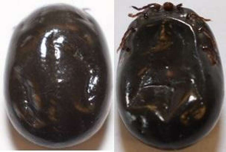
Ixodes scapularis (blacklegged or deer ticks)
The images below are of the Ixodes scapularis ticks, also known as blacklegged or deer ticks. From left to right, the male (M) with a dorsal scutum (also known as a shield on the hard-bodied tick) that covers the entire back on the male, the female (F) with only a portion of the back covered by the dorsal scutum, the nymph (N), and the larva (L).
Figure
Sourse: Procop GW, Church DL, Hall GS, et al. Koneman’s Color Atlas and Textbook of Diagnostic Microbiology. 7th edition. Philadelphia, PA: Wolters Kluwer Health, 2016.
Reported cases of babesiosis in the US1,2,22,23
Most cases of babesiosis in the US occur in seven states, five of which are located in the Northeast (MA, CT, RI, NY, and NJ) and two in the upper Midwest (MN and WI). The geographic range of babesiosis has expanded beyond these highly endemic areas and it is now reported all along the northeastern seaboard and inland, ranging from Maine to Maryland.
Sporadic cases of babesiosis have been reported in other areas of the US including the West Coast. Additionally, transfusion-associated cases of babesiosis can occur anywhere in the country. Congenital transmission of babesiosis has also been reported.
REFERENCES
1. Sanchez E, Vannier E, Wormser GP, Hu LT. Diagnosis, treatment, and prevention of Lyme disease, human granulocytic anaplasmosis, and babesiosis: a review. JAMA. 2016;315(16):1767–1777.
2. Vannier EG, Diuk-Wasser MA, Ben Mamoun C, Krause PJ. Babesiosis. Infect Dis Clin North Am. 2015;29(2):357–370.
3. Vannier E, Krause PJ. Human babesiosis. N Engl J Med. 2012;366(25):2397–2407.
4. Wormser GP, Dattwyler RJ, Shapiro ED, et al The clinical assessment, treatment, and prevention of Lyme disease, human granulocytic anaplasmosis, and babesiosis: clinical practice guidelines by the Infectious Diseases Society of America. Clin Infect Dis. 2006;43(9):1089–1134.
5. Centers for Disease Control and Prevention. Babesiosis surveillance—18 States, 2011. MMWR Morb Mortal Wkly Rep. 2012;61(27):505–509.
6. Levin AE, Krause PJ. Transfusion-transmitted babesiosis: is it time to screen the blood supply. Curr Opin Hematol. 2016;23(6):573–580.
7. Leiby DA. Babesiosis and blood transfusion: flying under the radar. Vox Sang. 2006;90(3):157–165.
8. Herwaldt BL, Linden JV, Bosserman E, Young C, Olkowska D, Wilson M. Transfusion-associated babesiosis in the United States: a description of cases. Ann Intern Med. 2011;155(8):509–519.
9. Scholtens RG, Braff EH, Healey GA, Gleason N. A case of babesiosis in man in the United States. Am J Trop Med Hyg. 1968;17(6):810–813.
10. Centers for Disease Control and Prevention. Notice to readers: final 2013 reports of nationally notifiable infectious diseases. MMWR Morb Mortal Wkly Rep. 2014;63(32):702–715.
11. Western KA, Benson GD, Gleason NN, Healy GR, Schultz MG. Babesiosis in a Massachusetts resident. N Engl J Med. 1970;283(16):854–856.
12. Diuk-Wasser MA, Vannier E, Krause PJ. Coinfection by Ixodes tick-borne pathogens: ecological, epidemiological, and clinical consequences. Trends Parasitol. 2016;32(1):30–42.
13. Herwaldt BL, McGovern PC, Gerwel MP, Easton RM, MacGregor RR. Endemic babesiosis in another eastern state: New Jersey. Emerg Infect Dis. 2003;9(2):184–188.
14. Thompson C, Spielman A, Krause PJ. Coinfecting deer-associated zoonoses: Lyme disease, babesiosis, and ehrlichiosis. Clin Infect Dis. 2001;33(5):676–685.
15. Paparone PW, Glenn WB. Lyme disease with concurrent ehrlichiosis. J Am Osteopath Assoc. 1994;94(7):568–570, 573, 577.
16. Hildebrandt A, Gray JS, Hunfeld KP. Human babesiosis in Europe: what clinicians need to know. Infection. 2013;41(6):1057–1072.
17. Wang G, Wormser GP, Zhuge J, et al Utilization of a real-time PCR assay for diagnosis of Babesia microti infection in clinical practice. Ticks Tick Borne Dis. 2015;6(3):376–382.
18. U.S. Food & Drug Administration. FDA approves first tests to screen for tickborne parasite in whole blood and plasma to protect the U.S. blood supply. 2018. http://www.fda.gov/newsevents/newsroom/pressannouncements/ucm599782.htm.
19. Facts and Comparisons. Drug. Facts and Comparisons 2013. St. Louis, MO: Wolters Kluwer Health; 2013.
20. Drugs.com. Quinine sulfate. 2017. http://www.drugs.com/monograph/quinine-sulfate.html.
21. Gelfand JA, Vannier EG. Clinical manifestations, diagnosis, treatment, and prevention of babesiosis. UptoDate. 2017. http://www.uptodate.com.
22. Joseph JT, Purtill K, Wong SJ, et al Vertical transmission of Babesia microti, United States. Emerg Infect Dis. 2012;18(8):1318–1321.
23. Centers for Disease Control and Prevention. Tickborne diseases of the United States. A reference manual for health care providers. 2017. http://www.cdc.gov/lyme/resources/TickborneDiseases.pdf.
____________________
**Comment**
According to Dr. Horowitz, an experienced LLMD (Lyme literate doctor), Babesia is a tenacious tick-borne infection that persists. Experience has shown him patients often need 9-12 months of treatment, a far cry longer than what is suggested here.
For treatment options, please see: https://madisonarealymesupportgroup.com/2016/01/16/babesia-treatment/
All patients of tick-borne infections need follow-up – years later from treatment.
While the research shows again and again the persistent symptoms of patients, the “powers that be” continue to treat this short-term and seemingly ignore the vast population out here struggling (and it’s far greater than the purported 5-10% of the patient population, I assure you).
The one drug, one disease paradigm also doesn’t work with most patients as we are often coinfected: https://madisonarealymesupportgroup.com/2017/05/01/co-infection-of-ticks-the-rule-rather-than-the-exception/ This link shows that 45% of tested ticks were coinfected and carried up to 5 different pathogens. This directly translates to human infection and a survey substantiates this: https://madisonarealymesupportgroup.com/2014/11/14/studies-show-why-its-tough-to-treat-lyme-and-co/ The most common co-infections in the LDo study were Babesia (32%), Bartonella (28%), and Ehrlichia (15%) while a study by Dr. Janet Sperling in Canada found that the most common were Bartonella (36%), Babesia (19%), and Anaplasma (13%).
Abstract
The potential for transmission of Babesia microti by blood transfusion is well recognized. Physicians may be unaware that products used for transfusion may be collected from geographically diverse regions. We describe a liver transplant recipient in South Carolina who likely acquired B. microti infection from a unit of blood collected in Minnesota. Also, one must be careful of the “history of tick bite,” as well, as many never see the tick or subsequent bite, and fail to get a rash. A nymphal tick is nearly impossible to see. Lyme/MSIDS is a CLINICAL diagnosis.
So much research begging to be done.