Piperacillin Kills Lyme Bacteria in Mice, Leaves Gut Microbiome Alone
https://www.lymedisease.org/piperacillan-kills-lyme/
Piperacillin kills Lyme bacteria in mice, leaves gut microbiome alone
From Northwestern University:
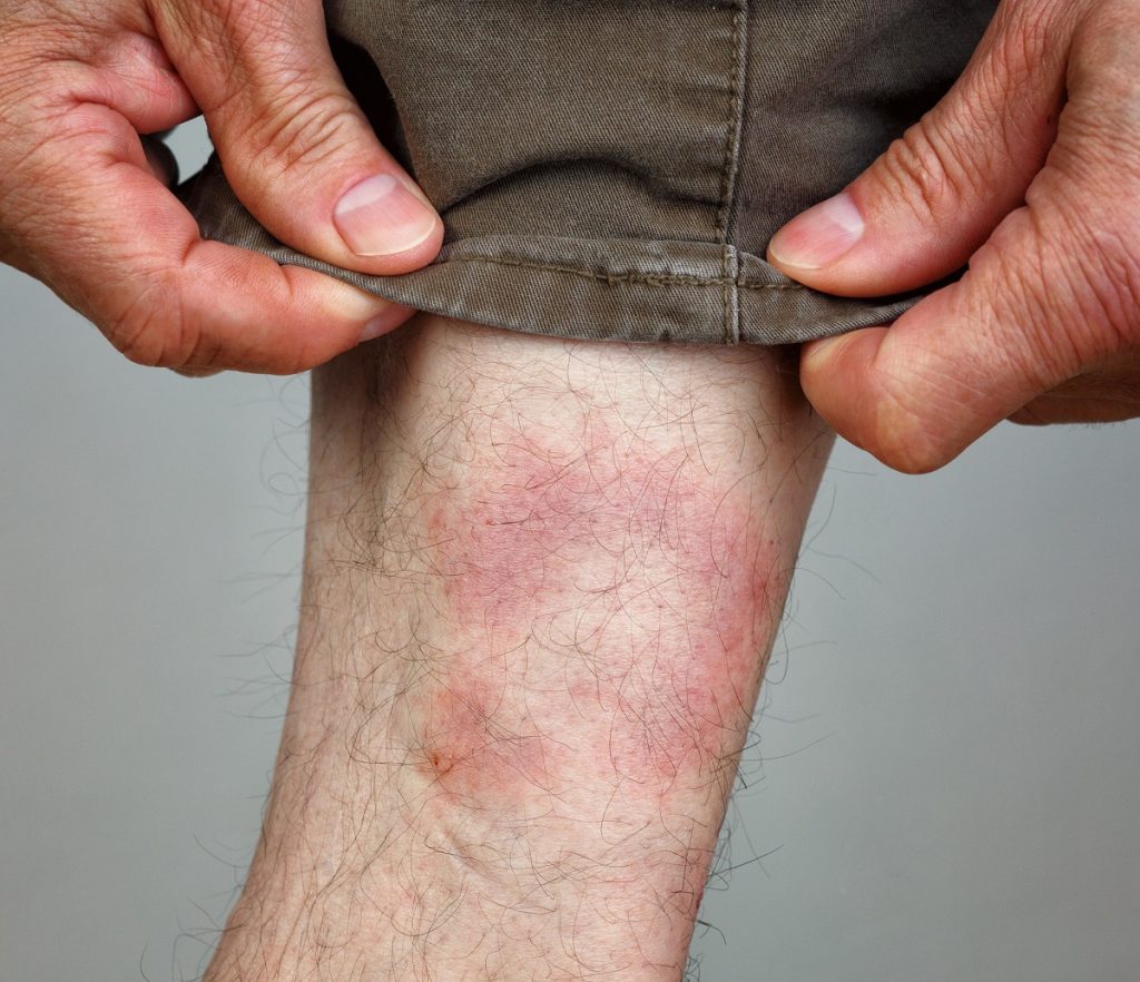
Lyme disease, a disease transmitted when deer ticks feed on infected animals like deer and rodents, and then bite humans, impacts nearly half a million individuals in the U.S. annually.
Even in acute cases, Lyme can be devastating; but early treatment with antibiotics can prevent chronic symptoms like heart and neurological problems and arthritis from developing.
Scientists from Northwestern University have identified that piperacillin, an antibiotic in the same class as penicillin, effectively cured mice of Lyme disease at 100-times less than the effective dose of doxycycline, the current gold standard treatment.
At such a low dose, piperacillin also had the added benefit of “having virtually no impact on resident gut microbes,” according to the study, in the journal Science Translational Medicine.
Doxycycline and other generic antibiotics, on the other hand, wreak havoc on the microbiome, killing beneficial bacteria in the gut and causing troubling side effects even as it kills the Borrelia bacteria that causes Lyme.
In addition to its negative impact on the gut, doxycycline also fails to help between 10 and 20% of individuals who take it, and it is not approved for use in young children — who are at the highest risk of tick bites, and therefore, of developing Lyme.
More effective, or at least more specific, treatment options are needed as climate change extends tick seasons and Lyme becomes more prevalent.
The need for customized medicine
“Powerful, broad-spectrum antibiotics that kill extracellular bacteria are seen as the most effective medication because physicians want to just kill the bacterium and don’t care how,” said Brandon L. Jutras, who led the research.
“This is certainly a reasonable approach, but I think the future for Lyme disease patients is bright in that we are approaching an era of customized medicine, and we can potentially create a particular drug, or a combination to treat Lyme disease when other fail. The more we understand about the various strains and species of Lyme disease-causing Borrelia, the closer we get to a custom approach.”
Jutras is an associate professor in the microbiology-immunology department of Northwestern University Feinberg School of Medicine, and a member of Northwestern’s Center for Human Immunobiology.
Jutras’s lab was recently named a Phase 3 winner in LymeX Diagnostics, the Steven & Alexandra Cohen Foundation’s $10 million competition to accelerate the development of Lyme disease diagnostics, and in 2021 he won the Bay Area Lyme Foundation Emerging Leader Award.
Piperacillin has already been FDA-approved as a safe treatment for pneumonia.
To reach the conclusion that the penicillin relative would be the most effective and targeted treatment, the team screened nearly 500 medicines in a drug library, using a molecular framework to understand potential interactions between antibiotics and the Borrelia bacteria.
Once the group had a short list of potentials, they performed additional physiological, cellular and molecular tests to identify compounds that did not impact other bacteria.
Prevents bacteria from growing
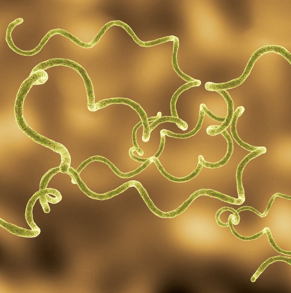
They found that piperacillin exclusively interfered with the unusual cell wall synthesis pattern common to Lyme bacteria, preventing the bacteria from growing or dividing and ultimately leading to its death.
Historically, piperacillin has been administered as part of a two-drug cocktail to treat severe strep infections because strep can break down beta-lactams (piperacillin’s class of antibiotics) unless accompanied by tazobactam, which is an inhibitor of the enzyme that inactivates piperacillin.
Jutras wondered if using the same two medications, rather than piperacillin alone, would be a more effective bacteria killer.
“Bacteria are clever,” Jutras said. “Strep and some other bacteria combat antibiotics by secreting beta-lactamases that inactivate piperacillin. We found the approach is totally irrelevant in the context of Lyme disease and another way that makes piperacillin more specific. Adding the beta-lactamase inhibitor doesn’t improve the therapy because Lyme Borrelia don’t produce beta-lactamase, but the cocktail does negatively impact the microbiome by becoming more broadly functional against beneficial residents.”
The study was supported by the Bay Area Lyme Foundation and United States Department of Agriculture (VA-160113), the Dennis Dean Research Grant (Virginia Tech), the National Institutes of Allergy and Infectious Disease (R01AI173256, R01AI178711), the Steven & Alexandra Cohen Foundation and the Global Lyme Alliance.
Click here for more about the study.
SOURCE: Northwestern University
_______________
**Comment**
A few points:
- Early treatment CAN prevent neurological problems, arthritis, & other chronic symptoms, but fails to do so in a subset of patients.
- Doxycycline does has a negative impact upon the gut, but far more than 10-20% go on to suffer long-term symptoms (chronic Lyme), with one researcher estimating the percentage to be more like 60%. A little factoid: the current research which comes up with 10-20% doesn’t include patients who are diagnosed and treated late, and this is somewhere between 30-40% of patients!
- Independent research has proven the climate has nothing to do with tick and disease proliferation. Further, the entire climate narrative is fraught with fraud and deceit and many experts continue to state there is no climate crisis, atmospheric CO2 emissions can not cause ‘global warming’, and that green energy policies have made the climate worse. Researchers really need to cease and desist with the climate mantra.
- But this ‘tell’ reveals that those doling out federal research grants hold all the cards, and researchers know they must comply with the narrative to get the dollars. These same public health ‘experts’ and politicians also own patents on the very things (drugs, tests, vaccines, etc) they are entrusted to protect the public from as well as set treatment guidelines.
- Researcher Kim Lewis out of Northeastern University has also identified compounds that are highly active and selective against Lyme disease in the mouse model. Going all the way back to 2015, he found hygromycin A to be highly effective against Lyme, yet here we are in 2025 with nadda.
- Lewis also proved what Dr. Burrascano clinically discovered – that by treating with antibiotics for a period and then stopping for a period (cycling) – if they did this four times, they discovered no bacteria in the petri dishes.
- Burrascano and Dr. Alan McDonald also proved patients can test negative but still be actively infected as well as the fact that dosage makes a difference as well. Mainstream research and medicine are clueless about these nuances and just continue to use a completely antiquated and faulty paradigm.
- This is why I hold little hope in any research that is federally funded. While advocates continue to bemoan lack of federal funding, I say good riddance. Nothing good ever comes from that quarter anyway. As long as federal funding is involved, the fraudulent Lyme narrative will taint everything that is done.
- Further, the federal government is complicit in tick research that purposely weaponized ticks to deliver deadly bacteria to be incapacitating and dropped them out of airplanes. Hello!
- Don’t believe me? Listen to Willy Burgdorfer, the “discoverer” of Lyme disease himself:
“The controversy in Lyme disease research is a shameful affair. I say that because the whole thing is politically tainted. Money goes to people that have for the past 30 years produced the same thing. Nothing. Serology or serology plus has to be started from scratch with people that don’t know beforehand the results of their research.
BOOM.
Sadly, the current research above is taking the same old tack that people are simply struggling with inflammation (PTLDS) – not an active infection. While this is always true, it is often only a partial truth, with active infection being the driver to the inflammation. In other words, treat the infection and symptoms get better or go away entirely. If only inflammation is treated, symptoms will continue until the infection(s) is/are dealt with. And this brings up another point entirely dismissed by mainstream research and medicine: this is commonly a polymicrobial issue – meaning more than one infectious organism is involved requiring yet more savvy, complex treatments.
Until these issues are addressed, I don’t want another dime of my money going to the same people that have done nothing for the past 40 years.
For more:
http:// Approx. 3 Min
Could Piperacillin Be the Lyme Breakthrough We Need?
Dr. Danial Cameron
May 6, 2025