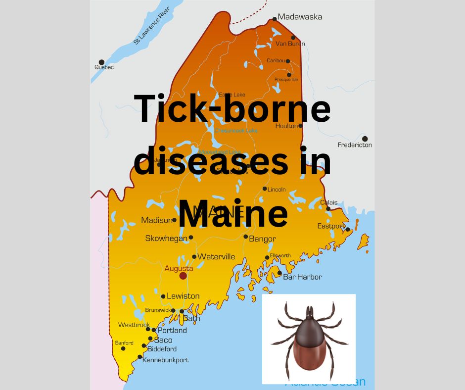
Anaplasma, Babesia odocoilei, and Lyme in Ticks – Found Widely Across Eastern Canada
https://www.jelsciences.com/articles/jbres1586.pdf
Tick-Borne Pathogens Anaplasma phagocytophilum, Babesia odocoilei, and Borrelia burgdorferi Sensu Lato in Blacklegged Ticks Widespread across Eastern Canada
John D Scott1 *, Elena McGoey2 and Risa R Pesapane2,3*
Corresponding author(s) John D Scott, Upper Grand Tick Centre, 365 St. David Street South, Fergus, Ontario N1M 2L7, Canada E-mail: jkscott@bserv.com DOI: 10.37871/jbres1586 Submitted: 13 October 2022 Accepted: 26 October 2022 Published: 27 October 2022 Copyright: © 2022 Scott JD, et al. Distributed under Creative Commons CC-BY 4.0
Abstract
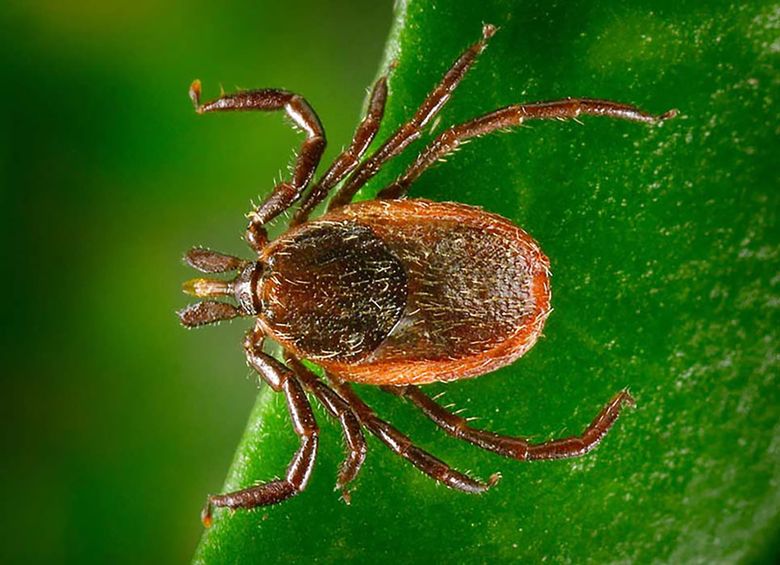
Blacklegged ticks, Ixodes scapularis, can transmit single or multiple infections during a tick bite. These tick-borne, zoonotic infections can become chronic and cause insidious diseases in patients.
In the present tick-pathogen study, 138 (48.9%) of 282 ticks collected from 17 sites in 6 geographic area in eastern Canada harbored various combinations of Borrelia burgdorferi sensu lato (Lyme disease), Anaplasma phagocytophilum (human anaplasmosis), and Babesia spp. (human babesiosis). Overall, 167 microbial infections were detected and, of these, 25 ticks had co-infections and two ticks had polymicrobial infections.
- the prevalence of Babesia spp. was 15.2%
- the ratio of Babesia odocoilei to Babesia microti was 41 to 1 with this sole B. microti being detected in Nova Scotia
- we provide the first documentation of B. odocoilei in the Maritimes
- Eastern Ontario had an infection prevalence for B. odocoilei of 25%―the highest among the areas surveyed in this study
- the predominant Babesia sp. was B. odocoilei
Based on our findings, health-care practitioners need to recognize that I. scapularis ticks removed from patients may be carrying multiple tick-borne pathogens. (See link for article)
_____________
For more:
- https://madisonarealymesupportgroup.com/2021/05/28/study-shows-babesia-odocoilei-is-pathogenic-to-humans/ Study found B. odocoilei in two of 19 participants. DNA amplicons from these two patients are almost identical matches with the type strains of B. odocoilei in GenBank. In addition, the same two human subjects had the hallmark symptoms of human babesiosis, including night sweats, chills, fevers, and profound fatigue. Based on symptoms and molecular identification, we provide substantive evidence that B. odocoilei is pathogenic to humans. Dataset reveals that B. odocoilei serologically cross-reacts with Babesia duncani.
- https://www.ncbi.nlm.nih.gov/pmc/articles/PMC3998201/ In Austria and Italy patients experienced a severe illness caused by EU1, a species closely related to B. odocoilei. InTaiwan it was (TW1) and in Korea (KO1). Human babesiosis is now reported from around the world. The study in this link states that reported human cases of babesiosis have been attributed, without strong molecular evidence to B. divergans: https://www.ncbi.nlm.nih.gov/pmc/articles/PMC3020600/
- https://madisonarealymesupportgroup.com/2019/04/01/bb-new-strain-of-babesia-found-in-tick-on-a-tropical-bird-in-canada/ First report of an Amblyomma inornatum tick cofeeding with a blacklegged tick, and first documentation of B. odocoilei in a tick parasitizing a bird.
- https://madisonarealymesupportgroup.com/2021/03/18/babesia-odocoilei-found-in-canadian-black-legged-ticks/ There are more than two Babesia spp. in North America that cause human babesiosis. This discovery signifies the first report of B. odocoilei causing human babesiosis.
- https://madisonarealymesupportgroup.com/2019/04/01/bb-new-strain-of-babesia-found-in-tick-on-a-tropical-bird-in-canada/
- https://madisonarealymesupportgroup.com/2021/10/06/detection-of-anaplasma-phagocytophilum-babesia-odocoilei-babesia-sp-borrelia-burgdorferi-sensu-lato-and-hepatozoon-canis-in-ixodes-scapularis-ticks-collected-in-eastern-canada/ H. canis was documented for the first time in Canada and, at the same time, demonstrates the first transstadial passage of H. canis in I. scapularis. Transstadial passage of Bbsl and B. odocoilei was also witnessed. A novel undescribed piroplasm (Babesia microti-like) was detected.