Archive for the ‘Ticks’ Category

Alpha-gal Syndrome – Symptoms, Diagnosis, & Treatment

https://www.lymedisease.org/alpha-gal-syndrome/

There is growing evidence that certain types of tick bites can trigger alpha-gal syndrome (AGS) a life-threatening allergy to red meat and meat-related products.

In some individuals, it appears tick bites can result in the sensitization to a carbohydrate known as galactose-alpha-1,3-galactose, or “alpha-gal” for short. This sugar molecule is found in most mammals you might be likely to eat, but not in fish or fowl.

Most recognized food allergies, such as to peanuts or shellfish, will prompt an immediate reaction after being consumed. That’s not the case with AGS, however, which can take up to eight hours (or even more) after exposure to produce a reaction.

Note: exposure to alpha-gal via inhalation, injected drugs or vaccines can cause an immediate reaction.

Alpha-gal syndrome - meats that contain alpha-gal

Examples of commonly consumed mammalian meats that contain alpha-gal include beef, pork, lamb, goat, venison and buffalo. Common foods that are derived from mammals include lard, milk, cream, ice cream, and cheese—although the majority of AGS patients do tolerate dairy products.

Personal products that use ingredients containing “hydrolyzed protein” (gelatin), lanolin, glycerin, collagen, or tallow are particularly problematic.

Additional products that can bring on an alpha-gal reaction are jello, gelatin capsules, certain medications, pig or cow heart valves, surgical mesh, certain vaccines and unlabeled “natural flavorings” in foods.

Some people with AGS also react to carrageenan, a common food additive made from red algae, which also contains alpha-gal. (So even being strictly vegan won’t necessarily protect you from AGS reactions.)

How are ticks involved in alpha-gal syndrome?

Alpha-gal meat allergy has been reported all over the world including Asia, Australia, Central America, Europe, Germany, Japan, South Korea, and the United States.

The tick species most often associated with Alpha-gal syndrome is the lone star tick

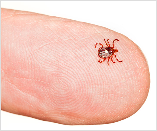
In the U.S., the tick species most often associated with AGS is the lone star tick (Amblyomma americanum) found throughout the South, East and parts of the Midwest. Recent research suggests that the blacklegged tick (Ixodes scapularis and Ixodes pacificus) may also be implicated in alpha-gal syndrome.

The Asian longhorned tick (Haemaphysalis longicornis), the primary trigger of AGS in Asia, has shown up in the US recently, but has yet to be implicated in AGS here. The Cayenne tick (Amblyomma cajennese) found in southern Texas and Florida has also been linked to AGS in Central America, but not yet in the U.S.

While no known pathogen has been linked to triggering AGS, more research is needed to understand the mechanism and the role that ticks play. Currently the thought is that the tick saliva plays a role in activating the allergy to alpha-gal.

Who’s at risk for AGS?

Alpha-gal syndrome is a much more common allergy in the U.S. today than it was a decade ago, with the number of laboratory-confirmed cases growing from 12 in 2009 to over 34,000 in 2019. Unfortunately, AGS has no insurance billing code (ICD code), nor is it a reportable illness to the CDC.

Experts agree alpha-gal syndrome is under-reported in geographic areas where tick bites are common.

Alpha-gal syndrome Lone Star Tick in the United States

Surveillance for IgE to alpha-gal. Percent positive rates are presented for IgE to alpha-gal within each of six regions in the United States, 2012-2013 (7300 samples). Diagonal white lines on the map represent the known geographic distribution of the lone star tick (Data and map, Viracor-IBT Laboratories; Tick Distribution, CDC).

For now, the biggest risk factor for AGS appears to be repeated bites by ticks that contain alpha-gal in their saliva and salivary glands. It is not understood why, but not everyone who is bitten by a tick containing alpha-gal will develop AGS.

While both children and adults can acquire AGS, most cases have been reported in adults.

Certainly, if a patient with recent tick exposure presents with sudden onset anaphylaxis and recurrent gastrointestinal symptoms, AGS should be considered.

Alpha-gal syndrome is a much more common allergy in the U.S. today than it was a decade ago, with the number of laboratory-confirmed cases growing from 12 in 2009 to over 34,000 in 2019.

What are the symptoms of alpha-gal syndrome?

The symptoms of alpha-gal syndrome are often delayed, making it much harder to pinpoint the trigger. Someone may wake up at 3 o’clock in the morning in the throes of serious allergic reaction, and have no idea it was brought on by a hamburger they ate the night before.

Symptoms can range from itching and stomach upset to breathing difficulty and full anaphylaxis. AGS reactions often start with itching of the palms of hands and soles of feet.

Common symptoms of AGS include:

  • 90% have skin symptoms: itching “pruritus,” flushing “erythema,” hives “urticaria” (swollen, pale red bumps or “wheals” on the skin), angioedema (swelling in deep layers below the skin)
  • 60% develop anaphylaxis (a potentially deadly reaction that can restrict breathing)
  • 60% have gastrointestinal symptoms (abdominal pain, diarrhea, acid reflux, cramping, vomiting)
  • 30-40% experience cardiac symptoms: rapid decrease in blood pressure (hypotension, POTS); palpitations (atypical chest symptoms)
  • 30-40% experience respiratory symptoms (wheezing, coughing, shortness of breath)
  • 20% of patients will have GI symptoms alone (may present like irritable bowel syndrome)
  • 3-5% develop mast cell activation syndrome
  • arthritis (rare)
  • mouth swelling, sores (rare)

How is AGS diagnosed?

If you experience symptoms after eating mammalian meat products, immediately notify your primary care physician or allergist. Unlike most tick-borne pathogens, the onset of AGS usually takes at least 4-6 weeks from the time of the tick bite. Complicating things further, about a third of patients do not recall a tick bite.

Your doctor should be able to determine if you have AGS based upon your clinical symptoms and a positive blood test: immunoglobulin E (IgE) to the oligosaccharide glactose-alpha-1,3 galactose (alpha-gal.)

In the U.S., Viracor is the main laboratory for AGS testing. The Viracor “specific IgE galactose-alpha-1,3-galactose” test can be taken at most commercial laboratories like Labcorp and Quest and shipped to Viracor.

Warning: The test for alpha-gal is often mistaken for “alpha-galactosidase” or “a-galactosidase A deficiency”—note these are the wrong tests! Because the test is so new, it is recommended to take the proper testing codes with you to the doctor and the laboratory. Click here to download and print a PDF on the proper testing codes for alpha-gal syndrome.

How is Alpha-gal syndrome treated?

There are currently no U.S. FDA-approved medications for the treatment of AGS. As with most allergies, the mainstay of management is avoidance of the allergen. Therefore, the best practice is to avoid exposure to:

  1. Mammalian meats
  2. Personal products containing mammalian derivatives
  3. Medical products containing mammalian proteins, derivatives or parts
  4. Medications containing mammalian proteins or derivatives

Knowing you must avoid mammalian products is only half the battle, as these products have worked their way into nearly every level of our modern life.

For instance, gelatin is the main ingredient of jellybeans, candy corn, marshmallows, puddings and the capsules of many medications. Chicken and turkey sausages may be stuffed in pork casings, lard (rendered pork fat) is found in many pre-made gravies, sauces, soups, candies, chips, fries, and more.

As with all serious allergies, it is important to have the proper diagnosis and be prepared with how to respond in the event of an emergency. Most allergists will recommend wearing a medical alert bracelet and carrying an EpiPen and an antihistamine with you at all times.

Avoiding alpha-gal hidden components

Mammalian proteins and parts can be found in many medications and medical products. . Because the source of many ingredients is not listed on product labels, your pharmacist may need to contact the manufacturer. Have your pharmacist ask specifically if it contains galactose-alpha-1,3-galactose, alpha-gal, mammalian meat, or any animal by-products.

Common sources of alpha-gal include:

  1. heart valve replacement derived from pig or cow,
  2. monoclonal antibodies (cetuximab)
  3. vaccines (zostavax, MMR and some flu),
  4. pancreatic enzyme replacement therapy,
  5. thyroid hormone replacement,
  6. fillers in medications (magnesium stearate, stearic acid, lactic acid, glycerin, gelatin, lactose)
  7. antivenom,
  8. protein powders,
  9. vaginal capsules
  10. heparin

Alpha-gal & co-infections

Ticks that carry alpha-gal are known to carry many other pathogens that can be simultaneously transmitted to humans. It is possible to acquire any of these other tick transmitted diseases and also have alpha-gal syndrome. It is also possible to have AGS alone.

Ticks that carry alpha-gal are known to carry many other pathogens

The lone star tick, the primary source of AGS in the U.S., is known to transmit the following diseases:

  • human monocytotropic ehrlichiosis (HME)
  • ehrlichiosis (Ehrlichia chaffeensis, Ehrlichia ewingii, and Panola Mountain ehrlichia)
  • Rocky Mountain spotted fever (RMSF)
  • tularemia (Francisella tularensis)
  • Heartland virus
  • Bourbon virus
  • Q fever
  • tick paralysis
  • STARI, an illness similar to Lyme disease, caused Borrelia lonestari
With alpha-gal recently discovered in blacklegged ticks, we may also begin to see an increase in AGS in patients with Lyme disease, anaplasmosis, babesiosis, ehrlichiosis, relapsing fever borreliosis, Powassan virus disease, and other diseases transmitted by these ticks.

How to prevent alpha-gal syndrome

For now, the best way to avoid getting AGS is to avoid tick bites. This means wearing tick repellent when working, hiking or playing in grassy or wooded areas where ticks are found. Protecting your pets and doing thorough tick checks after being outdoors is helpful.

If you are bitten by a tick, we suggest following these eight steps.

What to do if you have alpha-gal syndrome?

Learning you have an allergy to all mammalian products can be overwhelming. Because this is such a newly discovered condition there are few resources available.

When it comes to making medical decisions, it’s important to have a knowledgeable provider who understands the risks versus benefits of certain medications and procedures. Vaccines that contain gelatin are one of the riskier products, but if you need a rabies shot, for instance, your doctor may determine the benefits outweigh the risks and take the necessary steps to mitigate the adverse effects.

To learn more about the history, symptoms and how to diagnose alpha-gal syndrome listen to this interview with Dr. Scott Commins, of the University of North Carolina.

Additional help can be found at:

References:

  1. CDC | Alpha-gal allergy
  2. HHS | Alpha-Gal Syndrome Subcommittee Report to the Tick-Borne Disease Working Group
  3. Commins SP, Satinover SM, Hosen J, Mozena J, Borish L, Lewis BD, Woodfolk JA, Platts-Mills TA. (2009) Delayed anaphylaxis, angioedema, or urticaria after consumption of red meat in patients with IgE antibodies specific for galactose-alpha-1,3-galactose. J. Allergy and Clin Immunol 123(2):426-33. doi: 10.1016/j.jaci.2008.10.052.
  4. Commins, S. P., James, H. R., Kelly, L. A., Pochan, S. L., Workman, L. J., Perzanowski, M. S., Kocan, K. M., Fahy, J. V., Nganga, L. W., Ronmark, E., Cooper, P. J., & Platts-Mills, T. A. (2011). The relevance of tick bites to the production of IgE antibodies to the mammalian oligosaccharide galactose-α-1,3-galactose. J. Allergy and Clin Immunol, 127(5), 1286–93.e6. DOI: https://doi.org/10.1016/j.jaci.2011.02.019
  5. Commins SP (2020) Diagnosis & management of alpha-gal syndrome: lessons from 2,500 patients, Expert Review of Clinical Immunology, 16:7, 667-677, DOI: 10.1080/1744666X.2020.1782745
  6. Fiocchi A, Restani P, Riva E, Qualizza R, Bruni P, Restelli AR, Galli CL. (1995)  Meat allergy: I–Specific IgE to BSA and OSA in atopic, beef sensitive children. J Am Coll Nutr. 14(3):239-44. doi: 10.1080/07315724.1995.10718502. PMID: 8586772.
  7. Hamsten C, Tran TAT, Starkhammar M, Brauner A, Commins SP, Platts-Mills TAE, van Hage M. (2013) Red meat allergy in Sweden: association with tick sensitization and B-negative blood groups. J. Allergy and Clin Immunol. 132(6):1431-1434. doi: 10.1016/j.jaci.2013.07.050. Epub 2013 Oct 4. PMID: 24094548; PMCID: PMC4036066.
  8. Kuehn BM. (2018) Tick Bite Linked to Red Meat Allergy. JAMA. 23;319(4):332. doi: 10.1001/jama.2017.20802. PMID: 29362779.
  9. Mullins RJ, James H, Platts-Mills TA, Commins S.(2012) Relationship between red meat allergy and sensitization to gelatin and galactose-α-1,3-galactose. J. Allergy and Clin Immunol. 129(5):1334-1342.e1. doi: 10.1016/j.jaci.2012.02.038. Epub 2012 Apr 3. PMID: 22480538; PMCID: PMC3340561.
  10. Platts-Mills, TAE, Schuyler, AJ,Commins,SP, et. al ( 2018) Characterizing the Geographic Distribution of the Alpha-gal Syndrome: Relevance to Lone Star Ticks (Amblyomma americanum) and Rickettsia. J. Allergy and Clinical Immun 141;2. DOI: https://doi.org/10.1016/j.jaci.2017.12.470
  11. Wilson JM, Schuyler AJ, Workman L, Gupta M, James HR, Posthumus J, McGowan EC, Commins SP, Platts-Mills TAE.  (2019) Investigation into the α-Gal Syndrome: Characteristics of 261 Children and Adults Reporting Red Meat Allergy. J. Allergy and Clin Immunol Pract. 7(7):2348-2358.e4. doi: 10.1016/j.jaip.2019.03.031.

________________

**Comment**

I feel compelled to share the experiences of others as often it’s the only way we move forward in ‘Lymeland.’ In this case, many patients have been using ivermectin and fenbendazole with success. Make sure to work with your practitioner who knows everything you are taking as there can be synergystic effects and counter-indications for taking certain things together and/or due to medications/supplements for your pre-existing conditions. As always: this is never intended as medical advice or diagnosis.

In this case, a patient followed the advice of Dr. Makis and did two weeks of ivermectin and fenbendazole and then waited two weeks. She then consumed a 100% beef hamburger with no reactions. Prior to this treatment her life was extremely limited in that she couldn’t go out to eat or even attend family gatherings as she suffered with violent vomiting and anaphylaxis after ingestion, topical application, and even inhalation of any animal product.

While she does not mention specific dosages, the treatment was based upon Dr. Makis’ success treating Lyme disease in a similar fashion but using ivermectin and doxycycline. The protocol for that is

Ivermectin Cream applied topically on the tick bite

Doxycycline (100mg, 7 days a week for 30-60 days)

Ivermectin (1mg per 1kg body weight, 7 days a week for 30-60 days)

The rest of this protocol may also be beneficial, but not crucial for Lyme and Lone Star:

  • Tocotrienol and Tocopherol forms (all 8) of Vitamin E (400-800mg per day, 7 days a week). A product called Gamma E by Life Extension or Perfect E are both great.
  • Bio-Available Curcumin (600mg per day, 2 pills per day 7 days a week). A product called Theracurmin HP by Integrative Therapeutics is bioavailable.
  • Vitamin D (62.5 mcg [2500 IU] seven days a week).
  • CBD oil (1-2 droppers full [equal to 167 to 334 mg per day] under the tongue, 7 days a week) CBD-X: The most potent full spectrum organic CBD oil, with 5,000 milligrams of activated cannabinoids and hemp compounds CBD, CBN & CBG per serving.
  • Fenbendazole (300mg, 7 days a week) or in the case of severe turbo cancers up to 1 gram — for prophylaxis one 150mg tablet once or twice per week
  • Hydroxychloroquine (10mg/kg/day 7 days a week) – for prophylaxis one 200mg tablet once or twice per week
  • ImmunX immune support which also greatly increases the bioavailability of both Fenbendazole and Hydroxychloroquine (2 capsules per day)  for prophylaxis 2 capsules per day
  • Removing sugars and carbohydrates (cancer food) from your diet and replacing table sugar with a zero glycemic index, zero calorie, keto friendly rare sugar like AlluX

Predicting Current & Future Distribution of Western Black-legged Tick Across the Western US

https://journals.plos.org/plosone/article?id=10.1371/journal.pone.0244754

Predicting the current and future distribution of the western black-legged tick, Ixodes pacificus, across the Western US using citizen science collections

Published: January 5, 2021

https://doi.org/10.1371/journal.pone.0244754

Abstract

In the twenty-first century, ticks and tick-borne diseases have expanded their ranges and impact across the US. With this spread, it has become vital to monitor vector and disease distributions, as these shifts have public health implications. Typically, tick-borne disease surveillance (e.g., Lyme disease) is passive and relies on case reports, while disease risk is calculated using active surveillance, where researchers collect ticks from the environment. Case reports provide the basis for estimating the number of cases; however, they provide minimal information on vector population or pathogen dynamics. Active surveillance monitors ticks and sylvatic pathogens at local scales, but it is resource-intensive. As a result, data are often sparse and aggregated across time and space to increase statistical power to model or identify range changes. Engaging public participation in surveillance efforts allows spatially and temporally diverse samples to be collected with minimal effort. These citizen-driven tick collections have the potential to provide a powerful tool for tracking vector and pathogen changes. We used MaxEnt species distribution models to predict the current and future distribution of Ixodes pacificus across the Western US through the use of a nationwide citizen science tick collection program. Here, we present niche models produced through citizen science tick collections over two years. Despite obvious limitations with citizen science collections, the models are consistent with previously-predicted species ranges in California that utilized more than thirty years of traditional surveillance data. Additionally, citizen science allows for an expanded understanding of I. pacificus distribution in Oregon and Washington. With the potential for rapid environmental changes instigated by a burgeoning human population and rapid climate change, the development of tools, concepts, and methodologies that provide rapid, current, and accurate assessment of important ecological qualities will be invaluable for monitoring and predicting disease across time and space.

_____________________

**Comment**

Authors continue the debunked theory that the climate is behind tick proliferation.  Repeating something over and over does not make it true. This study was a complete waste of time and money and does nothing to help suffering patients.  Ticks are showing up worldwide in areas they shouldn’t be carrying pathogens they shouldn’t have.  Move on.  There’s much bigger fish to fry!

For more:  

Understanding Mycoplasma: Symptoms, Testing, & Treatment

https://rawlsmd.com/health-articles/mycoplasma-the-most-common-lyme-coinfection

Understanding Mycoplasma: Symptoms, Testing, and Treatment | RawlsMD

Mycoplasma: The #1 Lyme Coinfection + How to Outsmart It

by Dr. Bill Rawls
Updated 12/9/20

Mycoplasma is the stealthiest of all stealth microbes. It may be a major player in many chronic diseases associated with aging, but remarkably, most people — including most doctors — have limited awareness of it.

If you have Lyme disease, fibromyalgia, chronic fatigue syndrome, autoimmune disease, or possibly any other chronic illness, however, mycoplasma is a microbe you should know about.

Mycoplasma: The Master Manipulator

Mycoplasma is a parasite, meaning it can’t live without a host. And it’s the smallest of all bacteria: 4,000 of them can fit inside a single red blood cell in your body. By comparison, only 10-15 average-sized bacteria would fit in the same cell.

Unlike other bacteria, mycoplasmas don’t have a protective cell wall, creating an interesting survival strategy: They can shape-shift and fit into areas where other bacteria can’t go. For example, it also allows them to slip inside cells of the host. The lack of a cell wall makes mycoplasma resistant to some commonly prescribed classes of antibiotics like penicillins, which normally work by interrupting a bacteria’s cell wall so that when the bacteria divides, it falls apart.

More than 200 known types of mycoplasma (and probably many yet to be discovered) can infect animals and plants. There are at least 23 different varieties of mycoplasma that can infect humans. A few of them are considered harmless normal flora, but most have the potential to cause disease.

image split in three: tick, flea, and mosquito

Mycoplasma is spread by biting insects (ticks, mosquitoes, fleas, biting flies), sexual contact, contaminated food, and airborne droplets. Most everyone has been exposed to some form of mycoplasma. Several mycoplasma species have been closely associated with many chronic degenerative diseases like multiple sclerosis and Alzheimer’s disease, according to publications like the International Reviews of Immunology and the British Journal of Medical Practitioners, respectively.

Even beyond its tiny size, shape-shifting qualities, and proliferous nature, mycoplasma is a master at manipulating and outmaneuvering the host’s immune system. Half of its genetic makeup is devoted to that exclusive purpose.

While it has little ability to cause direct harm, it can use the host’s immune function to its advantage: Mycoplasma generates chronic low-grade inflammation and steals vital nutrients from the body.

In fact, everything that this stealthy microbe needs for survival — vitamins, minerals, fats, carbohydrates, and amino acids — must be scavenged from the host; it makes nothing itself. Mitochondria, which are the energy powerhouses of cells, are prime targets to sustain the microorganism, which helps explain why fatigue is always a factor in mycoplasma infections.

Mycoplasma favors infecting the cells of tissues that line different areas of the body. Common sites of infection include:

  • Nasal passages
  • Sinuses
  • Lungs
  • Lining of the intestinal tract
  • Genital tract
  • Vesicles inside the brain
  • Synovial lining of joints

They also commonly infect white blood cells, red blood cells, and brain tissue. Different mycoplasma has a preference for certain tissues, but all mycoplasma species possess the ability to infect any type of tissue and all organ systems.

The most common mycoplasma, Mycoplasma pneumoniae, has a preference for lung tissue. Initial infection with M. pneumoniae typically causes pharyngitis (sore throat), cough, fever, headache, malaise, runny nose — all the common symptoms of a basic upper respiratory infection.

man wrapped in blanket, coughing

If the person’s immune system is not full strength, the infection can progress to bronchitis and even pneumonia (about 20% of pneumonias). The type of pneumonia caused by mycoplasma, often called “walking pneumonia,” is rarely severe enough to result in hospitalization, though it can drag on for weeks or even months.

But even when those respiratory symptoms are cleared, it may not be the end of the story. That’s because after mycoplasma enters the body, it also infects white blood cells. And once inside a white blood cell, it can be carried to all parts of the body, infecting tissues and organs.

The potential for widespread infection is very much influenced by the status of the host’s immune function. If immune function is optimal, the microbe is contained after the initial infection, and no long-term harm occurs. Approximately 30-70% of people carry at least one species of mycoplasma without having symptoms. It essentially becomes like normal flora of the microbiome, which are the non-threatening microbes found on the skin, in the gut, and body cavities.

But most mycoplasma species aren’t normal flora, and they are just waiting for an opportunity to gain a foothold. If immune function slips for whatever reason, chronic, systemic infection becomes possible. Mycoplasma begins stealing vital nutrients and causing a wide range of symptoms that are unrelated to the initial infection. The general breakdown of tissues by stealth microbes like mycoplasma accelerates the aging process and is likely a primary factor in many, if not most, chronic degenerative diseases.

Stealth Characteristics of Mycoplasma

Stealth microbes are a stronger force together than when alone. In other words, mycoplasma may not be a problem unless another stealth microbe (or microbes) is present. Lyme disease may be a good example of this phenomenon.

image split in half: borrelia and mycoplasma

Mycoplasma is a common Lyme coinfection: It’s present in 75% or more of Lyme disease cases. Mycoplasma is known to be carried and spread by ticks, but it is also possible that mycoplasma is already present in the body when a bite from a tickcarrying borrelia — the primary bacteria associated with Lyme — occurs.

Immune dysfunction caused by the new tick-borne infection or possible other coinfection allows mycoplasma to proliferate and cause multi-systemic symptoms throughout the body. Many symptoms that occur in Lyme disease can be caused by mycoplasma, too.

Body Systems Affected by Chronic Mycoplasma

woman in bed, face in hand, tired

Mycoplasma infection may be localized to certain areas of the body (such as the lungs), or it can be more widespread and systemic. Parts of the body where symptoms can manifest include:

  • Joints: Mycoplasma commonly infects the synovial lining of joints, the lining protecting the joints. Ninety percent of people with rheumatoid arthritis test positive for mycoplasma in the synovial fluid.
  • Muscles: Muscle pain from breakdown of muscle fibers is common with systemic mycoplasma infection.
  • Heart: Mycoplasma can lead to inflammation of the heart, such as endocarditis, myocarditis, pericarditis.
  • Nerves: Mycoplasma scavenges fats from the myelin sheath covering nerve tissue. Not surprisingly, mycoplasma (and other microbes, including chlamydiaand borrelia) has been linked to multiple sclerosis and other neurodegenerative diseases, including ALS (Mycoplasma fermentans is most common) and Parkinson’s disease.

    Nerve involvement can be associated with neuropathic pain like burning and tingling in the hands and feet. Brain inflammation, contributing to insomnia, brain fog, depression, and anxiety, is common with systemic mycoplasma infection.

  • Immune system: Mycoplasma is a top candidate for explaining autoimmunity; it stimulates host self-damage, and it can live inside cells while simultaneously turning off the ability of the immune system to recognize the cell as abnormal.
  • Lungs: Mycoplasma in the lungs contributes to respiratory symptoms like sore throat, cough, fever, headache, malaise, runny nose, bronchitis, and pneumonia.
  • Digestive tract: Intestinal mycoplasma infection destroys villi — fingerlike projections in the small intestine that aid food absorption — and compromises the intestinal barrier. This allows accelerated damage by lectins in grains (especially wheat), beans, soy, nightshade vegetables, and dairy.

    Mycoplasma may contribute to leaky gut, or increased intestinal permeability. Severe mycoplasma intestinal infection can lead to nutritional deficiencies and weight loss. Infection of the gastric mucosa (stomach lining) can cause chronic gastritis with nausea and stomach discomfort.

  • Ears: Mycoplasma infection has been associated with hearing loss and ringing in the ears.
  • Eyes: The eyes may be impacted by mycoplasma with such issues as conjunctivitis, eye swelling, and vision loss.
  • Reproductive system: Research suggests mycoplasma has been found in ovarian cancer tissue. It may also contribute to interstitial cystitis, a bladder condition marked by severe pain and urinary frequency.
  • Blood: Mycoplasma has been found in the bone marrow of children with leukemia.

Diagnosing Mycoplasma and the Limitations of Testing

When it comes to testing, PCR (polymerase chain reaction) is the most accurate method for testing mycoplasma. It’s cost-effective and evaluates for the presence of mycoplasma’s genetic material, a test that’s easy, sensitive, and quick test to obtain at most laboratories.

However, PCR testing has limits because it only tests for a handful of mycoplasma species and primarily focuses on diagnosing acute respiratory or genital mycoplasma infections — not chronic, low-grade infections.

Female forensic technician collecting biological specimen in DNA

Another problem with diagnosing mycoplasma is that conventional science does not recognize chronic mycoplasma infections as being significant. Even though mycoplasma is commonly found in association with chronic degenerative diseases, it’s also found in one-third to two-thirds of any population without causing symptoms. In other words, it is assumed that mycoplasma just happens to be there but isn’t really a contributing factor in disease.

This type of thinking is simply a reflection of not understanding how stealth microbes operate. Mycoplasma does not cause disease unless it has an opportunity to do so. Individuals with a healthy immune system can harbor mycoplasma and suffer few ill effects. If immune function is disrupted by environmental factors or a coinfection with other stealth microbes, however, mycoplasma can definitely contribute to chronic disease.

When testing for mycoplasma, it is best to order a complete PCR mycoplasma panel, which will include:

  • M. fermentans
  • M. genitalium
  • M. hominis
  • M. penetrans
  • M. pneumoniae
  • M. synoviae
  • Ureaplasma urealyticum

But these are only the commonly-known species of mycoplasma; other lesser-known species could also be present.

Another problem with testing is that other stealth microbes can be associated with chronic infections with similar symptoms of mycoplasma infection, adding confusion to the clinical picture of what’s making a person ill. The list of knowns includes:

  • Yersinia enterocolitica
  • Chlamydophila pneumoniae
  • Chlamydia trachomatis
  • Campylobacter jejuni
  • Babesia
  • Bartonella
  • Ehrlichia
  • Anaplasma

Laboratories that test for mycoplasma include Medical Diagnostic Laboratories (MDL)and Armin Labs. Your healthcare provider may have additional recommendations for you.

But complete testing for the full range of all stealth microbes can cost hundreds or even thousands of dollars. Possibly the best course of action is assuming mycoplasma and other stealth microbes are there.

Stealth microbes only cause problems when immune function is suppressed. Addressing the causes of the underlying chronic immune dysfunction that allowed mycoplasma to flourish in the first place is the most effective solution for overcoming chronic infections.

Conventional Medical Solutions

The nature of mycoplasma makes it very resistant to conventional therapies. Many antibiotics target cell walls; since mycoplasma doesn’t have one, several classes of antibiotics are ineffective against the microbe. Some other antibiotics (doxycycline, erythromycin, clarithromycin, or azithromycin), block internal functions of bacteria and have some activity against mycoplasma, but activity is limited by the fact that mycoplasma bacteria only live inside cells where antibiotics have minimal penetration.

When it comes to chronic mycoplasma infections, the best approach is supporting the body’s natural healing potential.

Natural Solutions for Mycoplasma

Natural herbal therapy is the best therapeutic alternative for chronic mycoplasma. Herbs (especially medicinal mushrooms) work by:

  • Suppressing cytokine cascades
  • Reducing inflammation
  • Restoring normal immune function
  • Suppressing a wide range of covert pathogens

Consider the following herbs to get you started:

Cordyceps mushroom

Cordyceps (Cordyceps sinensis)

Native to Tibet, cordyceps is a medicinal mushroom that reduces cytokines and normalizes immune system functions. It is highly protective of cells, which reduces invasion by microbes.

Suggested dosage: 1-3 grams (1,000-3,000 mg) of whole mushroom cordyceps powder or 400-800 mg extract (standardized to >7% cordyceptic acid is preferred) two to three times daily.

Side effects: Mild nausea can occur, but in general, side effects are rare, even with higher doses. Allergic reactions are rare.

Chinese Skullcap purple flowers

Chinese Skullcap (Scutellaria baicalensis)

When combined with other herbs, Chinese skullcap has potent synergist properties. Additionally, it has strong antimicrobial and immunomodulating properties that are beneficial for suppressing mycoplasma and protecting tissues and organs infected with the microbe.

Suggested dosage: 400-1,000 mg two to three times daily. Root extract standardized to >30% baicalin is preferred. Note: American skullcap does not offer the same antimicrobial properties and should not be substituted.

Side effects: Gastrointestinal upset can occur, but side effects tend to be rare, even at high doses.

white Bidens flowers

Bidens (Bidens pilosa)

The herb offers potent antimicrobial and anti-inflammatory properties against mycoplasma, affecting mucous membranes of the body.

Suggested dosage: Bidens is most potent when prepared as an alcohol tincture. The dose may vary depending on the company, but tinctures are an excellent way to begin at a low dose and increase drops as tolerated.

Side effects: Some plants can be contaminated with heavy metals, so make sure you purchase the product from a reputable company that takes steps to minimize exposure. You should not take this plant if you are diabetic, as it can cause fluctuations in blood glucose or insulin levels.

Houttuynia white flower

Houttuynia (Houttuynia cordata)

Native to India and Nepal, houttuynia is a potent antiviral with activity also against mycoplasma.

Suggested dosage: The dose may vary depending on a company’s preparations.

Side effects: The herb can have a fishy smell but is otherwise well tolerated.

budding Anamu stem

Anamu (Petiveria alliacea)

Found in tropical, Amazonian regions of Central and South America, anamu offers excellent antimicrobial coverage for mycoplasma.

Suggested dosage: The daily dose of powdered herb is 1,000-2,000 mg twice daily.

Side effects: Note that anamu will give urine and feces a strong garlic-like odor. Generally, the herb is safe and well-tolerated, but it should be avoided in pregnancy.

Mullaca leaf berry

Mullaca (Physalis angulata)

Mullaca is another Amazonian herb with antimicrobial qualities to fight mycoplasma, and it works well as a complement to anamu. It can be found online as a loose powder (add it to smoothies or make your own capsules) or a tincture.

Suggested dosage: The daily dose for powdered herb is 1,000-2,000 mg twice daily.

Side effects: The herb is generally regarded as safe, however, it should be avoided during pregnancy or breastfeeding.

The Bottom Line

In addition to herbal therapy, the optimal path to recovery from chronic mycoplasma involves eliminating artificially-processed foods in favor of whole, nutrient-dense meals, reducing exposure to toxins, and managing chronic stress — all of which disrupt immune function and pave the way for stealth microbes to flourish. By minimizing these factors and implementing a comprehensive herbal therapy protocol, you can begin to curb chronic mycoplasma infections and support your body in the healing process.

Dr. Rawls is a physician who overcame Lyme disease through natural herbal therapy. You can learn more about Lyme disease in Dr. Rawls’ new best selling book, Unlocking Lyme.
You can also learn about Dr. Rawls’ personal journey in overcoming Lyme disease and fibromyalgia in his popular blog post, My Chronic Lyme Journey.

REFERENCES
1. K Waites and D Talkington, Mycoplasma pneumoniae and its Role as a Human Pathogen, Oct 2004, Clinical Microbiology Reviews
2. Hakkarainen, Turrunen, Miettinen, Kaitik, and Jannson, Mycoplasmas and Arthritis, Ann Rheu Dis, 1992, Oct 5 (11): p. 1170-1172
3. Baseman, Joel, et.al., Mycoplasmas: Sophisticated, Reemerging, and Burdened by Their Notoriety, CDC, Journal of Infectious Diseases, Vol 3, No.1, Feb 1997
4. Leslie Taylor, ND, Mycoplasmas – Stealth Pathogens (Review article), Jan 2001
5. Razin, Yogev, Naot, Molecular Biology and Pathogenicity of Mycoplasmas, Microbiol Mol Biol Rev, 1998, Dec; 62(4): p. 1094-1156
6. J Rivera-Tapia, N Rodriguez-Preval, Possible role of mycoplasmas in pathogenesis of gastrointestinal diseases, Rev Biomed 2006 17: 132-139
7. Berghoff, W, Chronic Lyme Disease and Co-infections: Differential Diagnosis, Open Neurol J., 2012, 6, p. 158-178
8. Gilroy, Keat, Taylor-Robinson, The Prevalence of Mycoplasma fermentans in patients with arthritides, Rheumatology, Vol 40 (12), p. 1355-1358
9. Zhang et al, Mycoplasma fermentans infection promotes immortalization of human peripheral blood mononuclear cells in culture, Blood 104 (13), p. 4252-4259
10. Walter Berghoff, Chronic Lyme Disease and Co-infections: Differential Diagnosis, Open Neurol J, 2012, 6: p. 158-178
11. Buhner S H, Healing Lyme Disease Coinfections, Healing Arts Press, Copyright 2013 http://www.cdc.gov/pneumonia/atypical/mycoplasma/index.html
12. Libbey JE, Cusick MF, Fujinami RS. Role of pathogens in multiple sclerosis. Int Rev Immunol. 2014;33(4):266-283. doi: 10.3109/08830185.2013.823422
13. BJMP 2009:2(4) 20-28
14. Huang S, Li JY, Wu J, Meng L, Shou CC. Mycoplasma infections and different human carcinomas. World J Gastroenterol. 2001;7(2):266-269. doi: 10.3748/wjg.v7.i2.266
_______________________
 
For more:  


Powassan Virus in Children

https://danielcameronmd.com/powassan-virus-in-children/

CASE REPORTS: POWASSAN VIRUS IN CHILDREN

powassan-virus-in-children

Powassan virus (POWV) is a tick-borne illness that can cause severe encephalitis. Animal studies have shown the virus can be transmitted to humans following a tick bite within 15 minutes. However, the cases described in a recent article “Powassan Virus Encephalitis Following Brief Attachment of Connecticut Deer Ticks” by Feder et al. “strongly suggest that infected ticks may also rapidly transmit POWV to people.” [1]

Powassan virus in children is not often reported. In this article, the authors describe two cases involving infants with tick bites who developed Powassan virus encephalitis. As the authors point out, their case report not only demonstrates that rapid transmission of POWV can occur, but it highlights the importance for parents/caregivers to follow tick bite prevention methods. In these cases, adults unknowingly exposed their children to ticks infected with Powassan virus.

Powassan virus encephalitis in two children

A 5-month-old child was hospitalized after experiencing fevers for two days, along with vomiting and facial twitching which progressed to seizures. Two weeks prior to the onset of symptoms, a tick was removed from the infant’s forehead. Test results for the Powassan virus were positive.

The second case involved a 2-month-old child who presented with a fever and listlessness for one day. “He then developed left sided focal seizures (rhythmic left arm twitching, facial deviation to the left, and tongue thrusting with lip smacking),” the authors write.

A tick was removed from the infant’s arm approximately two weeks before he was hospitalized. The parents believed the father or dog had brought a tick into the house following a walk outside. The tick was not engorged and had not fed for more than 24 hours, the authors report.

READ MORE: No neurologic damage in 3 children with Lyme disease and Powassan virus

“POWV infection was confirmed by a positive PRNT on both serum and CSF,” the authors explain.

“POWV infection of humans has been notable for the severity of both the acute disease and the long-term sequelae.”  In fact, chronic illness occurs in approximately 50% of patients, the authors report, with symptoms including hemiplegia [paralysis on one side of the body], wasting, personality changes, and headaches.

Adults unknowingly expose children

“The circumstances under which the 2 children reported here acquired infection require some comments that are pertinent to prevention of future cases,” the authors write.

Infants typically would not be exposed to tick bites. These cases demonstrate the importance in adopting tick bite prevention methods. Feder points out, “in both of our cases, parents presumably brought ticks into their homes after outdoor activities.”

In the first case, the father had been out walking in the woods and brushed off multiple ticks outside. He presumably brought a tick into the home. In the second case, a father had been walking outside with a dog. The family believed either the father or dog had brought the tick into the house.

Parents/caregivers should be educated about several preventative measures:

  • “Outdoor clothing may prevent access to skin, but the ticks may remain undetected and will crawl off the person when body heat is reduced, such as when a coat is removed.”
  • Parents should check for ticks more than once. “Because of searching for an optimal skin site, ticks will not immediately attach to a person.”
  • “Parents should be educated about the need to treat outdoor clothing with Permethrin, an effective mode of preventing tick bites … Contact with treated fabric will kill all ticks within 2 hours.”
  • “Parents should also be educated about the possibility that dogs could bring ticks into homes, and that these animals should be inspected after every outdoor exposure.”
  • “Most anti-tick preventives only work after a tick has attached to a dog [the tick needs to ingest the chemical], although there are collars that are impregnated with Permethrin or similar products that might repel or kill ticks.”

Editor’s Note:  Although the authors did not discuss treatment of the two infants, both recovered.

References:
  1. Feder HM, Telford S, Goethert HK, Wormser GP. Powassan Virus Encephalitis Following Brief Attachment of Connecticut Deer Ticks. Clin Infect Dis. 2020.

_____________________

**Comment**

Despite being told that all of this is “rare,” please remember that Powassan, along with numerous other tick-borne infections are not reportable illnesses; therefore, nobody has a clue about prevalence. Coppe Lab out of Wisconsin emphatically states Powassan is NOT rare:

For the last two years, Coppe Laboratories has dedicated a significant amount of time and resources to dispelling the myth that infection with Powassan virus, a virus transmitted by tick bite, is rare. The Centers for Disease Prevention and Control (CDC) reports only 100 cases of Powassan virus infection in the United States in the last 10 years. Indeed, that statistic gives the illusion that Powassan infection is rare. However, did you know that the only infections reported to CDC are those that are life-threatening, particularly cases causing severe inflammation of the brain like the case reported in LiveScience? Coppe has published three new papers in the last year that clearly show Powassan virus infection is not rare are at all,and until testing for this virus is included as part of tick-borne disease screening panels infections will continue to be underreported. Coppe’s Powassan Guide, which can be downloaded from the website, summarizes the findings from both tick and human Powassan prevalence studies, as well as defining the patient populations that would benefit most from Powassan testing.

For more:

Tick prevention:

https://madisonarealymesupportgroup.com/2019/04/12/tick-prevention-2019/

https://madisonarealymesupportgroup.com/2020/07/20/ticks-lyme-disease-information-for-families/

Remember, pets are called “Tick taxies” for a reason:  https://madisonarealymesupportgroup.com/2017/08/12/pet-owners-have-nearly-2-times-the-risk-of-finding-ticks/

Ticks Produce Antibiotic That Protects Them From Human Skin Bacteria

https://www.ucsf.edu/news/2020/12/419216/lyme-disease-ticks-produce-antibiotic-protects-them-human-skin-bacteria

Lyme Disease Ticks Produce Antibiotic That Protects Them From Human Skin Bacteria 

Gene Provides Protection During the ‘Really Risky Sport’ of Feeding on Blood

By Vicky Stein

Dec. 10, 2020

Ticks live dangerous lives, spending most of their time questing for a host across wildly different habitats and seasons. Once they encounter a reptile, bird, or a mammal like us, they become intimately connected with it – and all of its bacteria and viruses – for days on end. Though ticks are notorious for transmitting pathogens such as the Lyme disease bacterium, how does their immune system keep them safe from contracting pathogens themselves?

In a study published in Cell on Dec. 10, 2020, a research team led by UC San Francisco’s Seemay Chou, PhD, provides an answer to this mystery. The work, Chou said, reveals that ticks are exquisitely constructed blood-sucking machines, with immune systems specially tailored for this unique lifestyle. Their defense strategies are carried out both inside and outside their bodies, she said, killing even our resident microbes as they feed on us.

Five years ago, in work published in Nature, Chou and colleagues found a gene in tick DNA that produces a microbe-killing protein. In the new study, senior author Chou leveraged that discovery to show that, without the protection offered by this gene, ticks are vulnerable to infection with Staphylococcus, one of the most common types of “commensal” bacteria. These bacterial species carpet our skin surface, but generally don’t harm us.

“This is the first time anybody’s identified a natural pathogen of ticks, and established a mechanism for it,” said Chou, an assistant professor of biochemistry and biophysics at UCSF and Chan Zuckerberg Biohub Investigator whose work is supported by a Sanghvi-Agarwal Innovator Award. “Ticks pass more microbes to humans, livestock, and other animals than any other known arthropod, but now their own vulnerabilities are on the table.”

The tick gene in question, known as dae2, originally evolved in bacteria, where the protein it encoded worked as an offensive agent against other bacteria. Several hundred million years ago, right around the time that the ancestors of some of today’s ticks began feeding on blood, those ticks “stole” the gene, making it a part of their own genomes.

According to Chou, dae2 represents a rare example of so-called horizontal transfer of a gene from a bacterium to an animal, and the fact that this transfer occurred as blood feeding evolved might not be a coincidence.

“I’ve always wondered why blood-feeding is even a thing,” said Chou. Not only does blood take a lot of energy to process into useful food, but biting on and attaching to much larger animals “seems inherently like a really risky sport.” With a strong, dae2-enhanced immune system, she said, tick species could have flourished, expanding to fill their bloody ecological niche.

When she first began working with dae2 in Ixodes scapularis, the deer tick, Chou thought ticks’ acquisition of the gene must have something to do protection against tick-dwelling bacteria like B. burgdorferi, which causes Lyme disease in humans and other animals. In experiment after experiment, she and members of her lab tried to find a mechanism for the gene to inhibit this bacterium, until they had an epiphany.

It makes no sense for ticks to have acquired this immune effector to kill off the bacteria that it’s most notoriously known to stably associate with,” she said. So instead, the lab began the much more complicated process of looking for bacteria that ticks are not known to live with peaceably. When members of the lab, including co-first authors Beth M. Hayes, PhD, and Atanas D. Radkov, PhD, proposed exploring the idea that dae2 might protect against Staphylococcus bacteria, “I actually pooh-poohed the idea – I bet a beer against it,” said Chou.

But after the dae2 protein was introduced to a cloudy vial of cultured staph bacteria, Chou was shocked. “The tube went from murky to clear in like, a second,” she said. “I was kind of glad to have lost this bet. After two years of trying to figure out what was going on, it all started falling together.”

Now the team had a direction. As detailed in the Cell paper, the researchers embarked on a series of wide-ranging experiments, first comparing dae genes in a range of tick species with the bacterial tae genes from which they were originally derived. With these comparisons and high-resolution protein structures in hand, they used computer models of these proteins to compare their shape and orientation when they came in contact with molecules found in bacterial cell walls.

Next, they moved on to testing the proteins directly against actual molecules extracted from bacterial cell walls. While dae2 protein could quickly degrade this material, taken from common skin bacteria, Tae2 could not. Dae2 also killed a wide a range of bacteria, notably three very common species that are symbiotic partners of human skin.

The researchers then investigated whether dae2 could reach our skin. They found dae2 protein in tick salivary glands and saliva, and observed that, from there, the protein was transferred to the ticks’ blood meal hosts.

When Chou and the team cancelled out the effects of dae2 in a group of blood-fed ticks, using a technique known as RNA interference in some mice and immunizing other mice to the protein, they found higher levels of Staphylococcus bacteria than in ticks with the functioning protein. These ticks also stayed smaller and gained less weight than ticks with dae2.

“This is a new way of thinking about how ticks interact with microbes,” said Chou. Microbes borne by ticks cause disease in humans and animals worldwide, but that’s only half the story, she said. “Their commensal is our pathogen, and our commensal is their pathogen.”

Authors: Joining Chou, Hayes, and Radkov were Fauna Yarza, Sebastian Flores, Jungyun Kim, Ziyi Zhao, and Victoria Bowcut, all of UCSF; Katrina W. Lexa, of Denali Therapeutics, Liron Marnin and Joao H.F. Pedra of the University of Maryland School of Medicine; and Jacob Biboy and Waldemar Vollmer of Newcastle University.

Funding: The research was funded in part by grants from the NIH (R01AI132851, R01AI134696, R01AI116523), Research Councils UK (EP/T002778/1), the UCSF Program for Breakthrough Biomedical Research, and the Sandler Foundation. Additional support came from the Chan Zuckerberg Biohub, the Johnson & Johnson WiSTEM2D Award, the Pew Biomedical Research Foundation, and the Sangvhi-Agarwal Innovation Award. Yarza was supported by the National Science Foundation (1650113) and a grant to UCSF from the Howard Hughes Medical Institute through the James H. Gilliam Fellowships for Advanced Study program.

Disclosures: The authors declare no competing interests.

The University of California, San Francisco (UCSF) is exclusively focused on the health sciences and is dedicated to promoting health worldwide through advanced biomedical research, graduate-level education in the life sciences and health professions, and excellence in patient care. UCSF Health, which serves as UCSF’s primary academic medical center, includes top-ranked specialty hospitalsand other clinical programs, and has affiliations throughout the Bay Area.