
https://danielcameronmd.com/meningitis-and-radiculitis-lyme-disease/  Podcast Here

43-YEAR-OLD MAN WITH MENINGITIS AND RADICULITIS DUE TO LYME DISEASE

meningitis-lyme-disease

Hello, and welcome to another Inside Lyme Podcast. I am your host Dr. Daniel Cameron. In this episode, I will be discussing a unique case involving a 43-year-old man with neurological manifestations of Lyme disease including both meningitis and radiculitis.

The case was published in the journal Neurology International. [1] According to Dabiri and colleagues, the patient had a history of “scaly erythematous macular rash on his proximal medial upper and lower extremities.”

Within two weeks he presented with a broad range of symptoms “including cough, fever, anorexia, malaise, fatigue, myalgias, cervicalgia/neck stiffness with flexion and extension, mild photophobia, headache,”  the authors wrote.

The patient had extensive lab testing which revealed a mild abnormal liver function but no evidence of Lyme disease.  At the onset of symptoms, the patient refused to have a spinal tap.

Doctors presumed the man suffered from viral meningitis.

One month later, the patient developed progressive weakness, severe radicular lancinating pain, emotional lability along with depression and anxiety, an occasional action tremor in hands interfering with fine motor tasks, and tremor in his legs causing imbalance and instability.

Manifestations of the central nervous system (i.e, meningitis), as well as peripheral nervous system presentations (i.e., radiculitis) can occur in isolation or together.

Radiculitis or inflammation of the nerve root involving the peripheral nervous system (PNS) can lead to intractable pain, muscle denervation, and areflexia over one or a few adjacent dermatomes, wrote the authors.

At this point, results from a spinal tap were consistent with Lyme disease.  “A lumbar puncture was performed, and the patient’s cerebrospinal fluid (CSF) analysis showed lymphocytic pleocytosis with white blood cell count of 225 and elevated protein of 77 and decreased glucose 38,” the authors wrote.

The patient was treated with a 5-day course of doxycycline, followed by a month of intravenous ceftriaxone.

Approximately two weeks after starting treatment, the patient “noted his symptoms were significantly improved including resolution of the pain, weakness, constitutional and affective symptoms, while he still had some ambulatory difficulties.”

This podcast addresses the following questions:

  1. What is Lyme meningitis?
  2. What is Lyme radiculitis?
  3. Why is this case considered “unique”?
  4. CNS and PNS manifestations can occur in isolation or together?
  5. Can you discuss the patient’s symptoms of emotional lability, depression and anxiety?
  6. What is the significance of the rash?
  7. Initial testing for Lyme disease was inconclusive but follow-up tests were positive?
  8. Any significance to MRI and spinal tap results?
  9. What if the significance of a diagnosis of viral meningitis?
  10. What were the other symptoms that might have helped the diagnosis?
  11. Would clinical judgment to treat with antibiotics have been helpful?
  12. What are your thoughts regarding the course of treatment?
  13. Would it have been helpful to consider additional treatment for the remaining ambulatory difficulties?
    1. Thanks for listening to another Inside Lyme Podcast. You can read more about these cases in my show notes and on my website @DanielCameronMD.com. As always, it is your likes, comments, reviews, and shares that help spread the word about Lyme disease. Until next time on Inside Lyme.

Please remember that the advice given is general and not intended as specific advice as to any particular patient. If you require specific advice, then please seek that advice from an experienced professional.

Inside Lyme Podcast Series

This Inside Lyme case series will be discussed on my Facebook and made available on podcast and YouTube.  As always, it is your likes, comments, and shares that help spread the word about this series and our work. If you can, please leave a review on iTunes or wherever else you get your podcasts.

References:
  1. Dabiri I, Calvo N, Nauman F, Pahlavanzadeh M, Burakgazi AZ. Atypical presentation of Lyme neuroborreliosis related meningitis and radiculitis. Neurol Int. 2019 Dec 2;11(4):8318.
_____________________
https://madisonarealymesupportgroup.com/2019/03/17/first-case-of-b-corocidurae-in-native-european-presenting-as-meningitis-with-cranial-polyneuritis-cavernous-sinus-thrombosis/  European cases of B. crocidurae infection have been reported in travelers returning from endemic areas. We report the first autochthonous case in Europe of B. crocidurae infection, presenting as meningitis…..

https://www.hopkinslyme.org/news/treatment-delays-increase-risk-of-persistent-illness-in-lyme-disease/

Treatment Delays Increase Risk of Persistent Illness in Lyme Disease

Summary

In this retrospective study, one-third of Lyme disease patients report delayed treatment of greater than 30 days, and delayed treatment is associated with post-treatment Lyme disease (PTLD). Delays in treatment are shown to significantly impact patient outcomes and indicate that improved Lyme disease diagnosis and prevention strategies are needed.

Why was this study done?

The goals of the study were to analyze time-to-treatment in a sample of individuals treated for Lyme disease, including identifying risk factors for time to first medical contact and time under care. The study also evaluated associations between time-to-treatment and the development of post-treatment Lyme disease (PTLD). The study was done to determine if delays in treatment are significant to patient outcomes.

How was this study done?

A retrospective study was conducted of time-to-treatment among a general population sample of individuals treated for Lyme disease at Geisinger, a health system in Pennsylvania, the state with the most confirmed Lyme disease cases in the United States. Using electronic medical records and self-administered questionnaire data, respondents’ experiences were characterized by symptoms, care-seeking factors, diagnosis, and treatment. Associations between time-to-treatment and PTLD were examined, and risk factors were measured for delays in time to first medical contact and time under care.

Questionnaires were mailed to 5,314 adult patients who met previously described EHR-based criteria for Lyme disease between 2015 and 2017. Analyses focused on 778 respondents who reported that they were treated for Lyme disease within the past 5 years and reported a rash and/or a positive blood test for Lyme disease. Time-to-treatment was measured in days as the sum of two time windows: time to first medical contact and time under care. Logistic regression was used to evaluate factors associated with delayed time-to-treatment in each time window (>14 days vs. ≤14 days) and the association between total time-to-treatment (>30 days vs. ≤30 days) and PTLD.

What were the major findings?

In the sample of Lyme disease patients studied, delays in medical contact and care were identified in one-third of individuals and delays were associated with a higher risk for post-treatment Lyme disease. In the sample,

  • 25% had time to first medical contact greater than 14 days
  • 21% had time under care of a medical professional greater than 14 days
  • 31% had a total time-to-treatment greater than 30 days

Factors positively associated with delayed time to first medical contact included being uninsured and attributing initial symptoms to something other than Lyme disease. Diagnoses between November and April, and the absence of rash were positively associated with delays in contact and care. First medical contact in an emergency department or “other” setting showed delays in care.

Individuals whose treatment was delayed, defined as time-to treatment >30 days, had 2.26 times the odds of developing PTLD as those who were treated within 30 days of symptom onset.

What is the impact of this work?

Despite post-treatment Lyme disease occurring in an estimated 10-20% of Lyme disease cases, no peer-reviewed studies have evaluated the role of time-to-treatment in PTLD. This retrospective study indicates timely treatment may be important in preventing PTLD and other long-term consequences of Lyme disease.

Strategies to ensure more timely treatment of Lyme disease should include educational campaigns targeting patients and healthcare providers. Education could improve the recognition of the varied clinical presentations of Lyme disease and the erythema migrans rash and underscore the limitations of diagnostic tests. Although urgent care clinics can help patients be seen earlier, there is a clear need to improve disease recognition and diagnosis and reduce time-to-treatment in this setting. Another opportunity to reduce time-to-treatment is to build awareness among patients and medical providers of the risk of Lyme disease throughout the year in endemic regions.

Education, diagnostics, and prevention approaches are needed and should address the risk factors for treatment delays and aim to reduce both the time before and after contacting a medical professional.

This research was supported by:

funded by the Steven & Alexandra Cohen Foundation

Publication Information

Hirsch AG, Poulsen MN, Nordberg C, Moon KA, Rebman AW, Aucott JN, Heaney CD and Schwartz BS (2020) Risk Factors and Outcomes of Treatment Delays in Lyme Disease: A Population-Based Retrospective Cohort Study. Front. Med. 7:560018. doi: 10.3389/fmed.2020.560018

Johns Hopkins Lyme Disease Research Center

The Johns Hopkins Lyme Disease Research Center is focused on patient-based research in all manifestations of Lyme disease. Our goal is to translate our pioneering research into improved patient care, education, and health outcomes.

USE OF THIS SITE

All information contained within the Johns Hopkins Lyme Disease Research Center website is intended for educational purposes only. Physicians and other health care professionals are encouraged to consult other sources and confirm the information contained within this site. Consumers should never disregard medical advice or delay in seeking it because of something they may have read on this website.

_____________________

**Comment**

Although this study is helpful for demonstrating that delays in diagnosis and treatment are bad, there are a number of concerns:

  1. The continued use of outdated and stringent criteria such as the EM rash and blood serology testing. Reality has proven a huge subset of patients meet neither.
  2. Electronic medical records and self-administered questionnaire data are only as effective as the people are filling them out, the questions being asked, and the objectivity of the person compiling the data.
  3. The PTLD label is deceptive and assumes ongoing symptoms are not caused by persistent/chronic infections.

The major findings; however, were helpful in showing that more than half were not obtaining treatment until 14-30 days.  This is important in the light of this mouse study which demonstrated Bb in the brain within a week of infection.

The factors of being uninsured and uneducated on tick-borne illness led to delayed time to 1st medical contact as well as those being seen between Nov-April and the absence of rash (both of which really should be under the banner of being uneducated about tick-borne illness as you can be infected in any month and getting the rash is highly variable).

https://journals.plos.org/plospathogens/article?id=10.1371/journal.ppat.1009256

A murine model of Lyme disease demonstrates that Borrelia burgdorferi colonizes the dura mater and induces inflammation in the central nervous system

journal.ppat.1009256.g003

Fig 3. B. burgdorferi in the dura mater are extravascular and motile.  Multiphoton image of ex vivo dura mater from C3H mouse after infection with GFP-Bb_297 for 7 days. B. burgdorferi is shown in green; collagen (second harmonics) shown in blue. Imaging parameters: Wavelength = 910 nm, pixel resolution = 135. See S1 Movie for image series movie showing spirochete motility.  https://doi.org/10.1371/journal.ppat.1009256.g003

Timothy Casselli, Ali Divan, Emilie E. Vomhof-DeKrey, Yvonne Tourand, Heidi L. Pecoraro, Catherine A. Brissette

Published: February 1, 2021

https://doi.org/10.1371/journal.ppat.1009256


Abstract


Lyme disease, which is caused by infection with Borrelia burgdorferi and related species, can lead to inflammatory pathologies affecting the joints, heart, and nervous systems including the central nervous system (CNS). Inbred laboratory mice have been used to define the kinetics of B. burgdorferi infection and host immune responses in joints and heart, however similar studies are lacking in the CNS of these animals. A tractable animal model for investigating host-Borrelia interactions in the CNS is key to understanding the mechanisms of CNS pathogenesis. Therefore, we characterized the kinetics of B. burgdorferi colonization and associated immune responses in the CNS of mice during early and subacute infection. Using fluorescence-immunohistochemistry, intravital microscopy, bacterial culture, and quantitative PCR, we found B. burgdorferi routinely colonized the dura mater of C3H mice, with peak spirochete burden at day 7 post-infection. Dura mater colonization was observed for several Lyme disease agents including B. burgdorferi, B. garinii, and B. mayonii. RNA-sequencing and quantitative RT-PCR showed that B. burgdorferi infection was associated with increased expression of inflammatory cytokines and a robust interferon (IFN) response in the dura mater. Histopathologic changes including leukocytic infiltrates and vascular changes were also observed in the meninges of infected animals. In contrast to the meninges, we did not detect B. burgdorferi, infiltrating leukocytes, or large-scale changes in cytokine profiles in the cerebral cortex or hippocampus during infection; however, both brain regions demonstrated similar changes in expression of IFN-stimulated genes as observed in peripheral tissues and meninges. Taken together, B. burgdorferi is capable of colonizing the meninges in laboratory mice, and induces localized inflammation similar to peripheral tissues. A sterile IFN response in the absence of B. burgdorferi or inflammatory cytokines is unique to the brain parenchyma, and provides insight into the potential mechanisms of CNS pathology associated with this important pathogen.

___________________

**Comment**

Technically one could say this isn’t the brain.  It’s the outer layer called the meninges – of which, the dura mater is one layer.  Regardless, this inflammation caused by infections can cause swelling, pain, and so much more.  It’s also why some Lyme/MSIDS patients have been diagnosed with Chiari.

Important excerpts:

Overall, we report that B. burgdorferi routinely colonizes the meninges in laboratory mice during early and subacute infection, and induces similar localized inflammatory gene expression profiles as other peripheral tissues as well as histopathological changes.

Conclusion:

Overall, the findings reported in this study are significant, as the lack of a tractable animal model has hindered our understanding of host-pathogen interactions in the CNS during B. burgdorferi infection. Our results provide insight into potential mechanisms of CNS pathologies associated with Lyme disease, and describe a model system that will allow for future studies evaluating the bacterial, host, and environmental factors that can contribute to the severity of CNS involvement during B. burgdorferi infection. Such studies are critical for the development and implementation of novel prophylactic and therapeutic interventions for this important disease.

The discussion section mentioned something that’s always interested me: the location of the tick bite or injection site.  The authors state that although they were able to “readily culture spirochetes from the blood of all mice at day 7 post-infection, dura spirochetes were rarely detected in mice inoculated in the footpad, and spirochete burdens were dramatically reduced in mice inoculated in the dorsal lumbar skin compared to thoracic skin.”  Further, dissemination of Bb happens in more ways than via blood and that these other ways, such as through the lymphatic system, may contribute to increased early colonization of the dura mater in mice.  This was only true for early infection and by 28 days, Bb in the dura mater were comparable regardless of the inoculation site.

This article reveals what patients have been experiencing for decades: heads that feel as if they were going to explode.  I wondered if there would ever be a day without a headache.  I personally found that Minocycline was one of the most productive antibiotics for this.  


https://healthimpactnews.com/2021/the-shaky-science-behind-the-deadly-new-strains-of-sars-cov-2/  9-Minute Video Here

The Shaky Science Behind the “Deadly New Strains” of Sars-Cov-2

Feb. 18, 2021

by Rosemary Frei
Off-Guardian

ACCORDING TO WHAT WE HEAR FROM OFFICIALS AND THE MAINSTREAM MEDIA, THE NEW VARIANTS ARE THE MOST DANGEROUS AND UNPREDICTABLE BEINGS SINCE OSAMA BIN LADEN.

Everyone needs to stay safe from these invisible but murderously mighty microbes by shunning contact with the unwashed, unmasked and unvaccinated.

But is that drastic approach — which is accompanied by severe curtailment of civil liberties and constitutional rights — warranted?

It turns out that the case for the variants’ contagiousness and dangerousness centres largely on the theoretical effects of just one change said to stem from a mutation in the virus’s genes.

And, as I’ll show in this article, that case is very shaky.

I also have an accompanying nine-minute ‘explainer’ video (Highly recommend. Please see link at top of page)

That one change is known as N501Y — scientific shorthand for the substitution of one protein building block (amino acid) for another at position 501 in the part of the virus called the spike protein.

Specifically, position 501 lies in the portion of the spike protein that’s responsible for the intimate coupling between the virus and cells that lets the virus slip inside and multiply.

[Note that any such amino-acid switcheroo is correctly called a change, not a mutation. Mutations occur only in genes. For some reason many scientists and scribes who ought to know better are mistakenly calling N501Y and other amino-acid changes ‘mutations.’ ]

A very preliminary study published Dec. 22, 2020, suggested that N501Y also is present in the South African variant named 501Y.V2. And another very preliminary study, published January 12, 2021, asserted it was also present in the new strain emerging from the Brazilian jungle, dubbed P.1.

On top of that, the South African variant is being reported as evading immunity and B.1.1.7 sharing this escape route. And scientists are depicting new variants with N501Y on board as spreading very fast. Some say they make herd immunity impossible, so every single person on earth has to be vaccinated. The models also suggest B.1.1.7 is up to 91% deadlier than the regular novel coronavirus.

(Yet so far it seems the main basis for officials saying it’s more deadly is shown in the minutes of the Jan. 21, 2021 meeting of an influential UK committee called New and Emerging Respiratory Virus Threats Advisory Group [NERVTAG ]. There, they cite modeling papers which haven’t yet been published – which means that until they’re published there’s no way to check their work.)

THREE NON-PEER-REVIEWED THEORETICAL-MODELING PAPERS WHICH CATAPULTED VARIANTS INTO THE SPOTLIGHT

Public-health officials, politicians and the mainstream media around the world turned their collective headlights on the variants right after the publication of three theoretical-modeling papers on B.1.1.7, a variant originating in the U.K. The first was a Technical Briefing by Public Health England published Dec. 21 (it’s the first of an ongoing series of reports on the variant authored by people working at the agency and at other institutions), the second a paper published Dec. 23 by a mathematical-modeling group at the London School of Hygiene and Tropical Medicine, and the third a theoretical-modeling manuscript posted Dec. 31 by a large group of UK scientists.

None of the three papers was checked over for accuracy by objective observers – a process called ‘peer review.’ Nonetheless, all three were portrayed as solid science by many scientists, politicians, public-health officials and the press.

(I reached out for comment to Public Health England, as well as to the first author of the second paper Nicholas Davies, and to the London School of Hygiene and Tropical Medicine. The only reply I received was from a media-relations person at Public Health England; she told me no one was available for an interview.)

(Neil Ferguson was a co-author of the first and third papers. The UK government has relied on Ferguson’s mathematical modeling for many years. This is despite his work turning out to be highly inaccurate time after time. He  also supposedly stepped down from his government-advisory role last May after being caught secretly meeting with his married lover during a time when it was illegal to make contact with anyone outside of one’s household, thanks in large part to his modelling. But he was quickly restored to positions of influence. In an article and accompanying video coming out next week, I describe the connections and conflicts of interest surrounding Ferguson and the modeling papers’ other authors.)

WHAT EFFECT IS N501Y SAID TO HAVE?

In N501Y, the amino acid that’s swapped out at position 501 in the spike protein is asparagine; by scientific convention it’s represented by the letter ‘N.’ The amino acid that’s swapped in in its place is tyrosine, and it’s represented by the letter ‘Y.’ Hence ‘N501Y.’

Position 501 in the amino-acid sequence sits in the part of the spike protein that protrudes from the surface of the virus. Specifically, it’s said to lie in the region of the spike protein that latches or ‘binds’ to the mechanism that is the gatekeeper for whether the virus can enter the cell. That gate-keeping mechanism is known as the ‘ACE2 receptor.’

This region of the spike protein – known as the ‘receptor binding domain’ (RBD) — binds to the gate keeping mechanism, the ACE2 receptor. When the RBD and the ACE2 receptor bind, the cell membrane, which is the circular barrier between the area outside the cell and the cell contents, opens up and allows the virus to enter.

N501Y is posited to make the spike protein bind tighter to the ACE2 receptor. Influential theoreticians have performed mathematical modeling based on this hypothesis. This modeling suggests that this tighter binding allows the virus to enter more easily, and that therefore this makes the virus more transmissible.

Yet as far as I’ve been able to find, there is still no concrete, direct proof of this. And note that epidemiological data cannot be used to definitively detect the effect of an amino-acid in a virus. Only experiments involving direct observation of the virus’s interaction with the body can determine that.

The main evidence that the top three theoretical-models cite as proof of stronger bonding between the N501Y form of the novel coronavirus and the RBD is from just three scientific manuscripts, and these describe experiments with the virus in mice or petri dishes, not observation of whether in fact the variants are truly more contagious or more deadly.

DETAILS OF THE THREE PAPERS THAT UNDERPIN THE ASSERTION THAT N501Y BOLSTERS CONTAGIOUSNESS

One of those three papers was published Sept. 25, 2020, in Science. It describe experiments involving involving six rounds of division of the virus in mice.

The researchers found a large amount of the virus in the mice lungs right from the first round of division. Based on this, they pronounced the virus to have “enhanced infectivity.” However, they didn’t actually test whether the virus is  more transmissible/contagious – that is, whether it moves from mouse to mouse more easily.

They performed ‘deep sequencing’ and reported that they found the N501Y change in the ‘mouse-adapted’ virus. Next they did ‘structural remodeling’ on it and wrote that this analysis…

suggested that the N501Y substitution in the RBD of SARS-CoV[-2] S protein increased the binding affinity of the protein to mouse ACE2.

All of this is very different than direct observations of the variant virus’s behaviour in mice or humans.

The second paper was posted on bioRχiv on Dec. 21, 2020. It describes an “engineered decoy receptor for SARS-CoV-2.” The complicated series of molecular-biological manoeuvers in vitro were performed that is hard to follow and understand – there is no ‘Methods’ section laying out the details and sequence of what they did; rather, the researchers’ approach to their experiments is scattered across all sections of the paper including in the accompanying Supplementary Material. This is many steps removed from real-life situations. The authors conclude from their manoeuvers that laboratory-mutated novel coronavirus with the N501Y mutation seems to bind more tightly to their ‘engineered decoy’ form of the RBD receptor than the RBD receptor that normally occurs in nature.  (The idea, it seems, is that this ‘engineered decoy’ could be injected into people with the goal of getting the new variant to bind to it rather than to cells, thereby stopping it from gaining entry into cells and reproducing.)

bioRχiv is an online-only journal. (It’s pronounced ‘bioarchive’; that’s because the Greek letter χ is pronounced ‘kai.’ I presume the letter χ is used in the journal’s title because the χ2 [‘chi-square’] test is a widely used form of statistical analysis in scientific papers.) The journal has the tagline ‘The Preprint Server for Biology.’ ‘Preprint’ means non-peer-reviewed. bioRχiv focuses entirely on Covid-19-papers and is sponsored by the Chan Zuckerberg Initiative. It has a sister publication medRχiv that also focuses on Covid-19,

The Initiative is the creation of Facebook head Mark Zuckerberg and his wife Priscilla Chan. Facebook has been among the very active censors of information including scientific papers that diverge from the official narrative about Covid.

The third paper  was posted on the website of the online journal bioRχiv on June 17, 2020, and then in Cell on Sept. 3, 2020.

Like the other two papers, it is extremely removed from direct observation of the virus’s behaviour in live animals or humans. In fact, the third paper doesn’t even use human or animal cells. It involves a ‘yeast-surface-display platform’ as a basis for performing ‘deep mutational scanning’ of the novel coronavirus’s RBD. That ‘platform’ is an artificial structure the paper’s authors constructed for measuring binding between antibodies and various RBD regions containing an array of mutations.

According to this paper, the N501Y amino-acid change results in stronger binding of the virus to the RBD.

However, the papers’ authors state in the last section of their paper that:

It is important to remember that our maps define biochemical phenotypes of the RBD, not how these phenotypes relate to viral fitness. There are many complexities in the relationship between biochemical phenotypes of yeast-displayed RBD and viral fitness.

Translation: “Just because our biochemistry experiments showed that the presence of N501Y or other changes in the RBD seems to make the RBD bind tighter to the ACE2 receptor, we don’t know whether any of these changes make the virus more ‘fit’/transmissible.”

And note also that one of the authors of the third paper, Allison Greaney, is quoted as saying in an August 2020 article from the Fred Hutchison Cancer Research Center where she and several of the other authors work, that:

The virus already has a ‘good enough’ ability to bind to ACE2. There’s no reason to believe that going beyond that level will make it more pathogenic or transmissible…[b]ut the RBD may be able to tolerate a number of mutations.

As another note, the third paper was first published in bioRχiv and then published three months later in the peer-reviewed journal Cell. In Cell the paper is labelled ‘Elsevier-Sponsored Documents’ (see image below) (Elsevier is the publishing empire that owns Cell, among hundreds of other journals). I couldn’t find anything online about what ‘Sponsored’ means, nor about what or who sponsored this particular paper; and I couldn’t find any other papers with this designation. So I emailed Cell’s PR manager John Caputo on the evening of Jan. 18 and followed up by leaving him a voicemail message on Jan. 19. I haven’t heard back from him.
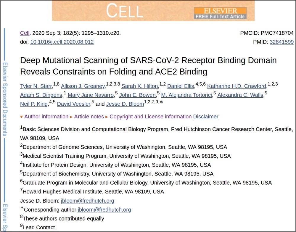
‘Deep Mutational Scanning of SARS-CoV-2 Receptor Binding Domain Reveals Constraints on Folding and ACE2 Binding’ (Tyler N. Starr et al.)

A BRIEF WORD ABOUT ANOTHER AMINO-ACID CHANGE IN B.1.1.7

I’ll quickly turn to another of the key changes said to be present in B.1.1.7. This change, the deletion of three amino acids was described in a paper published on the website of medRχiv on November 13, 2020. (Earlier in this article I mention that medRχiv is a creation of the Chan Zuckerberg Initiative.)

The mutation purportedly makes B.1.1.7 invisible to one of the three key functions of the polymerase chain reaction (PCR) test. That function is detection of the gene that has the genetic code for one of the two main spike proteins on the outer surface of the novel coronavirus.

However, that conclusion is based on only sequencing of the virus in a mere six people who tested positive for the novel coronavirus. On top of that, the paper was not subjected to scrutiny by other scientists (a process known as ‘peer review’) before it was published.

In addition, the Covid diagnoses of those six people were themselves determined by PCR. And PCR has been shown to have a very high rate of false positives — that is, to very frequently give a positive result in people who in fact do not harbour the novel coronavirus at all.

The authors of that paper themselves conclude that:

this result should be interpreted with caution. As a limited number of samples with the S-negative profile [i.e., tests that were positive for two of the three portions of the PCR test but not for the third, S-gene, portion] were sequenced, we could not exclude the presence of other S mutations associated with this profile…. Moreover we could not determine whether the deletion affected the primer or other probe-binding region as their coordinates were not available.

It’s a good bet that similar sleights of hand are behind the new wave of papers and headlines focusing on the amino-acid change dubbed E484K.

WHAT’S THE LESSON FROM ALL THIS?

That the pronouncements about the dire danger posed by the new variants aren’t based on solid science.

They appear to be aimed more at scaring the public into submitting to harsher and longer restrictions than helping to create truly evidence-based policies.

So follow the golden rules. Read the primary scientific-paper sources. Analyze them and think for yourself. Don’t let your reasoning be swept away by the 24-7, fear-filled news cycle.

Rosemary Frei has an MSc in molecular biology from the Faculty of Medicine at the University of Calgary, was a freelance medical writer and journalist for 22 years and now is an independent investigative journalist. You can watch her June 15 interview on The Corbett Report, read her other Off-Guardian articles follow her on Twitter and read her website here.

https://www.nytimes.com/2007/01/22/health/22whoop.html

Credit…Jon Gilbert Fox for The New York Times

Dr. Brooke Herndon, an internist at Dartmouth-Hitchcock Medical Center, could not stop coughing. For two weeks starting in mid-April last year, she coughed, seemingly nonstop, followed by another week when she coughed sporadically, annoying, she said, everyone who worked with her.

Before long, Dr. Kathryn Kirkland, an infectious disease specialist at Dartmouth, had a chilling thought: Could she be seeing the start of a whooping cough epidemic? By late April, other health care workers at the hospital were coughing, and severe, intractable coughing is a whooping cough hallmark. And if it was whooping cough, the epidemic had to be contained immediately because the disease could be deadly to babies in the hospital and could lead to pneumonia in the frail and vulnerable adult patients there.

It was the start of a bizarre episode at the medical center: the story of the epidemic that wasn’t.  (See link for article)

__________________

**Comment**

Testing fiascos are part and parcel of the CDC’s MO.

For more: