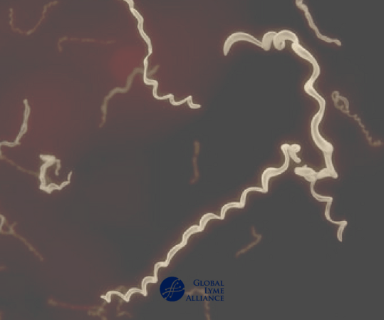
Tick-Borne Relapsing Fever (TBRF) is caused by spirochetes in the genus
/Borrelia/. Very limited information exists on the incidence of this
disease in humans and domestic dogs in the United States. The main
objective of this study is to evaluate exposure of dogs to /Borrelia turicatae/, a causative agent of TBRF, in Texas.
To this end, 878 canine serum samples were submitted to Texas A&M
Veterinary Medical Diagnostic Laboratory from October 2011 to September
2012 for suspected tick-borne illnesses. The recombinant Borrelial
antigen glycerophosphodiester phosphodiesterase (GlpQ) was expressed,
purified, and used as a diagnostic antigen in both ELISA assays and
Immunoblot analysis. Unfortunately, due to significant background reaction, the use of GlpQ as a diagnostic marker in the ELISA assay was not effective in discriminating dogs exposed to /B/. /turicatae/. Nevertheless, immunoblot assays showed that 17 out of 853 samples tested were considered to be seropositive, which constitutes 1.99% of all Texas
samples tested in this study.
The majority of positive samples were from central and southern Texas.
Exposure to TBRF spirochetes may be seasonal, with 70.59% (12 out of 17)
of the cases detected between June and December. In addition, 2 out of the 17 sero-reactive cases (11.76%) showed reactivity to both /B/. /burgdorferi/ (causative agent of Lyme disease) and /B/. /turicatae/ (a causative agent of TBRF).
This is the first report of TBRF sero-prevalence in companion animals in an endemic area. Our findings further indicate that /B/. /turicatae/ is maintained in domestic canids in Texas in regions where human disease also occurs, suggesting that domestic dogs could serve as sentinels for this disease.
______________
**Comment**
I predict much more to come. Texan Lyme/MSIDS patients – we stand with you.
FIVE POSTDOCTORAL SCIENTISTS WILL FOCUS ON CHRONIC LYME
GREENWICH, Conn (Dec. 5, 2017)—Global Lyme Alliance (GLA), the leading 501(c)(3) dedicated to conquering Lyme and other tick-borne diseases through research, education and awareness, today announced the awarding of its first-ever fellowships to five young postdoctoral scientists whose work focuses on Post-Treatment Lyme Disease Syndrome (PTLDS) or chronic Lyme.
The three-year fellowships, made possible with the support of Deborah and Mark Blackman, will support five recent Ph.D. graduates with specific interest in understanding whether persistence of the bacteria Borrelia burgdorferi, the Lyme disease pathogen, or host evasion mechanisms are responsible for the continued symptoms experienced by patients treated for Lyme disease.
“The new fellows are hard-working and brilliant young scientists with fresh ideas who will tackle the mechanistic underpinnings of PTLDS,” said Mayla Hsu, Ph.D., GLA’s director of research and grants. “We’re delighted to be able to support these researchers at the beginning of their careers.”
The five are:
George Aranjuez, Ph.D., University of Central Florida, is studying the molecular mechanisms that Borrelia uses to survive during mammalian infection and how it evades the immune system.
Ashley Groshong, Ph.D., University of Connecticut, is examining the link between Borrelia protein metabolism and its ability to form persister cells thus evading antibiotic assault.
Matthew Muramatsu, Ph.D., University of Texas-Southwestern, is exploring how the genetics of persister Borrelia differ from that of replicating bacteria. His work will focus on how the transcription signals that start the persister pathway are regulated.
Bijaya Sharma, Ph.D., Tufts University, is studying whether immune deficiency is related to continued symptoms in Borrelia-infected mice. Her work explores the genetic factors in Borrelia that underlie bacterial persistence.
Xuran Zhuang, Ph.D., University of Maryland, will use tick microinjection to study the growth of persister bacteria and its genetic pathways in samples she recovers from patients.
The “Blackman-GLA Postdoctoral Fellowships” will total $1,125,000. Each fellow will receive $75,000 per year, for each of the three years, including travel expenses to Lyme disease conferences and an invitation to GLA’s annual closed-door scientific symposium, where they will participate in scientific discussions, present their findings and meet with program donors Deborah and Mark Blackman.
All applicants were required to submit a detailed scientific proposal for expert review. Annual reports on progress of project milestones will be required before second and final installments will be awarded.
“Supporting young researchers at the beginning of their careers shows the commitment of GLA and the Blackmans to nurture the development of a cadre of experts in the Lyme disease field,” said Scott Santarella, GLA’s CEO. “We also hope that the findings of these scientists will be potentially broadly applicable to other infectious diseases.”
In addition to the fellowships, GLA awards grants each year to researchers working on promising projects that best fulfill the organization’s goal to improve diagnostic testing and uncover more effective treatment protocols. Proposals received for the 2017-18 research grant cycle represented a broad range of interests ranging from tick ecology to co-infections, from new treatment approaches to basic biology of Borrelia infection in the mouse model, in both its acute and chronic stages. The 2017-18 grantees will be announced before the end of the year.
Global Lyme Alliance (GLA) is the leading 501(c)(3) dedicated to conquering Lyme disease through research, education and awareness. GLA has gained national prominence for funding the most urgent and promising research in the field, while expanding education and awareness programs for the general public and physicians. Learn more at GLA.org.
Interview With Professor Laane About the Suppression of Microscopy for Lyme Diagnostics
Written by Huib Kraaijeveld
Around 2003 the WHO encouraged research into microscopy as a direct test for the Borrelia spirochete, the pathogen causing Lyme disease. When a promising new and simple technique was discovered in 2013, it was however violently attacked. Not on the science itself, which is the normal procedure in science, but personally. Now retired professor microbiology Morten Laane was fired after he gave a lecture at a scientific conference in 2014. Moreover, his laboratory was closed down, the website of the scientific journal was hacked and the article disappeared. An exclusive interview.
“If experts treated airplane accidents in the same manner as an average medical scientist studies disease, I would book my next travel to the USA by ship.”
Professor Laane
Courtesy of Under Our Skin
Who is professor Laane?
Born in July 1940, in the small city of Toensberg, Morton Laane grew up during the second World War. “My mother visited her sister in the city of Bergen the ninth of april 1940 the day war broke out in Norway. They survived the severe attacks by the German warships. My mother travelled along the coast by a Norwegian local ship to the small city of Toensberg where the family lived close to the Oslo fjord. This was not a very smart idea, as several ships were attacked and destroyed by German submarines.
My father was in Finmark, the arctic part of Norway, together with a substantial number of Norwegian soldiers as a medical officer. When I was born, my mother, who had professional training in handling weapons, sat alone with a hidden gun in a small flat close to the German headquarter across the street. When the free artic part capitulated a couple of months after the start of the German invasion, he was first arrested by the Germans, later released and went back to Toensberg.
In 1947 my father showed me how to detect the Syphilis spirochete by very simple microscopy. Numerous sailors lived in this city known for its substantial commercial fleet. Back then, Syphilis infection was not uncommon in sailors.
My father had a small, old Leitz brass microscope of high quality. A tiny sample from the patient was mixed with an equal part of drawing ink on the glass slide made for microscopy. Drawing ink consists of extremely tiny black particles (coal). They do not penetrate into the bacteria. Normally they are almost invisible. Light pass through the unstained bacteria against a total black background.
This started my interest for microscopy. Later my father specialised in psychiatry. He had no use of his microscope and I got it as a gift. Looking back, almost everything in my life started as a hobby.
I became scientific assistent in general experimental genetics. My boss, Dr. Øistein Stromnaes worked with the fungus Penicillium. Nobody had seen its chromosomes before, but I discovered a simple method to count them in the light microscope.
So I was offered a research fellowship a the University of Bergen, I continued my research there and my PhD dissertation took place in 1971. The committee evaluated my theses as very good and suggested that I might have the qualifications of a personal professor position for life time and further develop new methods in genetics.
One of my discoveries regarding Penicillium brought me in contact with Lynn Margulis, then Lynn Sagan, wife of the famous Carl Sagan. Her research on cellular evolution and endosymbiosis led to the understanding that the energy-converting organelles, the mitochondria and chloroplasts, are actually bacterial symbionts living inside eukaryotic cells. This discovery earned her the reputation of being the most famous scientist in evolutionary biology after Charles Darwin.
My discovery was connected with her spirochete research, which focused on the symbiotic nature of the relationship between spirochetes and their hosts, in particular the noted ability of these bacteria to form dormant “round bodies” that are capable of reactivation. Nobody in the world knew more about spirochetes than Lynn.*
Later I discovered a somewhat similar structure in the slime mould (Physarum) which was much used for experimental research. I got in touch with Professor Ivar Giaever, a Nobel laureate in Physics and we cooperated for many years supervising Master and Ph.D. Students.”
Microscopic Lyme diagnostics
Microscopy is considered the Golden Standard in diagnostics for Syphilis. Given his background and expertise in microscopy, it made sense to Laane to try and find a method to detect the Borrelia in infected blood.
Laane: “My coworkers and I had published a series of papers in the Norwegian journal ‘Biolog’ regarding a problematic sheep disease called “alveld”. The papers contained unusual and spectacular images of possible toxic blue green algae (bacteria) that were suspected to be connected with this disease.
The Biological Institute then asked for cooperation regarding Lyme infections in order to compare microscopy and molecular tests. The Institute was responsible for administration regarding formal permissions to do medical research. It turned out that no reliable data were obtained from the molecular studies. The institute was responsible for this part. Microscopy showed, however, characteristic spirochetes in a number of patients together with other organisms such as Babesia.
The syphilis and Borrelia spirochetes share many properties. They cause permanent infections in humans which cause long-lasting, multi-stage diseases. They exist in more than one form – sometimes in the long spirochete form, but also in a round “cyst-like” form. The spirochete form can be made visible by simple methods and observed with a microscope.
Borrelia spirochetes in dry smears and in isotonic solutions appear as very thin cells, only 0.2 micrometers wide, and as very long – in the range of 50 to 100 micrometers. These are visible above the resolution limit for light microscopy – due to what we call light interference – by slight defocusing. In dry preparations, a focus error of more than 1/1000 mm renders them invisible under the microscope. In wet preparations, the cells swell substantially.
My colleague and I published the results of our part of the project due to our firm belief that microscopy is useful for detecting these infections. Beforehand we were assured by the Institute that formal permissions were granted by the health authorities. It turned out that the Institute, somehow had forgot to point out in a formal enough manner that a substantial part of the project included microscopy by us!
Our project leader was educated as a medical doctor, and had later became a biologist. He was also Head of the Institute to which the project was connected. We, my colleague and I, got a copy of the first page of the application accepted and signed by the Health authorities. We did not, unfortunately, see the entire application before it was too late, as the project description was incomplete in that it did not express without any doubts that about half of the project involved extensive use of microscopy to analyze samples.
The project leader knew of my cooperation with Oeystein Brorson, the extremely clever technician who was able to grow Borrelia from infected blood samples. We had agreed that the application should be written to state that extensive microscopy was needed for the project as a way to confirm the results of molecular tests.
The Institute received around 2 million euro (NOK 20 mln) and bought two DNA machines. The test project was thus an official Institute project, not an application by a group of three scientists. In hindsight, we should have asked to see the complete application, but we did not and went on the trust for our project leader. This turned out to be a mistake, as we were later blamed for not reading the regulations for medical research and additional legal papers.
The trust that caused us to not read the full application was labelled as “intention,” to mean we intended to subvert the regulations when we embarked on this project. This is simply not true.
Silenced for speaking
After publishing the 2013 article ‘A simple method for the detection of live Borrelia spirochetes in human blood using classical microscopy techniques’, professor Laane was invited to give a lecture at the 2014 Norvect conference in Oslo. An English patient saved the pdf, so you can still read it, via the link provided.
I was present at that conference and still remember how nervous he was. The reason was that several medical professors complained to his university. He was threatened with losing his job, if he would speak at the conference.
In fact, he did not literally speak – as you can see in the movie below – but used performing arts to show the slides of the spirochetes. Professor Laane was fired anyway and his laboratory was closed down.
Laane: “As for being forbidden to speak, Waldemar Broegger, a Geology professor in the late half of the nineteenth century was forbidden to speak about Charles Darwin’s evolution theory. Lynn Margulis was originally ridiculed for her theories on the evolutionary origins of mitochondria and chloroplasts and later received numerous awards for this discovery. As both of these scientists were found to be right and later became famous, I feel I am in good company.
The 2013 publication of ‘A simple method for the detection of live Borrelia spirochaetes in human blood using classical microscopy techniques’ in the journal Biological and Biomedical Reports resulted in much opposition by conservative medical doctors and some scientists, most of whom had little or no microbiology experience working with spirochetes in a laboratory environment.
The article and research was criticized because the cellular objects we were observing were presumed to be “artifacts,” meaning objects of some other origin that just magically appeared in our samples. It’s extremely important to point out that all of our principal research was performed under highly controlled conditions, and our results were confirmed using more than one microscopy method. The critics of our work have yet to explain how these supposed “artifacts” we observed were able to reproduce and even move like spirochetes, which we observed them doing, and even more importantly, the critics have failed to explain how an “artifact” could possibly contain nucleic acids from Borrelia.
Yet, in that same year Dr. Alan MacDonald found out independently the same as me regarding how to detect chronic Lyme infections in human blood. Before this the assumed borrelia bacteria had been found by a Norwegian microscopist, who had an extremely ill son from Lyme.
MacDonalds arguments in these two videos (part one and part two) are brief, but very much to the point. He has later developed methods to detect single Borrelia bacteria direct in a microscope slide by exact molecular methods.
Oeystein Brorson is also mentioned by MacDonald. He was the researcher who grew all known strains of Borrelia in Norway, sent me samples and cooperated with Lynn Margulis and me, until he got disabled due to disease and had to leave his job.
After my lab was closed down, also the website of the scientific journal that published our article was so severely hacked that it stayed offline for three years. Once it came back up, our article had dissapeared.
The hacking of the Journal ‘Biological and Biomedical Reports’ seems to be done with a person knowing more than my closest enemies. And was someone with access to a quite advanced compilator system. Why this was done might well have been political or personal prestige.
I have never seen this happening on any topic in science before.”
Proof or propaganda?
In 2016 the Norwegian Health Department (FHI) published the study ‘Validate or falsify: Lessons learned from a microscopy method claimed to be useful for detecting Borrelia and Babesia organisms in human blood’. It stated,
“microscopy by the LM-method identified structures claimed to be Borrelia- and/or Babesia in 66% of the blood samples of the patient group and in 85% in the healthy control group”.
The TV2 reporter in the infamous documentary ‘Deceit or Borrelia’ misquoted this sentence by claiming that “it was proven that Dark Field microscopy produces 85% false positives”.
There are however suspicions that the Norwegian study was set up to discredit professor Laane’s work, using contaminated blood.
Laane:
“Officially, on their website The National Institute of Public Health claimed that the only known case of Babesia in Norway was a veterinarian that have had his spleen removed, and else that Babesia was unknown in the Norwegian population.
But I found a faked sample of Babesia. It was blood from a cow (or ox). It was mixed with human blood from a socalled control person and sent to me by mail.
Of course, this sample clotted impossible to see anything in the sample by microscopy except lumps of erytrocytes and fibrine fibers. If I had got the original sample unmixed, I would have found them at once. The FHI showed me a few images from a preparation they had made themselves, BEFORE it was mixed with human blood. The Babesia images there were the same as I found in several so-called control samples.
When I showed them, they looked a little worried – like school children not telling the truth to their teacher – but they would not admit that control samples contained Babesia merozoites.”
Borrelia stained with the Mysterud-Laane method, blue color.
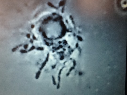
Live biofilm of Borrelias developing in a mixture of blood and sodium citrate (this patient was tested positive also by molecular methods by American laboratory).
Serology
In the introduction to their 2016 article, the FHI authors also state that Lyme serology has a 70-90% sensitivity in the earliest stages and a sensitivity of almost 100% in later stages, which suddenly seems to become the new mantra in several countries. This seems to have been taken from a 2016 article written by IDSA and CDC authors, which was criticised for using circular logic.
Laane:
“A recent report presented in Norwegian newspapers claim that inaccurate science, especially in medical topics, is common. Norwegian Professor Oeyvind Oesterud has written several short articles in Aftenposten, Norway’s largest newspaper about science and popular science papers.”
He points out that false conclusions are favoured by both journals and media on the criteria that discoveries should be spectacular. Unless they are not, they are not accepted. Also the official evaluation system for research grants favours this. Many of these discoveries can not be repeated. But it generates much money for the universities.
Such scientists believe in accepted science. Any deviation shown by experiment may be interpreted as incorrect or false. One may wonder why they publish at all. Regarding deviating disease data they are often neglected – according to what the medical scientist was taught as a student by his professor. The deviating results are never studier further and ignored as “errors”. Up to about half of recent medical papers may contain this.
The result is delayed progress, sometimes with big consequences for severely ill patients. Patients that could be saved. Disease problems are very complicated and there is need for research listening to what both the patients tell and meticulous analysis of unexpected data.
As a final thought:
“Travel by modern airplanes is now very safe. If experts treated airplane accidents in the same manner as an average medical scientist studies disease, I would book my next travel to the USA by ship.” Professor Laane
Exception or a pattern?
The European Union has recently provided a 2 million euro grant to a cooperation of three parties to develop a better Lyme test, as the current one is considered imperfect. At best.
So why are there several indication of the active repression and sabotage of new, better and direct ways to diagnose Lyme? Is professor Laane’s story unique or showing a doisturbing pattern?
“This never happened before on any topic in the history of science in Norway”, Laane said in his interview with the makers of ‘Under Our Skin Emergence’. Yet also in other countries scientists have been attacked for working on promising new and direct Lyme tests.
In 2014, French lab director Schaller was fined was fined with paying 280,000 euro to the government and was sentenced to nine months in jail. Also in 2014 the American Centers for Disease Control and Prevention (CDC) publicly attacked the credibility of Advanced Laboratory Services, which had developed a culture-based test for Lyme disease diagnosis. The basis for the CDC’s attack has since been proven wrong, yet the CDC has never retracted the article in which the errant criticism was made.
A direct and ‘no false-positive’ DNA test for Lyme was no longer made available to the general public, after its inventor was fired from a Connecticut hospital in 2010. And recently media attacked the tests of specialised labs in Germany, using undercover reporters and twisted patients’ stories, claiming they were either not ‘FDA approved’. As professor Ahern explained in her recent interview, none of the tests promoted by the CDC or your national Health agencies are ‘FDA approved’.
The next interview with Dr. Lee will look into what happened around the development and public accessibility of the DNA diagnostics mentioned above. It will be published very soon.
* Professor Laane and I will work together on a future article about the work of Lynn Margulis, as too few people seem to have heard of her important work.
Professor Andrea Swei prepares to pull a tick off a western fence lizard.
East Coast gets the limelight, but California’s ticks play by different rules
Lots of things probably come to mind when you think of Lyme disease, but California likely isn’t one of them. Most of the human cases of Lyme — and most research studies on it — happen in the northeastern U.S. But new work from San Francisco State University Biology Professor Andrea Swei and her former graduate student Jessica Kwan shows that the West Coast may have a thing or two to teach the East about the disease.
“The West Coast has been largely ignored,” said Swei. “And I think that’s a mistake.”
For Swei, California’s lower prevalence of Lyme is an opportunity to probe how the bacteria that causes the disease spreads differently on the West Coast. One leading explanation has to do with the different hosts that Lyme-carrying ticks prefer. In the Northeast, young ticks like to feed on the blood of small mammals like mice and chipmunks. In California, however, they have another favorite: western fence lizards. These lizards have unique blood that actually kills off the Lyme bacteria present in ticks, essentially un-infecting them.
That got Kwan thinking. If the Lyme disease bacteria dies when its host feeds on a lizard, what happens to all the rest of the tick’s microbial residents? After all, the whole community of microbes in a tick — what scientists call its “microbiome” — could affect whether Lyme-causing bacteria thrive.
The team, with Kwan at the helm, captured western black-legged ticks from around Marin County both by dragging nets through vegetation and by pulling them off lizards. They also raised some in the lab. The team then tested tens of millions of snippets of bacteria DNA from the ticks and organized these snippets by how similar they were to one another. That gave them an idea of the diversity of microbial life that was living in each tick, and how that diversity changed over the ticks’ lives.
They found that ticks harboring a less diverse community of microbes were more likely to be infected with the Lyme pathogen, and that those microbe communities actually became less diverse as the ticks fed and grew.This came as a surprise — both findings ran counter to research done back east. “We were thinking in the East Coast model,” explained Kwan. California ticks, apparently, play by different rules.
The results also hint at a more complicated role for western fence lizards in the transmission of Lyme. “They’ve been touted as this secret weapon against the Lyme disease bacteria,” Swei explained. But if they make the whole microbiome of a feeding tick less diverse, that tick may actually be more likely to harbor and spread the bacteria that causes the disease.
Swei and her team plan to keep on probing the unique California Lyme disease system. Armed with new data, they’ll keep investigating this more complicated picture of the western fence lizard’s role. They even plan to look at how different strains of the same species of Lyme-causing bacteria could change how the disease spreads. “There’s a lot we can still learn from the system here,” says Swei.
Please know that Andrew Spielman’s tick maps have been used for decades against patients. Doctors look at the flawed maps and proclaim, “Lyme isn’t here because the tick isn’t here.” That is getting blown out of the water on a daily basis.
“When I started working in this area, I was told, point blank, Lyme disease was not in the South, and human Lyme disease could not occur — there were no ticks and no germs,” he recalls. But he was not convinced that the blacklegged ticks in the North and South differed much — or that Spielman’s discovery represented a separate species at all.
In a series of experiments from 1989-1990, Oliver demonstrated that so-called Northern deer ticks (dammini) and the blacklegged ticks (scapularis) found up and down the East Coast bit exactly the same animals in the lab. In 1992, he showed that even ticks from widely separated areas like Georgia and Massachusetts were genetically too similar to be different species. And breeding ticks from the North and South in the lab, Oliver demonstrated that a series of matings produced reliably fertile offspring — a crucial test of species boundaries.
Oliver’s definitive experiments blew up the idea that dammini was a new or separate species. The name dammini was dropped from the scientific literature. But Spielman’s framework — restricting Lyme to the Northern ticks — remained intact.
Vertical vs. horizontal transmission of the microbiome in a key disease vector, Ixodes pacificus
Kwan JY, Griggs R, Chicana B, Miller C, Swei A. Vertical vs. horizontal transmission of the microbiome in a key disease vector, Ixodes pacificus. Mol Ecol. 2017;00:1–12. https://doi.org/10.1111/mec.14391
First published:
Abstract
Vector-borne pathogens are increasingly found to interact with the vector’s microbiome, influencing disease transmission dynamics. However, the processes that regulate the formation and development of the microbiome are largely unexplored for most tick species, an emerging group of disease vectors. It is not known how much of the tick microbiome is acquired through vertical transmission vs. horizontally from the environment or interactions with bloodmeal sources. Using 16S rRNA sequencing, we examined the microbiome of Ixodes pacificus, the vector of Lyme disease in the western USA, across life stages and infection status. We also characterized microbiome diversity in field and laboratory-collected nymphal ticks to determine how the surrounding environment affects microbiome diversity. We found a decrease in both species richness and evenness as the tick matures from larva to adult.When the dominant Rickettsial endosymbiont was computationally removed from the tick microbial community, we found that infected nymphs had lower species evenness than uninfected ticks, suggesting that lower microbiome diversity is associated with pathogen transmission in wild-type ticks. Furthermore, laboratory-reared nymph microbiome diversity was found to be compositionally distinct and significantly depauperate (lacking in numbers or species) relative to field-collected nymphs. These results highlight unique patterns in the microbial community of I. pacificusthat is distinct from other tick species. We provide strong evidence that ticks acquire a significant portion of their microbiome through exposure to their environment despite a loss of overall diversity through life stages. We provide evidence that loss of microbial diversity is at least in part due to elimination of microbial diversity with bloodmeal feeding but other factors may also play a role.