Archive for the ‘Testing’ Category

Ticked Off: New Brighton Tattoo Artist Joins Event to Stamp Out Lyme Disease

https://www.timesonline.com/entertainmentlife/20190126/ticked-off-new-brighton-tattoo-artist-joins-event-to-stamp-out-lyme-disease

NEW BRIGHTON —

Josh DeLay is ticked off about Lyme disease and rightfully so.

In 2017, Pennsylvania topped the nation with 9,250 confirmed cases and 2,650 probable, according to the federal Centers for Disease Control and Prevention, almost as many as found in New England states combined.

DeLay, 35, of Beaver Falls, owner of DLay ’n Ink Tattoos and Body Piercing in New Brighton, is among 35 tattoo artists across the country participating in Ink to End Lyme, an event to increase awareness and raise money to treat and cure the tick-borne illness now detected in all 50 states and the District of Columbia. The majority of cases are concentrated in the Northeast and upper Midwest.

DeLay said he will donate all proceeds from tattoos and body piercings Jan. 30 to Lyme research. He’s freed his schedule from 10 a.m. to 9 p.m. to accommodate walk-in customers. On an average day, he has four to five appointments and charges $85 an hour.

This is the third year for Ink to End Lyme, sponsored by nonprofit Lyme Warrior — a team of people with chronic Lyme disease working to find better treatment, testing and understanding of the illness. DLay’n Ink is one of three tattoo shops in Pennsylvania participating. The other two are near Philadelphia.

DeLay learned of the event through Facebook when tagged by a friend last month. A few days later, a client told him she was just diagnosed with the disease. DeLay thought that was more than coincidence and took it as a sign to get involved.

“If you can help, you should. It’s one day of my time,” he said, adding that he’s supported fundraisers to help animals, veterans and community projects such as the ongoing one in Beaver Falls to raise money to repair and reopen Tigerland Wave Pool.

When first diagnosed with diabetes years ago, he didn’t have health insurance.

“I was going to the clinic in Beaver Falls. Thank God, they had a (funding) program set up and I was able to get insulin,” he said.

Funding should be available to advance research for Lyme disease, too, he said.

“It’s a shame. There should be a cure. Why isn’t there?” he said.

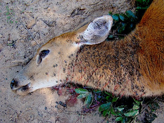
The past month, DeLay’s Googled tattoo flash cards — designs printed on paper or cardboard to give customers ideas — of Lyme-related images and has also been hand drawing his own. Images range from the simplicity of the lime-green Lyme disease awareness ribbon, a bandaged lime, to more intricate drawings of disease-carrying animals — white-tailed deer, mice, chipmunks, gray squirrels, opossums and raccoons.

Of course, customers don’t have to be inked with Lyme-related tattoos. They can choose whatever design they want.

But customers must be 18 or older. In Pennsylvania, it’s unlawful to tattoo anyone under 18 without the presence and consent of a parent or guardian. Tattoo artists who violate the law face a third-degree misdemeanor charge, which can lead to a $100 fine or up to three years in prison.

Lyme Warrior founder Lauren Lovejoy of Blacksburg, Va., said it took years before she was correctly diagnosed with the disease and she slowly became sicker and sicker. A self-described workaholic, she found herself mostly bedbound.

“I was so lightheaded, shaky, and generally weak, that I had issues functioning at my office job,” she wrote on the Lyme Warrior website. “I went to doctor after doctor, but after another month full of missed days of work, I had to acknowledge that I could not function. I spent almost every day in bed.”

A tattoo devotee, she organized artists around the country to help her cause in the education of Lyme disease and her mission to raise funds. Last year, $12,000 was raised.

In Mid-Atlantic states, including Pennsylvania, blacklegged ticks infected with the bacterium Borrelia burgdorferi spread the disease to humans by attaching themselves to the body — usually hard-to-see areas such as scalp, groin and armpits — and then biting flesh.

Annually, the number of people diagnosed with Lyme disease in the United States approximates 300,000, according to the CDC.

Symptoms include fever, headache, fatigue, muscle and joint pain and characteristic bull’s-eye patterned rash. Untreated, infection can spread to joints, heart and nervous system. If treated with antibiotics early, people usually recover, the CDC said.

Most cases of Lyme are missed, according to Lyme Warriors. Standard testing is only 20 to 30 percent accurate. Lovejoy said she was misdiagnosed twice.

The Pennsylvania Department of Health’s 2017 Lyme Disease Report indicated Beaver County had 211 cases. Allegheny County had 432, and Lawrence County had 82. Butler County had the most with 658.

DeLay thinks development of wilderness areas forcing humans and animals to share habitats helps spread Lyme and other tick-borne illnesses.

“We’re getting closer to the woods, and the woods are getting closer to us,” he said. “I heard about coyotes in Beaver Falls yesterday. Someone saw a coyote. If you think about it, they’re cutting trees down everywhere. Look at Chippewa (Township). It used to be all woods.”

Lovejoy also blames climate change and global warming.

And something even more alarming, she said: “an asexual tick that doesn’t need a partner to reproduce.”

The invasive Asian longhorn tick was discovered in Pennsylvania’s Centre County last year.

_________________

**Comment**

If you find an attached tick, GET TO THE DOCTOR RIGHT AWAY!  Do not take a “wait and see approach.”  Everyone admits that treating for this promptly makes all the difference.  Why wait?  It’s not worth the gamble.  If it were me, I’d demand prophylactic treatment.  For more on all things Lyme:  https://madisonarealymesupportgroup.com/2016/02/13/lyme-disease-treatment/

  • Take a picture of the tick
  • Put it in a sealed baggie and stick it into the freezer
  • Take pictures of any rashes.  They are notorious for coming and going.  By the time you get to the doctor it might be gone.

It’s true that most cases are missed and that testing is abysmal, but the information on waiting for a fever and rash are just as deluded.  

Fewer than 50% recall a rash and many rashes are “atypical.”  

Dr. Klinghardt has stated that a bulls-eye rash only happens on subsequent tick bites – not the first one.  One thing’s for sure –

IF you have an EM rash, YOU HAVE LYME, period.

For prevention:  https://madisonarealymesupportgroup.com/2017/05/11/tick-prevention-and-removal-2017/

https://madisonarealymesupportgroup.com/2018/05/27/study-conforms-permethrin-causes-ticks-to-drop-off-clothing/

https://madisonarealymesupportgroup.com/2018/04/03/fire-good-news-for-tick-reduction/  Burning gave a 98% reduction in ticks.

Lastly, do your reading before considering a tattoo.  Tattoos are not without significant health risks:  https://www.medicaldaily.com/tattoos-affect-your-health-long-term-side-effects-ink-has-your-immune-system-404404  In brief:

  • Tattoo ink can be toxic & contain carcinogens (including metals)
  • Tattoos can lead to errors in medical treatment (MRI burns & swelling, tattoo ink appearing as malignant cells)
  • Tattoos can cause infections (HIV, hepatitis C, staph, mycobacteria)
  • They can lead to tattoo-induced skin disorders (sarcoidosis, lichen planis)
  • They can cause allergic reactions
  • They can cause scarring
  • They can cause granulomas (bumps to encapsulate foreign substances such as ink particles)
  • Lyme/MSIDS patients are already in an epic battle for their health.  Purposely adding anything that assaults the body isn’t wise.
 “Tattoo ink has risk of infection”

https://youtu.be/AHlyK5W7rIw  (Click on this link to learn more) 

Approx. 45 Sec

Published on Sep 28, 2012

Nearly 40 individuals across the US exhibited unusual rashes after receiving tattoos. After an investigation, the Food and Drug Administration (FDA) and Centers for Disease Control (CDC) confirmed 22 skin infections spanning over four states in the past year. The symptoms are linked to tattoo ink contaminated with M. Chelonae. This cousin of tuberculosis entered the ink via contaminated water used during the distillation process.


Ehrlichia Found in Argentinian Marsh Deer For the 1st Time

https://www.ncbi.nlm.nih.gov/m/pubmed/30619709/

Evidence of Ehrlichia chaffeensis in Argentina through molecular detection in marsh deer (Blastocerus dichotomus).

Guillemi EC, et al. Int J Parasitol Parasites Wildl. 2019.

Abstract

Vector-borne pathogens are responsible for serious emerging diseases and have been widely described in wildlife. Ehrlichia chaffeensis causes the zoonotic “monocytic ehrlichiosis” in humans, is transmitted by the tick Amblyomma americanum and its reservoir host is the white-tailed deer (Odocoileus virginianus) in North America. Little is known about the native reservoir and the tick vectors involved in the transmission cycle in South America. We report here the detection of E. chaffeensis in a study on marsh deer (Blastocerus dichotomus) mortality in Argentina, in different time periods between 2007 and 2016. Four deer, from two distinct populations, were positive for E. chaffeensis through molecular methods. Additionally, the variable-length PCR target (VLPT) region of positive samples was genotyped. Our results provide the first evidence of E. chaffeensis in autochthonous Cervidae from Argentina, contributing to uncover the distribution of this tick-borne infection in South America.

1-s2.0-s2213224418300889-gr3_lrgFig. 3. Dead marsh deer with high tick burden.

____________________

More on Ehrlichia:  https://madisonarealymesupportgroup.com/2018/12/02/everything-thats-known-about-ehrlichiosis/

https://www.lymedisease.org/ehrlichiosis-tick-borne-disease-no-one-heard/  The author brings up a valid point about the potential of there being undiagnosed Ehrlichia behind a ME/CFS diagnosis in a subset of patients since it infects white blood cells and the mitochondria.  The article also gives helpful percentages of symptoms and the following information:

  • Fever/chills and headache (majority of cases)

  • Fatigue/malaise (over two-thirds of cases)

  • Muscle/joint pain (25% – 50%)

  • Nausea, vomiting and/or diarrhea (25% – 50%)

  • Cough (25% – 50%)

  • Confusion or brain fog (50% of children, less common in adults)

  • Lymphadenopathy (47% – 56% of children, less common in adults)

  • Red eyes (occasionally)

  • Rash (approximately 60% of children and 30% of adults)

OTHER MODES OF TRANSMISSION

Ehrlichia chaffeensis has been shown to survive for over a week in refrigerated blood. Therefore these bacteria may present a risk for transmission through blood transfusion and organ donation. It has also been suggested that ehrlichiosis can be transmitted from mother to child, and through direct contact with slaughtered deer. (14, 15)

https://madisonarealymesupportgroup.com/2018/10/02/north-carolina-ehrlichia-often-overlooked-when-tick-borne-illness-suspected/

https://madisonarealymesupportgroup.com/2018/07/24/oklahoma-ehrlichiosis-central/

https://madisonarealymesupportgroup.com/2018/03/09/dogs-ehrlichiosis/


1st Detection of Rickettsia africae & Tropical Bont Tick Found in France

https://www.ncbi.nlm.nih.gov/m/pubmed/30656468/

First molecular detection of Rickettsia africae in a tropical bont tick, Amblyomma variegatum, collected in Corsica, France.

Cicculli V, et al. Exp Appl Acarol. 2019.

Abstract

Here we report the first detection of Amblyomma variegatum, a tick species of medical and veterinary importance, and the first molecular evidence of a pathogen, Rickettsia africae, both new to Corsica (France). In August 2018, an ixodid tick with an unusual morphology was removed from the ventral part of a cow’s whole skin in a slaughterhouse located in the village of Ponte-Leccia (Haute-Corse). The tick was morphologically identified as an adult male of A. variegatum. This result was confirmed by 16S rDNA sequence analysis with a close relative being a sequence from Senegal showing 99% nucleotide identity. We tested the tick for Ehrlichia and Rickettsia. The tick was positive to Rickettsia and the corresponding sequence matched with R. africae. There is little or no risk of the introduction and establishment of a viable population of A. variegatum in Corsica by migrating birds. However, if it did, it could produce major economic losses for livestock production. Further studies and sustained surveillance are indicated, not only focusing on this species of tick and this rickettsia, but also on other microorganisms of veterinary and medical importance that might be transmitted in Corsica and other Mediterranean islands.

__________________

**Comment**

http://entnemdept.ufl.edu/creatures/livestock/ticks/tropical_bont_tick.htm  The tropical bont tick is considered one of the most detrimental of the tick species present in Africa and now the Caribbean (CaribVet 2011b, Stachurksi and Lancelot 2006).

Now it’s been found in France.

Of human health concern, it is implicated as a potential vector for Crimean-Congo hemorrhagic fever virus (CCHF)Yellow Fever virus (CDC 2001, Merck 2011), & African Tick Bite Fever (Rickettsia africae),which results in fevers, headaches and swollen lymph nodes (Parola et al. 1999).

It has had a huge effect on the livestock industry, primarily through its transmission of heartwater disease, Ehrlichia ruminantium (formerly Cowdria ruminantium) (Allan et al. 1998, CaribVet 2011b, OIE 2009, Parola et al. 1999) and their association with dermatophilosis, Dermatophilus congolensis (Allan et al. 1998, Barré and Garris 1990, CaribVet 2011b, Merck 2011).

It’s also been implicated as a vector or potential vector for several diseases to include Dugbe virus, and Jos virus (Merck 2011). In the Caribbean, only heartwater disease and dermatophilosis have been detected in the hosts and have demonstrated clinical symptoms.

The dispersal of tropical bont tick is associated with the migration patterns of the cattle egret, Bubulcus ibis (OIE 2009, Barré et al. 1995, Pegram and Eddy 2002). The birds transport the larvae and the nymphs of the tick (Deem 1998). Because the non-feeding stages of the life cycle occur within the environment and not on the host, the transport of infested vegetation and litter is a potential source for dispersion as well (Alderink and McCauley 1988, Barré et al. 1995).

The adult ticks are found on various domesticated species such as camels, cattle, goats, sheep and even dogs (Deem 1998, Merck 2011). The ticks also are found on various species of wildlife throughout the distribution range, but the adults are generally found on the larger mammals (Barré et al.1995, Yonow 1995).

Biological controls (birds, parasitic wasps, nematodes, bacteria, and fungi) have not been very successful in eradicating the tick.

tropical_bont_tick02Adult male tropical bont tick, Amblyomma variegatum Fabricius. Photograph by Alan Walker, University of Edinburgh.

tropical_bont_tick03Adult female tropical bont tick, Amblyomma variegatum Fabricius. Photograph by Richard Matthews and Alan Walker, University of Edinburgh.

Other ticks in other places have been found to carry CCHF (it can also be spread human to human):  https://madisonarealymesupportgroup.com/2018/08/19/monster-ticks-found-in-germany-threaten-europe-with-deadly-disease-crimean-congo-fever/

https://madisonarealymesupportgroup.com/2018/06/14/crimean-congo-the-asian-ebola-virus/


Remember Deer Keds? Study Shows Bartonella Causing Deer Ked Dermatitis in Humans

https://www.ncbi.nlm.nih.gov/pmc/articles/PMC525279/

. 2004 Nov; 42(11): 5320–5323.
PMCID: PMC525279
PMID: 15528732

Isolation of Bartonella schoenbuchensis from Lipoptena cervi, a Blood-Sucking Arthropod Causing Deer Ked Dermatitis

ABSTRACT

Bartonella schoenbuchensis, which commonly causes bacteremia in ruminants, was isolated from the deer ked Lipoptena cervi and was shown to localize to the midgut of this blood-sucking arthropod, causing deer ked dermatitis in humans. The role of B. schoenbuchensis in the etiology of deer ked dermatitis should be further investigated.

________________

**Important Take-aways**

  • Deer Ked incidental infestation in humans is well documented
  • Skin tests with deer red whole-body extracts were positive in ALL patients
  • Testing showed both immediate & delayed reactions
  • 57% of patients had elevated serum immunoglobulin E (IgE) levels
  • Deer keds appear to be an ideal vector for efficient transmission
  • The risk for transmission to humans is apparent
  • B. schoenbuchensis is most closely related to B bacilliformis, an important human pathogen also transmitted by a fly (Lutzomyia verrucarum)
  • Five variants were found – some of which may pose a larger risk than others
  • Clinical scenario of deer led dermatitis resembles a primary manifestation of Cat Scratch disease caused by B. henselae.
  • A positive delayed-type hypersensitivity skin test, like that characteristically observed for B. henselae antigens in cat scratch disease (), was also reported for all cases of deer ked dermatitis when whole deer ked extracts were used for the skin test (). Also, C3 deposits in dermal vessels like those described for deer ked dermatitis () are consistent with infection by vasculotropic bartonellae (). Taken together, certain clinical and histological characteristics of deer ked dermatitis are reminiscent of human infection by bartonellae, indicating that these pathogens should be considered possible etiological agents of deer ked dermatitis.

In summary, our study has provided evidence that deer keds collected from roe deer and red deer in Germany are commonly infected by B. schoenbuchensis. Furthermore, we have shown that B. schoenbuchensis colonizes the midgut of these arthropods and that this pathogen can be cultured at high titers from surface-sterilized arthropods. Our data suggest an important risk for the transmission of B. schoenbuchensis or related bartonellae to humans by the bite of an infected deer ked and suggest that a potential role of bartonellae in the etiology of deer ked dermatitis should be investigated further.

___________________

**Comment**

Some of you will remember this: http://danielcameronmd.com/swarming-deer-flies-quickly-expose-people-lyme-disease-anaplasmosis/  Entomologists corrected Dr. Cameron and he published, “RETRACTION: STILL NO EVIDENCE THAT DEER FLIES OR DEER KEDS TRANSMIT B. BURGDORFERI OR A. PHAGOCYTOPHILUM.”

Yet, this 2018 study shows the deer ked does carry Bb and Anaplasma:  https://madisonarealymesupportgroup.com/2018/10/04/deer-fly-lyme-carrying-ectoparasite-on-the-move/  Although rarely reported in the United States, this vector (Lipoptena cervi, i.e. deer ked), recently has been shown to carry Borrelia burgdorferi and Anaplasma phagocytophylum from specimens collected domestically.

In 2016, Bb and Anaplasma was found via PCR in a Pennsylvanian deer led:  https://www.ncbi.nlm.nih.gov/pubmed/27860010

In 2017, Bartonella spp. was found in Polish deer ked:  https://www.ncbi.nlm.nih.gov/pubmed/29037227

Besides, Bb and Anaplasma, Bartonella has also been found in Norwegian Deer Flies: https://madisonarealymesupportgroup.com/2018/10/02/bartonella-found-in-deer-flies-deer-moose/  Bartonella, a huge player in Lyme/MSIDS, was found in 85% pools of adult wingless deer ked (n = 59). Two Bartonella lineages were identified based on phylogenetic analysis of the gltA gene and ITS region sequences.

Research is now desperately needed to connect these potential dots of how Lyme/MSIDS patients are acquiring Bartonella and other TBI’s.  We need transmission studies done on many, many vectors.  The one used by entomologists to downplay other vectors is 30 years old:  https://www.ncbi.nlm.nih.gov/pubmed/?term=3170711

And even it shows Bb infection or antibodies in various horse flies & mosquitoes.

https://madisonarealymesupportgroup.com/2017/04/18/bartonella-vectors/

https://madisonarealymesupportgroup.com/2018/11/07/are-mosquitoes-transmitting-lyme-disease/

https://madisonarealymesupportgroup.com/2016/07/23/german-study-finds-borrelia-in-mosquitos/


Study Shows Diminished Pathogen-specific Antibody Production in Coinfected Mice Contributing to Persistent Infection

https://www.ncbi.nlm.nih.gov/pubmed/30619263

Age-Related Differential Stimulation of Immune Response by Babesia microti and Borrelia burgdorferi During Acute Phase of Infection Affects Disease Severity.

Abstract

Lyme disease is the most prominent tick-borne disease with 300,000 cases estimated by CDC every year while ~2,000 cases of babesiosis occur per year in the United States. Simultaneous infection with Babesia microti and Borrelia burgdorferi are now the most common tick-transmitted coinfections in the U.S.A., and they are a serious health problem because coinfected patients show more intense and persisting disease symptoms. B. burgdorferi is an extracellular spirochete responsible for systemic Lyme disease while B. microti is a protozoan that infects erythrocytes and causes babesiosis. Immune status and spleen health are important for resolution of babesiosis, which is more severe and even fatal in the elderly and splenectomized patients.

Therefore, we investigated the effect of each pathogen on host immune response and consequently on severity of disease manifestations in both young, and 30 weeks old C3H mice.

At the acute stage of infection, Th1 polarization in young mice spleen was associated with increased IFN-γ and TNF-α producing T cells and a high Tregs/Th17 ratio. Together, these changes could help in the resolution of both infections in young mice and also prevent fatality by B. microti infection as observed with WA-1 strain of Babesia. In older mature mice, Th2 polarization at acute phase of B. burgdorferi infection could play a more effective role in preventing Lyme disease symptoms. As a result, enhanced B. burgdorferi survival and increased tissue colonization results in severe Lyme arthritis only in young coinfected mice. At 3 weeks post-infection, diminished pathogen-specific antibody production in coinfected young, but not older mice, as compared to mice infected with each pathogen individually may also contribute to increased inflammation observed due to B. burgdorferi infection, thus causing persistent Lyme disease observed in coinfected mice and reported in patients.

Thus, higher combined proinflammatory response to B. burgdorferi due to Th1 and Th17 cells likely reduced B. microti parasitemia significantly only in young mice later in infection, while the presence of B. microti reduced humoral immunity later in infection and enhanced tissue colonization by Lyme spirochetes in these mice even at the acute stage, thereby increasing inflammatory arthritis.

___________________
**Comment**
Glad to see more work done on the polymicrobial nature of Lyme/MSIDS as most of us out here in Lyme-land struggle with numerous pathogens, not just Lyme (borrelia).

Key Quote:  Our findings recognize that microbial infections in patients suffering from TBDs do not follow the one microbe, one disease Germ Theory as 65% of the TBD patients produce immune responses to various microbes.”

Another problem:  

83% of all commercial tests focus only on Lyme (borrelia), despite the fact we are infected with more than one microbe.

https://madisonarealymesupportgroup.com/2018/11/17/investigating-disease-severity-in-an-animal-model-of-concurrent-babesiosis-lyme-disease/  These findings suggest that B. Burgdorferi coinfection attenuates parasite growth while B. Microti presence exacerbates Lyme Disease-like symptoms in mice.

https://madisonarealymesupportgroup.com/2018/10/02/1st-documented-case-of-girl-with-blood-stream-infection-with-bartonella-with-coinfection-of-another-bartonella-strain/

https://madisonarealymesupportgroup.com/2017/05/01/co-infection-of-ticks-the-rule-rather-than-the-exception/  Our study reveals high pathogen co-infection rates in ticks, raising questions about possible co-transmission of these agents to humans or animals, and their consequences to human and animal health. We also demonstrated high prevalence rates of symbionts co-existing with pathogens, opening new avenues of enquiry regarding their effects on pathogen transmission and vector competence.

https://madisonarealymesupportgroup.com/2018/10/11/babesia-found-in-patient-with-persistent-symptoms-following-lyme-treatment/  Because the Ixodes scapularis tick can harbour and transmit multiple parasites simultaneously, the possibility of coinfection should be considered in any patient not responding to appropriate initial medical therapy.

To date, ticks can transmit 18 and counting pathogens – ALL as devastating as Lyme: https://madisonarealymesupportgroup.com/2017/07/01/one-tick-bite-could-put-you-at-risk-for-at-least-6-different-diseases/

https://madisonarealymesupportgroup.com/2017/10/28/lyme-wars-part-5-coinfections/  (Click on NBC link for new story.  Approx 5 Min.)  All tests came back negative.  Don’t be fooled.  This stuff ISN’T RARE!  Dr. Phillips discusses how Bartonella isn’t even on the radar and is often confused with Lyme as symptoms overlap greatly.