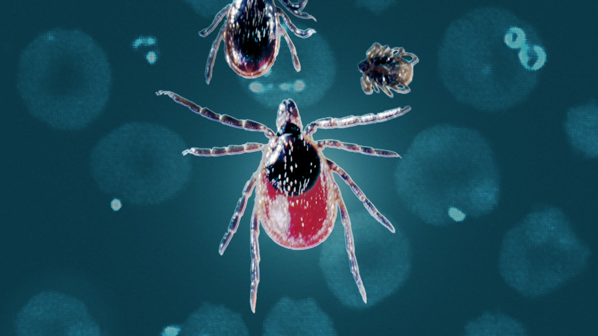
Best Herbal Antibiotic Plans for Lyme, Bartonella, and Babesia
https://treatlyme.com/guide/best-herbal-antibiotics-for-lyme-bartonella-babesia/
Best Herbal Antibiotic Plans for Lyme, Bartonella, and Babesia
By Dr. Marty Ross

Science Meets Buhner for Best Herbal Antibiotic Options
History Speaks
Historically, most herbal antibiotic regimens for used tick-borne infections are based on the writings and experience of master herbalist Stephen Buhner. His work is science related. However, most of the herbal antibiotics he recommends do not have actual studies showing they work in the lab or in humans for killing specific tick-borne infections. For instance, he recommends Andrographis to kill Borrelia based on science showing it kills another spirochete called Leptospirosis. And Buhner recommends Sida Acuta to address Babesia because it is used as an antimalarial, even though there is no research showing it works for Babesia.
Buhner’s writings occurred before the discovery of persister Borrelia (Lyme) and Bartonella which I describe below. So, his writings did not specifically address how to deal with these hibernation forms of germs.
Enter Science
Over the last few years, researchers are rushing to find new ways to kill the terrible Bs (Borrelia, Bartonella, and Babesia). Some of the interest in looking at herbal medicine options is the discovery of hibernating persister growth states of Borrelia and Bartonella that do not respond to classic herbal medicines or prescription regimens that target growing states of these germs. Out of this laboratory work, we now know that Buhner’s Andrographis does not work against Borrelia, but many other agents do.
In 2023 Shor and Schweig published their review of newer laboratory studies showing which herbal medicines work in the lab to kill the growing, persister, and biofilm states of Borrelia and Bartonella. This work also reveals numerous agents that can kill Babesia. Table 1. below is drawn from the Shor-Schwieg article. My table is more limited than the one published in their paper but focuses on what I have found clinically to be the most relevant herbal antibiotics.
Table 1. Herbal Antibiotic Actions

How to Interpret Table 1
- About G P B. Borrelia and Bartonella exist in growing states, hibernation states, and biofilm communities. The growing states are also called active states. The hibernators are also called persisters or stationary states. Biofilms are mostly known as biofilms. I prefer to use the terms growing (G), persister (P) and biofilms (B) while Shor and Schweig refer to active, stationary, and biofilm states. Keep this in mind if you review their article and more extensive table.
- About Blank. In some instances, a blank space in the table means the research did not look to see if an herbal agent actually addresses the identified problem. For instance, Zhang and colleagues showed that cinnamon, clove, and oregano oils kill Borrelia biofilms, but their research did not look at whether these herbal oils help Bartonella biofilm. Given the similarity of biofilm structures, cinnamon, clove and oregano oils may actually be good agents against Bartonella biofilms.
- About Sida Acuta and Houttuynia. Buhner recommends Sida Acuta and Houttuynia to address Bartonella. He also recommends Sida Acuta for Babesia. These key herbal antibiotics are not included in my table or the work of Shor-Schweig because there was no research conducted looking at these agents. This does not mean they do not work, but based on science, we do not know. (See link for article)
_____________
**Comment**
The article gives numerous treatment options for each pathogen. We can be extremely thankful to have all of this information in an easy to find and use format which is supported by science.
For more:
- https://madisonarealymesupportgroup.com/2024/08/16/antibiotics-vs-herbs-one-docs-experience/
- https://madisonarealymesupportgroup.com/2025/01/31/herbs-for-bartonella-babesia/
- https://madisonarealymesupportgroup.com/2021/05/07/john-hopkins-study-5-herbs-that-can-kill-babesia/
- https://madisonarealymesupportgroup.com/2017/10/13/oregano-cinnamon-and-clove-found-to-have-high-anti-persister-activity-for-bb/
- https://madisonarealymesupportgroup.com/2020/02/22/evaluation-of-natural-botanical-medicines-for-activity-against-growing-and-non-growing-forms-of-b-burgdorferi-in-vitro/
- https://madisonarealymesupportgroup.com/2023/07/20/lyme-disease-the-pursuit-of-a-clinical-cure/
- https://madisonarealymesupportgroup.com/2016/02/13/lyme-disease-treatment/
- https://madisonarealymesupportgroup.com/2016/01/16/babesia-treatment/
- https://madisonarealymesupportgroup.com/2016/01/03/bartonella-treatment/