STAT reported Thursday that a Chicago hospital treating severe Covid-19 patients with Gilead Sciences’ antiviral medicine remdesivir in a closely watched clinical trial is seeing rapid recoveries in fever and respiratory symptoms, with nearly all patients discharged in less than a week.
What does this early peek at the data tell us?
What does it not tell us?
And what comes next with clinical trials of remdesivir?
STAT senior writer Adam Feuerstein and Matthew Herper brief you on what’s known so far, and take questions from STAT Plus subscribers, in a live video chat.
The live chat will stream below starting at 1 p.m. ET on Friday, April 17.
_____________________
**Comment**
Interestingly, China wants a national patent on it. This would mean if granted, they would exclusively control the drug. Chinese authorities state they won’t seek a “compulsory license,” which means nations can override the patent in national emergencies. This is frightening.
Lyme disease is a multisystem infection caused by Borrelia burgdorferi that mainly affects the joints, the heart, and the nervous system. Neurological complications usually manifest in untreated patients and present as meningitis, cranial neuropathies, and radiculoneuritis. The authors present the case of a 48-year-old male who developed loss of vision in the right eye over a period of two months. On physical examination a relative afferent pupillary defect of the right eye was noted. Visual evoked potential test revealed delayed P100 latency bilaterally, confirming a bilateral optic neuropathy. The analysis of the cerebrospinal fluid (CSF) showed a lymphocytic meningitis. After an extensive work-up, a diagnosis of Lyme neuroborreliosis with meningitis and optic neuritis was made. The patient was treated with antibiotics and showed gradual improvement. The follow-up brain MRI revealed a mild T2 hyperintensity on the right optic nerve with gliosis, sequelae of the inflammatory process.
Lyme disease should always be considered in patients from endemic areas with nonspecific symptoms. The diagnosis of neuroborreliosis is challenging, but prompt identification and treatment can prevent the development of complications and sequelae.
________________
**Comment**
Again, just because there isn’t thousands of cases in the literature – doesn’t mean this is rare. It’s only rare because so many go undiagnosed and testing misses over half of all patients. If you type in eye or optic in the search bar on this website, you will quickly determine eye issues with Lyme is not rare at all.
How T. gondii infection causes seizures, and psychiatric illness for some
Summary: Study shows how Toxoplasma parasitic infections promote the loss of inhibitory signaling in the brain by altering the behavior of microglia.
Source: Virginia Tech
Think about traffic flow in a city – there are stop signs, one-way streets, and traffic lights to organize movement across a widespread network. Now, imagine what would happen if you removed some of the traffic signals.
Among your brain’s 86 billion neurons are the brain’s own version of stop signals: inhibitory neurons that emit chemicals to help regulate the flow of ions traveling down one cell’s axon to the next neuron. Just as a city without traffic signals would experience a spike in vehicle accidents, when the brain’s inhibitory signals are weakened, activity can become unchecked, leading to a variety of disorders.
In a new study published in GLIA on March 11, Virginia Tech neuroscientists at the Fralin Biomedical Research Institute at VTC describe how the common Toxoplasma gondii parasite prompts the loss of inhibitory signaling in the brain by altering the behavior of nearby cells called microglia.
The Centers for Disease Control and Prevention estimates that 40 million Americans have varying levels of Toxoplasma infection, although most cases are asymptomatic. Commonly passed to humans via exposure to farm animals, infected cat litter, or undercooked meat, the parasitic infection causes unnoticeable or mild, to flu-like symptoms in most healthy people. But for a small number of patients, these microscopic parasites hunker down inside of neurons, causing signaling errors that can result in seizures, personality and mood disorders, vision changes, and even schizophrenia.
“After the initial infection, humans will enter a phase of chronic infection. We wanted to examine how the brain circuitry changes in these later stages of parasitic cyst infection,” said Michael Fox, a professor at the Fralin Biomedical Research Institute and the study’s lead author.
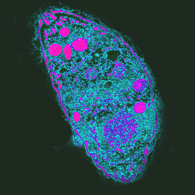
The parasite forms microscopic cysts tucked inside of individual neurons.
“The theory is that neurons are a great place to hide because they fail to produce some molecules that could attract cells of the immune system,” said Fox, who is also director of the research institute’s Center for Neurobiology Research.
Fox and his collaborator, Ira Blader, recently reported that long-term Toxoplasma infections redistribute levels of a key enzyme needed in inhibitory neurons to generate GABA, a neurotransmitter released at the specialized connection between two neurons, called a synapse.
Building on that discovery, the scientists revealed that persistent parasitic infection causes a loss of inhibitory synapses, and they also observed that cell bodies of neurons became ensheathed by other brain cells, microglia. These microglia appear to prevent inhibitory interneurons from signaling to the ensheathed neurons.
“In neuropsychiatric disorders, similar patterns of inhibitory synapse loss have been reported, therefore these results could explain why some people develop these disorders post-infection,” Fox said.
Fox said the inspiration for this study started years ago when he met Blader, a collaborating author and professor of microbiology and immunology at the University at Buffalo Jacobs School of Medicine and Biomedical Sciences, after he delivered a seminar at Virginia Tech. Blader studied Toxoplasma gondii and wanted to understand how specific strands of the parasite impacted the retina in mouse models.
Working together, the two labs found that while the retina showed no remarkable changes, inhibitory interneurons in the brain were clearly impacted by the infection. Mice – similar to humans – exhibit unusual behavioral changes after Toxoplasma infection. One hallmark symptom in infected mice is their tendency to approach known predators, such as cats, displaying a lack of fear, survival instincts, or situational processing.
“Even though a lot of neuroscientists study Toxoplasma infection as a model for immune response in the brain, we want to understand what this parasite does to rewire the brain, leading to these dramatic shifts in behavior,” Fox said.
The parasite forms microscopic cysts tucked inside of individual neurons. The image is in the public domain.
Future studies will focus on further describing how microglia are involved in the brain’s response to the parasite.
Among the research collaborators is Gabriela Carrillo, the study’s first author and a graduate student in the Translational Biology, Medicine, and Health Program. Previously trained as an architect before pursuing a career in science, Carrillo chose this topic for her doctorate dissertation because it involves an interdisciplinary approach.
“By combining multiple tools to study infectious disease and neuroscience, we’re able to approach this complex mechanistic response from multiple perspectives to ask entirely new questions,” Carrillo said. “This research is fascinating to me because we are exposing activated microglial response and fundamental aspects of brain biology through a microbiological lens.”
The study’s other contributing authors include Valerie Ballard, a Roanoke Valley Governor’s School high school student; Taylor Glausen, a graduate student working in Blader’s laboratory at the University at Buffalo; Zack Boone, a Virginia Tech undergraduate student; Cyrus Hinkson, a fourth-year Virginia Tech Carilion School of Medicine student; and Elizabeth Wohlfert, an assistant professor of microbiology and immunology at the University at Buffalo.
Source: Virginia Tech Media Contacts:
Whitney Slightham – Virginia Tech Image Source:
The image is in the public domain.
Toxoplasma infection induces microglia‐neuron contact and the loss of perisomatic inhibitory synapses
Infection and inflammation within the brain induces changes in neuronal connectivity and function. The intracellular protozoan parasite, Toxoplasma gondii, is one pathogen that infects the brain and can cause encephalitis and seizures. Persistent infection by this parasite is also associated with behavioral alterations and an increased risk for developing psychiatric illness, including schizophrenia. Current evidence from studies in humans and mouse models suggest that both seizures and schizophrenia result from a loss or dysfunction of inhibitory synapses. In line with this, we recently reported that persistent T. gondii infection alters the distribution of glutamic acid decarboxylase 67 (GAD67), an enzyme that catalyzes GABA synthesis in inhibitory synapses. These changes could reflect a redistribution of presynaptic machinery in inhibitory neurons or a loss of inhibitory nerve terminals. To directly assess the latter possibility, we employed serial block face scanning electron microscopy (SBFSEM) and quantified inhibitory perisomatic synapses in neocortex and hippocampus following parasitic infection. Not only did persistent infection lead to a significant loss of perisomatic synapses, it induced the ensheathment of neuronal somata by myeloid‐derived cells. Immunohistochemical, genetic, and ultrastructural analyses revealed that these myeloid‐derived cells included activated microglia. Finally, ultrastructural analysis identified myeloid‐derived cells enveloping perisomatic nerve terminals, suggesting they may actively displace or phagocytose synaptic elements. Thus, these results suggest that activated microglia contribute to perisomatic inhibitory synapse loss following parasitic infection and offer a novel mechanism as to how persistent T. gondii infection may contribute to both seizures and psychiatric illness.
Toxoplasmosis causes many mental issues and psychiatrist E. Fuller Torry believes that 75% of schizophrenia is associated with infections, with Toxo a significant portion.
Nawrocki and his colleague first discussed this case in the Air Medical Journal in 2018.
A 15-year-old boy experienced an episode of exertional syncope while at a trampoline park. Syncope is a temporary loss of consciousness.
He had a history of an attention-deficit/hyperactivity disorder.
His friends immediately called 911. They were not sure how long he was unconscious.
The boy was pale with a heart rate of 300 beats per minute, according to the medics. His EKG showed a wide complex tachyarrhythmia. That means that the heart is fast, and the QRS parts of his EKG were wide.
He was given a dose of amiodarone, but the ventricular tachycardia remained a problem. Amiodarone is a medicine used to treat and prevent irregular heartbeats.
At the hospital, he was dizzy and was short of breath. His heart rate remained between 290 and 300. His blood pressure dropped to 66/30 mm Hg.
He required synchronized cardioversion. Synchronized cardioversion is a procedure similar to electrical defibrillation. Synchronized cardioversion uses low energy synchronized with the heart.
His EKG changed to a third-degree heart block. That occurs when there is a complete block of impulses from the atrium to the ventricle. The atrium is the top of the heart, and the ventricle is the bottom. He had pacing pads placed.
He was flown to a cardiac intensive care unit (ICU) at a children’s tertiary care hospital via rotary-wing aircraft staffed with two flight nurses.
He continued to have a complete heart block with a fast rhythm.
CARDIAC LYME DISEASE
Doctors suspected Lyme disease and prescribed intravenous ceftriaxone. The diagnosis of Lyme disease was confirmed by “anti-Lyme titers” and Western blot tests.
His low heart rate and low pressure continued. The doctors inserted a transvenous pacemaker to control the heart rate.
He had three additional episodes of ventricular tachycardia. He was cardioverted twice.
But after several days of intravenous antibiotics, the boy’s heart block gradually resolved, and the pacemaker was removed.
He completed 28-days of IV treatment. He remains well on a one-year follow-up, according to the authors.
The authors note that conduction problems have been reported previously in untreated Lyme disease. The list of conduction abnormalities includes first and second-degree AV block, ST and T wave changes, prolonged QT, junctional tachycardia, and complete heart block.
There was no mention of conduction problems in patients who have been treated. I have not seen conduction problems in patients I have treated.
What can we learn from these cases?
Cardiac Lyme disease or more commonly known as Lyme carditis, can lead to life-threatening rhythm disturbances.
The conduction problems with cardiac Lyme disease can rapidly change.
Antibiotic treatment was helpful.
A pacemaker was necessary in this case. The doctor was able to remove the pacemaker after treatment with antibiotics.
What questions do these cases raise?
How often does cardiac Lyme disease occur?
Are there cases of cardiac Lyme disease cases that are not diagnosed?
Were there any warning signs of cardiac Lyme disease that would have allowed treatment before the young man collapsed?
4. Would the doctors have been able to recognize cardiac Lyme disease early before the need for hospitalization, air transport, and cardioversion?
TREATING TICK-BORNE DISEASE IN MY PRACTICE
In my practice, each individual requires a careful assessment. That is why I order a broad range of blood tests for other illnesses in addition to tick-borne infections. I also arrange consultations with specialists as needed.
Many patients are complex, as highlighted in this Inside Lyme Podcast series.
We need more doctors with skills recognizing Lyme carditis. We hope that professionals evaluating individuals with Lyme carditis can use this case to remind them to look for tick-borne illnesses and treat accordingly.
Inside Lyme Podcast Series
This Inside Lyme case series will be discussed on my Facebook and made available on podcast and YouTube. As always, it is your likes, comments, and shares that help spread the word about this series and our work. If you can, please leave a review on iTunes or wherever else you get your podcasts.
Sign up for our newsletter to keep up with our cases.
References:
Nawrocki PS, Poremba M. A 15-Year-Old Male With Wide Complex Tachyarrhythmia. Air Med J. 2018;37(6):383-387.
Summary: University of Melbourne researchers are conducting trials to see if intravenous administration of zinc chloride will help combat some of the effects of COVID-19. Previous studies have shown zinc is effective at slowing the rate of other respiratory infections, such as SARS.
Source: University of Melbourne
A world-first trial will see researchers from Austin Health and the University of Melbourne use intravenous zinc to fight the symptoms of coronavirus (COVID-19).
The trial will be led by Dr Joseph Ischia from Austin Health, along with Dr Oneel Patel from the Department of Surgery at the University of Melbourne, who has a long history of investigating the protective effects of intravenous zinc against organ damage induced by lack of oxygen.
Dr Ischia said COVID-19 is especially dangerous because it replicates inside a patient’s body which can lead to respiratory conditions like bronchitis and pneumonia.
“If COVID-19 enters a patient’s lungs then they often need to be placed on a ventilator to help their breathing and, in severe cases, COVID-19 can cause multiple organ failure and brain injury due to a lack of oxygen,” Dr Ischia said.
Dr Patel said studies have shown that zinc is very effective at slowing the rate that similar viruses such as SARS (Severe Acute Respiratory Syndrome) and common cold (a type of coronavirus) replicate in the body.
“Our published studies have also shown that high doses of zinc can protect vital organs such as the heart, kidneys and liver against the damage caused by a lack of oxygen,” Dr Patel said.
The clinical trial has been fast-tracked to test whether receiving a daily injection of zinc chloride will benefit patients with coronavirus.
“There is currently no specific treatment available for patients who have COVID-19 and are at high risk of respiratory failure, which means this study has the potential to have an enormous positive impact on their clinical outcomes,” Dr Ischia said.
“Importantly, we hope to show that we can save lives by limiting the impact of the symptoms. We are expecting to have preliminary results of the trial available after only seven days so we will know very quickly how effective this treatment is.”
The trial is the culmination of a rapid collaboration between surgeon scientists as well as intensive care, infectious diseases and respiratory medicine doctors at Austin Health, working with the Australian pharmaceutical firm, Phebra.
The clinical trial has been fast-tracked to test whether receiving a daily injection of zinc chloride will benefit patients with coronavirus. The image is adapted from the University of Melbourne news release.
Phebra Chief Executive Officer Dr Mal Eutick said intravenous (IV) zinc injections, manufactured at Phebra’s multi-purpose sterile injectables plant in Sydney, would be used in the trial.
“Zinc has been proven to be effective in treating severe pneumonia and other viruses although not COVID-19 to date. This trial is an extraordinary opportunity to discover if IV zinc can help us respond to the current pandemic,” Dr Eutick said
“If successful this could save lives and with this trial we should know in a short time frame. In particular, it could be very important for those high risk elderly patients and also help reduce the level of general anxiety in the community.”
However, both Dr Ischia and Dr Eutick warned of the need to manage the risk of zinc overdose for patients.
“Zinc can be toxic and it will be carefully administered as part of the trial to ensure patients are safe,” Dr Ischia explained.
About this COVID-19 research article
Source: University of Melbourne Media Contacts:
Media team – University of Melbourne Image Source:
The image is adapted from the University of Melbourne news release.