Archive for the ‘Bartonella’ Category

Cat Scratch Disease: Vet Suffers Extreme Fatigue For a Decade After Catching Rare, Severe Case of Bartonella Infection (That Isn’t Rare)

https://www.newsweek.com/cat-scratch-disease-vet-suffers-extreme-fatigue-decade-after-catching-rare-severe-case-1444715

CAT SCRATCH DISEASE: VET SUFFERS EXTREME FATIGUE FOR A DECADE AFTER CATCHING RARE, SEVERE CASE OF BARTONELLA INFECTION

A vet has been left suffering with extreme fatigue for almost a decade, after she caught an infection from a cat scratch which caused symptoms so severe she thought she was going blind or had a brain tumor.

A flea-infested cat scratched Victoria Altoft, 41, from the county of Somerset in south west England, while she was at work in the fall of 2010, PA Real Life reported.

Weeks later, Altoft’s muscles and joints were in pain and she was hit by night sweats, leading her to assume she had the flu. She was “utterly exhausted” and took the uncharacteristic decision to take two weeks off work.

“I just couldn’t get out of bed,” she told PA Real Life. As time passed, her joints swelled up, which her doctor put down to post-viral inflammation.

But Altoft became worried when her vision started to blur. She went for an emergency eye appointment, and medics thought her symptoms could be caused by a brain tumor or the condition multiple sclerosis, which affects the central nervous system.

Tests revealed she was suffering a rare Bartonella infection, and doctors prescribed her with antibiotics: the treatment given to serious cases of the condition.

black cat kitten pet animal stock getty
Cat scratch disease can be passed on by infected pets. GETTY

The bacteria is carried by infected fleas which live on animals like cats or dogs. Lice and sandflies are also vectors of the Bartonella group of bacteria which can cause cat scratch disease, as well as Carrion’s disease (only found in the Andes Mountains), and trench fever (most often present in people who live in areas of poverty with poor sanitation).

In most people, cat scratch disease doesn’t require treatment and fades by itself in between two to four months. But severe cases require antibiotic treatment.

Symptoms materialize several days or weeks after the bacteria invades the body. After three to 10 days, a painless raised red spot might appear on the skin where the infection passed through the skin. Over time this may become filled with fluid, with a crust forming before it heals. The lymph nodes near the site of infection might become swollen, red and hot to the touch, and puss-filled. Other symptoms include a general feeling of illness, headache, fatigue, and fever and—less often—sore throat and weight loss.

It took a year for her sight to return to normal. Altoft told PA Real Life she still suffers from fatigue despite being scratched in 2010.

“To this day, it’s difficult to know exactly what the long-lasting effects of contracting Bartonella are, as there is so little research, but I know I’m not the same now as I was before it happened,” she said.

Altoft is working with the Big Flea project run by the University of Bristol and the pharmaceutical company MSD Animal Health, who are researching the parasites which affect dogs and cats in the U.K.

The vet urged pet owners to take flea infestations seriously as they can pose a serious threat to human health.

____________________

**Comment**

First off, Bartonella is NOT RARE.

Second, someone PLEASE cut the nails on that cat!

For many, many people Bartonella is NOT something that, “fades by itself in between two to four months.”

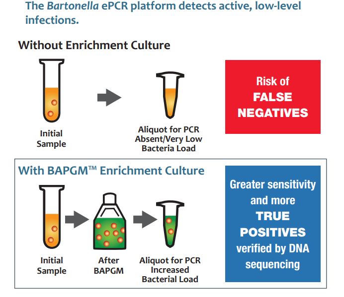
Bartonella is a particularly tenacious infection that can cause so many symptoms it boggles the mind. Couple it with Lyme disease and you are one sick dog. Throw in Babesia, and you are in bed for a long, long time.

https://madisonarealymesupportgroup.com/2019/04/24/human-bartonellosis-an-underappreciated-public-health-problem/Excerpt from full-text

KNOWN DISEASES CAUSED BY BARTONELLA INFECTIONS INCLUDE:
  • Carrion’s disease
  • cat-scratch disease
  • chronic lymphadenopathy
  • trench fever
  • chronic bacteraemia
  • culture-negative endocarditis
  • bacilliary angiomatosis
  • bacilliary peliosis
  • vasculitis
  • uveitis [1,2,4,6,7,9,10,11].
RECENTLY, BARTONELLA INFECTIONS HAVE BEEN LINKED TO MORE DIVERSE MANIFESTATIONS SUCH AS:
  • hallucinations
  • weight loss
  • muscle fatigue
  • partial paralysis
  • pediatric acute-onset neuropsychiatric syndrome (PANS)
  • other neurological manifestations [6,8,10].

Regarding vectors, it’s far more than fleas, lice, and sandflies:

Bartonella spp. are zoonotic pathogens transmitted from mammals to humans through a variety of insect vectors including the sand fly, cat fleas, and human body louse [4,5]. New evidence suggests that ticks, red ants, and spiders can also transmit Bartonella [15,16,17,18]. Bed bugs have been implicated in the transmission cycle of B. quintana and have been artificially infected [19]. B. quintana was found in bed bug feces for up to 18 days postinfection [19]. The diversity of newly discovered Bartonella species, the large number and ecologically diverse animal reservoir hosts, and the large spectrum of arthropod vectors that can transmit these bacteria among animals and humans are major causes for public health concern.

Regarding ticks….

3.3 Arachnids (Spiders &Ticks)

Over the last 10 years, the topic of ticks transmitting Bartonella species has been widely debated. Evidence exists to support the transmission of Bartonella through many different species of ticks.

Ixodid ticks, also known as hard ticks, appear to be the main type of tick associated with these bacteria. Tick cell lines have been used to show that Bartonella can replicate and survive within:

  • Amblyoma americanum (Lone Star Tick)
  • Rhipicephalus sanguineus (Brown Dog Tick)
  • Ixodes scapularis cells [77] (Deer Tick)

In California, questing ticks of

  • Ixodes pacificus (Western Black legged Tick)
  • Dermacentor occidentalis (Pacific Coast Tick)
  • Dermacentor variabilis (American Dog Tick)

were collected when in the adult and nymphal stages and tested for Bartonella by PCR for the citrate synthase gene. [78]. All types of ticks were found to contain Bartonella DNA, although in varying percentages and locations.These data alone do not prove that ticks can transmit Bartonella spp. Bacteria; however, the results do show Bartonella DNA occurring naturally in these wild ticks.

I know researchers are currently working on the link between Bartonella and cancer. Recently a young boy was diagnosed with schizophrenia but was found to have Bartonella:  https://madisonarealymesupportgroup.com/2019/03/24/cat-scratch-disease-caused-teens-schizophrenia-like-symptoms-report-says/

All you have to do is type “Bartonella” into the search bar on this website and let your fingers do the walking.  Bartonella is HUGE and quite common.

https://madisonarealymesupportgroup.com/2018/05/07/fox-news-bartonella-is-the-new-lyme-disease/

More on Bartonella: https://madisonarealymesupportgroup.com/2016/01/03/bartonella-treatment/

It’s a killer:  https://www.ncbi.nlm.nih.gov/pmc/articles/PMC3044516/#!po=1.02041

Look at the pictures of what it did to this woman:  https://madisonarealymesupportgroup.com/2019/05/28/woman-wakes-up-with-black-eye-swollen-face-after-cat-scratch-that-left-her-on-iv-drip-for-four-days/I assure you – this would not have faded on its own….

Lastly, Dr. Ericson has incredible imaging showing Bartonella surviving around tissues where a PIC line pumped antibiotics directly into the body:  https://madisonarealymesupportgroup.com/2019/02/27/advanced-imaging-found-bartonella-around-pic-line/

Trust me.  You don’t want this.


Other Arthropod-Borne Bacteria Causing Nonmalarial Fever in Ethiopia

https://www.ncbi.nlm.nih.gov/pubmed/31184993/

2019 Jun 10. doi: 10.1089/vbz.2018.2396. [Epub ahead of print]

Arthropod-Borne Bacteria Cause Nonmalarial Fever in Rural Ethiopia: A Cross-Sectional Study in 394 Patients.

Abstract

Bacterial arthropod-borne pathogens are a common cause of fever in Africa, but their precise impact is unknown and usually underdiagnosed in the basic rural laboratories of low-resourced African countries. Our aim was to determine the prevalence of arthropod-borne bacterial diseases causing fever among malaria smear-negative patients in a rural hospital located in Ethiopia. The study population included patients aged 2 years or older; referred to Gambo Rural General Hospital (West Arsi, Ethiopia), between July and November 2013, for fever or report of fever in the previous 48 h; attending the outpatient department; and testing negative for malaria by Giemsa-stained thin blood smears. We extracted DNA from 394 whole blood samples, using reverse line blot assays of amplicons to look for bacteria from the genera: Anaplasma, Bartonella, Borrelia, Coxiella, Ehrlichia, Francisella, and Rickettsia.

Thirteen patients showed presence of DNA for these pathogens: three each by Borrelia spp., the Francisella group (F. tularensis tularensis, F. tularensis holartica, and F. novicia), Rickettsia bellii, and Rickettsia Felis, and one by Bartonella rochalimae. Thus, in this rural area of Africa, febrile symptoms could be due to bacteria transmitted by arthropods. Further studies are needed to evaluate the pathogenic role of R. bellii.

_________________

**Comment**

What if some of this is mosquito-borne as well? We frankly don’t know because the transmission studies are screaming to be done.

https://madisonarealymesupportgroup.com/2018/11/07/are-mosquitoes-transmitting-lyme-disease/

https://madisonarealymesupportgroup.com/2018/02/12/wolbachia-laced-mosquitoes-being-released-why-lyme-msids-patients-might-be-negatively-affected/


Retinal Vessel Occlusion Caused by Bartonella Infection

https://www.ncbi.nlm.nih.gov/pmc/articles/PMC6236082/

. 2018 Nov 19; 33(47): e297.
Published online 2018 Oct 29. doi: 10.3346/jkms.2018.33.e297
PMCID: PMC6236082
PMID: 31044568

A Case of Retinal Vessel Occlusion Caused by Bartonella Infection

An external file that holds a picture, illustration, etc. Object name is jkms-33-e297-g003.jpg
An external file that holds a picture, illustration, etc. Object name is jkms-33-e297-g004.jpg

A 29-year-old female visited the emergency room with sudden visual loss in the right eye started on the same day. She had been suffering from fever for two days. A best-corrected visual acuity (BCVA) was 0.5/0.7 in the Snellen chart. Fundus examination (Fig. 1) showed multiple retinal hemorrhages. Severe vascular sheaths around the optic disc area were present in the right eye. Candle-wax-dripping sign in the superior hemisphere were found in the left eye.

An external file that holds a picture, illustration, etc. Object name is jkms-33-e297-g001.jpg

On systemic examination, erythema of the lower extremities (Fig. 1C) and right inguinal lymph node enlargement were discovered. With systemic doxycycline (100 mg) and gentamicin (90 mg) administration, fever subsided after three days. Bartonella infection was confirmed after 10 days with in-house indirect immunofluorescent assay (IFA) analysis (immunoglobulin G; cutoff points for seropositive titer at 1:64). Lymph node biopsy showed necrotizing granulomatous lymphadenitis (Fig. 1D). On the same day, the BCVA decreased to hand motion in the right eye. When asked, she could not specify when the vision loss began. The candle-wax-dripping sign in the left eye had progressed to vascular sheath with flame-shaped hemorrhages. Fluorescein angiography shows a rack of filling of the retinal arteries. Blocked fluorescence by retinal hemorrhage was found in the whole area of right eye and in the superotemporal quadrant of left eye. Inner-retinal hyper-reflectivity of the right eye and cystoid macular edema in the left eye were revealed (Fig. 2). The impression was central retinal artery and vein occlusion for the right eye and branch retinal artery and vein occlusion for the left eye, associated with severe vasculitis secondary to Bartonella infection. The patient was treated with a systemic methylprednisolone 500 mg, anticoagulant (Enoxaparin sodium 60 mg) and Rifampin (300 mg). Three month after disease onset, the BCVA in the right eye improved to 0.1. For photographs and medical records that consisted possible identification of the patient, a consent form was obtained from the patient for use of publication.

An external file that holds a picture, illustration, etc. Object name is jkms-33-e297-g002.jpg

ACKNOWLEDGMENTS

The authors thank Professor Jin-Soo Lee, Department of Internal Medicine, Inha University School of Medicine, for his help with the in-house IFA analysis.

Footnotes

Funding: This research was supported by the Bio & Medical Technology Development Program of the National Research Foundation of Korea (NRF), funded by the Korean government, the Ministry of Science and ICT (MSIP) (NRF-2017M3A9E2056458).

Disclosure: The authors have no potential conflicts of interest to disclose.

Contributed by

Author Contributions: Conceptualization: Woo M, Kim SW. Investigation: Woo M, Ahn S. Writing – original draft: Woo M. Writing – review & editing: Ahn S, Song JY, Kim SW.

References

1. Kwon HY, Im JH, Lee SM, Baek JH, Durey A, Park SG, et al. The seroprevalence of Bartonella henselae in healthy adults in Korea. Korean J Intern Med. 2017;32(3):530–535. [PMC free article][PubMed] []
____________________

Breaking Down the Baronella Spp. ePCR Triple Blood Draw: How Does It Work?

https://www.galaxydx.com/bartonella-epcr-steps/

Breaking down the Bartonella spp. ePCR Triple Blood Draw: How does it work?

ArminLabs (EliSpot) With Dr. Schwarzbach – Podcast

http://www.betterhealthguy.com/episode93

Why You Should Listen

In this episode, you will learn about EliSpot testing and the various testing options available through ArminLabs in Germany.

Watch The Show

Listen To The Show

Find The Show On…

About My Guest

My guest for this episode is Dr. Armin Schwarzbach.  Armin Schwarzbach, MD, PhD is a medical doctor and a specialist in laboratory medicine from the laboratory ArminLabs in Augsburg, Germany.  Dr. Schwarzbach began by studying biochemistry at Hoechst AG in Frankfurt, Germany and pharmacy at the University of Mainz in Germany in 1984. In 1985 he studied medicine for 6 years at the University of Mainz and finished his MD in 1991.  Dr. Schwarzbach developed the worldwide first Radioimmunoassay (RIA) for human Gastric Inhibitory Polypeptide from 1986 – 1991, getting his PhD in 1992.  He is member of the Swiss Association for tick-borne diseases, the German Association of Clinical Chemistry and Laboratory Medicine, and the German Society for Medical Laboratory Specialists.  He is an Advisory Board member of AONM London, England, and Board member of German Borreliosis Society, and Member and former Board Member of the International Lyme and Associated Diseases Society (ILADS) and has served as an expert on advisory committees on Lyme Disease in England, Australia, Canada, Ireland, France, and Germany.  Dr. Schwarzbach is the founder and CEO of ArminLabs in Augsburg, Germany and has specialized in diagnostic tests and treatment options for patients with tick-borne diseases for over 20 years.

Key Takeaways

  • What is an EliSpot?
  • What organisms can be tested for using EliSpot technology?
  • How specific is the EliSpot in testing for Borrelia, Bartonella, Babesia, and other organisms?
  • Does the state of the immune system matter when considering EliSpot results?
  • Which infections are the most persistent?
  • Can the EliSpot be used to track progress or success of treatment?
  • What is Yersinia and where might it be encountered?
  • Can EliSpot testing be used in newborns and infants?
  • What role do viruses such as EBV, CMV, Coxsackie, and others play in chronic illness?
  • Can Mast Cell Activation Syndrome be triggered by viruses?
  • Why are Mycoplasma and Chlamydia so important to explore?
  • Why is IgA testing a promising new direction in laboratory medicine?
  • Is CD57 helpful clinically?
  • What microbes are more commonly associated with specific medical conditions?
  • How common are Rickettsial organisms?
  • What is “Post Lyme Syndrome”? Is it real?

Connect With My Guest

http://arminlabs.com

Disclaimer

The content of this show is for informational purposes only and is not intended to diagnose, treat, or cure any illness or medical condition. Nothing in today’s discussion is meant to serve as medical advice or as information to facilitate self-treatment. As always, please discuss any potential health-related decisions with your own personal medical authority.