I’m planning to present an hour long slideshow to educate people on what Lyme disease (MSIDS) really is, the ongoing controversy about how to diagnose, and treat it, many of the issues keeping people from becoming well, and a general over view of tick-borne illness. This is a great refresher course as well as a great opportunity to educate family and friends interested in learning more.
There will be plenty of time for Q & A afterward as well as time for support and help.
The Ad Hoc Patient and Physician Coalition against IDSA Proposed Lyme Guidelines
SEP 7, 2019 —
From: CARL TUTTLE <runagain@comcast.net>
To: tickbornedisease@hhs.gov
Cc: (91 Undisclosed recipients)
Date: September 7, 2019 at 8:48 AM
Subject: The Ad Hoc Patient and Physician Coalition; Comments of the IDSA Proposed Lyme Guidelines September 6, 2019
To the Tick-Borne Disease Working Group;
Please follow the link below to see comments to the IDSA Proposed Lyme Guidelines. These comments are endorsed by members around the world because what has been established here in the United States has adversely affected public health globally.
The link provided is from my personal Dropbox storage area.
The Ad Hoc Patient and Physician Coalition
Comments of the IDSA Proposed Lyme Guidelines September 6, 2019
About the Ad Hoc Patient and Physician Coalition: The Ad Hoc Patient and Physician Coalition consists of patients and physicians who are concerned that the proposed Lyme disease guidelines of the Infectious Diseases Society of America (IDSA), American Academy of Neurology (AAN), and American College of Rheumatology (ACR) will further restrict access to care and harm patients by leaving them undiagnosed and undertreated.The coalition consists of over 75 organizations, from 8 countries: the US, Canada, Australia, UK, France, Netherlands, Germany, Belgium (including LymeDisease.org, the national Lyme Disease Association, Bay Area Lyme Foundation among others) and the International Lyme and Associated Diseases Society, which represents clinicians who treat Lyme disease nationwide. (List of signatories attached.)
These comments were authored by Lorraine Johnson, JD|MBA, CEO LymeDisease.org and Dr. Elizabeth Maloney, who represents the International Lyme and Associated Diseases Society. The comments were filed on August 8, 2019 and revised on September 6, 2019.
Page 56
Ad Hoc Patient and Physician Coalition Members As of September 6, 2019
The following 77 organizations have endorsed the Ad Hoc Patient and Physician Coalition Comments of the IDSA Proposed Lyme Guidelines. (List updated Sept. 6, 2019)
Countries represented: United States, Canada, Australia, France, United Kingdom, Netherlands, Germany, and Belgium.
Alabama Lyme Disease Association
Arkansas Lyme Coalition
Arkansas Lyme Foundation
Association Québécoise de la Maladie de Lyme (Canada)
Bay Area Lyme Foundation
BC Lyme (British Columbia, Canada)
Brookfield/Wolfeboro New Hampshire support group
Borreliose-Bund, Germany
Canadian Lyme Disease Foundation
Central Massachusetts Lyme Foundation
Charles E. Holman Morgellons Disease Foundation
Chronic Lyme Disease Support and Protest UK.
ChroniLyme (Lyon, France)
Colorado Tick-Borne Disease Awareness Association
Dean Center for Tick Borne Illness, Massachusetts
Dutch Lyme Association (Lymevereniging), Netherlands
Florida Lyme Disease Association
France Lyme, Association de Lutte Contre les Maladies Vectorielles à Tiques (France)
Howard County Lyme Awareness Group, Maryland
Hudson Valley Lyme Disease Association
Illinois Lyme Association
International Lyme and Associated Diseases Society
Kentucky Lyme Disease Association
Le Droit de Guerir (“The right to heal”), France
Litchfield County Lyme Network (Connecticut)
Living with Lyme, London/Middlesex (Canada)
LivLyme Foundation
Lyme Action Network
Lyme Alliance of the Berkshires
Lyme Association of Greater Kansas City
Lyme Center, Chico, California
LymeDisease.org
Lyme Disease Association (US)
Lyme Disease Association of Australia
Lyme Disease Association of Southeastern Pennsylvania
Lyme Disease Association, Rhode Island Chapter
Lyme Disease Education & Support Groups of Maryland
Lyme Disease UK
Lyme Hope (Canada)
Lyme-int Association (Belgium)
Lyme Newport (Rhode Island)
Lyme Ontario (Canada)
Lyme Stats 57
LymeConnection.org
LymeLight Foundation
Madison Lyme Support Group
Massachusetts Lyme Legislative Task Force
Memphis Tennessee Lyme Network
Midcoast Lyme Disease Support & Education
Military Lyme Support
MSIDS, Inc, Australia
NE Ohio Lyme Foundation
New Brunswick Lyme Disease Association Inc.- LymeNB (Canada)
New Jersey Lyme Resource
New York City Lyme Disease Support Group
OnLyme-Aktion.org (Action Alliance Against Tick-borne Diseases), Germany
Ontario Lyme Alliance (Canada)
PA Lyme Resource Network (Statewide)
PA Lyme Resource Network, Bucks County
PA Lyme Resource Network, Columbia County
PA Lyme Resource Network, Lehigh Valley
Patient Centered Care Advocacy Group
Pennsylvania Lyme Disease Awareness Committee of Chester-Delaware County Farm Bureau (PLDAC)
Penticton, BC area Lyme Group (Canada)
Sam’s Spoons Foundation for Lyme Support & Education
Southern Arizona Lyme Disease Association
Southern Oregon Lyme Disease
Texas Lyme Alliance
Tick Wise Education, Inc. (501c3)
Time for Lyme, ASBL (Belgium)
TxLyme Yahoo Online Support Group (Texas)
Utah Coalition for Lyme Disease
Vernon Lyme Disease Support Group, Vernon, BC (Canada)
Patients with Bartonella species infection (bartonellosis) complain of a variety of nonspecific vision problems that can affect every function of the eye. Making diagnosis and treatment decisions even more difficult, these problems can be caused by a variety of other pathogens and diseases. Fortunately, many peer-reviewed case publications, especially in ophthalmology journals, are available that describe both common and uncommon eye symptoms caused by bartonellosis.
Documented links between Bartonella species infection and vision problems focus on Bartonella henselae (cat scratch disease) and Bartonella quintana (trench fever). B. henselae is associated with contact with animals and vectors, especially cats and fleas, while B. quintana is associated with body lice. Additional species implicated include B. elizabethae and B. grahamii.
Overview
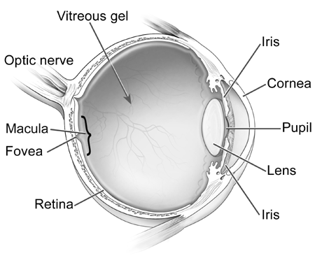
The eye consists of structures that focus light on nerve receptors at the back of the eye, nerves that feed into the optic nerve which connects to the brain, and a blood supply that connects through a central artery and vein into the body’s blood circulation. Bartonellosis can affect each of these parts of the eye.
Patients with bartonellosis-related eye problems may have symptoms in other organs as well that can help clarify whether the eye symptoms are caused by bartonellosis. Ophthalmologists and other physicians look for these additional signs because all of the eye conditions that can be caused by bartonellosis can be caused by a variety of bacteria and viruses, as well as other health conditions including autoimmune disorders.
It can be difficult to narrow down the possible causes of bartonellosis. Sometimes patients can’t remember any events that would have exposed them to Bartonella species. Other times the event, such as a cat scratch, may have occurred weeks to a month or more before symptoms appear and the patient may not think there is a connection. It can take detailed questioning by physicians to identify the possibility of Bartonella species exposure.
Structural Eye Symptoms
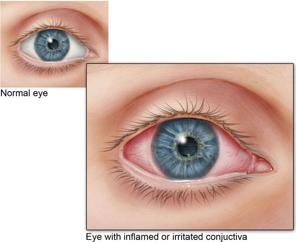
The most common way bartonellosis is seen in the eye is called Parinaud oculoglandular syndrome (POGS). About five percent of patients with acute cat scratch disease have this syndrome, which is characterized by follicular conjunctivitis (pink eye) with swollen lymph nodes nearby. It is often accompanied by a fever, and there may be bumps on the eyelid. Other symptoms known to be caused by bartonellosis, such as endocarditis (swelling of the inner lining of the heart), may also indicate that bartonellosis should be suspected.
Conjunctivitis results in easily seen inflammation of the usually white part of the eye (Source: CDC).
Inflammation of the middle layer of the structure that surrounds the eyeball is called uveitis. Uveitis causes redness of the eye and can cause light sensitivity, pain and floaters. Uveitis is sometimes associated with bartonellosis.
While case reports of eye symptoms caused by bartonellosis generally describe a sudden-onset condition, one case report describes a woman who had symptoms of bartonellosis in various body organs for more than five years, including chronic conjunctivitis (pink eye). She had multiple tests and treatments over that time including a Bartonella species test that was positive but considered nonspecific. It was only after other treatments didn’t work that antibiotics were administered. The antibiotics resolved her various symptoms.
Neurological Eye Symptoms
Neuroretinitis, an inflammation of the optic nerve head, occurs in about 2% of people with cat scratch disease (acute Bartonella henselae infection). Two-thirds of cases of neuroretinitis are caused by bartonellosis.
Neuroretinitis is usually characterized by sudden, complete vision loss and swelling that creates a star pattern in the macula (the light-sensitive tissue at the back of the eye that feeds information into the optic nerve). Though this is the typical case of neuroretinitis caused by bartonellosis, it can vary greatly. It can cause changes such as seeing odd shapes or colors Furthermore, case reports have included people who lose their vision with no other symptoms, have blurry vision with a headache, and more.
Treatment can usually, but not always, restore vision, but it can take months to resolve and there can still be long-term consequences. Complications can also occur. In one case, a child was diagnosed with neuroretinitis. Treatment was started six weeks after the diagnosis, but his vision in one eye got worse. After treatment, a full-thickness macular hole was discovered. The hole was monitored and resolved after six months.
Vascular Eye Symptoms
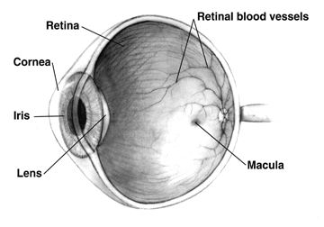
The eye has an important network of tiny blood vessels that provide nourishment to the tissue, but unnecessary growth of new capillaries can lead to a range of symptoms such as vision problems. Vasoproliferation (irregular growth of new blood vessels) may be more common in immunocompromised people, such as those being treated with chemotherapy products. These symptoms can be observed on the skin and in the liver and spleen and may also occur in the eye.
Tiny blood vessels in the eyes supply nutrients to cells. In the above image, larger retinal blood vessels can be seen (Source: NIH).
Vasoproliferative symptoms seem to be caused by vascular endothelial growth factor (VEGF) stimulated by bartonellosis. More research on the relationship between VEGF and bartonellosis is needed. Meanwhile, anti-VEGF agents have been used to treat vasoproliferative eye symptoms.
Conclusion
Bartonellosis can affect every part of the eye, and symptoms can be sudden and severe. Diagnosis and treatment decisions are complicated by other pathogens and diseases that can cause similar symptoms. It is important for patients and physicians to be aware of any prior animal or insect exposure that may indicate Bartonella infection. Considering additional systemic symptoms of bartonellosis may also help to clarify the diagnosis.
Learn more about bartonellosis and the testing that Galaxy Diagnostics offers here.
References
Kalogeropoulos, C. et al. (2011). Bartonella and intraocular inflammation: A series of cases and review of literature. Clinical Ophthalmology, 5, 817-829. doi:10.2147/OPTH.S20157 https://www.ncbi.nlm.nih.gov/pmc/articles/PMC3130920/
Woo, M. et al. (2018). A case of retinal vessel occlusion caused by Bartonella infection. Journal of Korean Medical Science, 33(47), e297. doi:10.3346/jkms.2018.33.e297 https://www.ncbi.nlm.nih.gov/pmc/articles/PMC6236082/
Fairbanks, A. M. et al. (2019). Treatment strategies for neuroretinitis: Current options and emerging therapies. Current Treatment Options in Neurology, 21(8), 36. doi:10.1007/s11940-019-0579-0 https://www.ncbi.nlm.nih.gov/pubmed/31278547
Michel, Z. et al. (2019). Multimodal imaging of two unconventional cases of Bartonella neuroretinitis [epub ahead of print]. Retinal Cases & Brief Reports. doi:10.1097/ICB.0000000000000893 https://www.ncbi.nlm.nih.gov/pubmed/31348120
Gunzenhauser, R. C. et al. (2019). The development and spontaneous resolution of a full-thickness macular hole in Bartonella henselae neuroretinitis in a 12-year-old boy. American Journal of Ophthalmology Case Reports, 15, 100515. doi:10.1016/j.ajoc.2019.100515 https://www.ncbi.nlm.nih.gov/pubmed/31341998
Toumanidou, V. et al. (2017). Neuroretinitis secondary to Bartonella henselae in a patient with myelinated retinal nerve fibers: Diagnostic dilemmas and treatment. Ocular Immunology and Inflammation, 27(3), 396-398. doi:10.1080/09273948.2017.1409357 https://www.ncbi.nlm.nih.gov/pubmed/29283743
Mabra, D. et al. (2018). Ocular manifestations of bartonellosis. Current Opinion in Ophthalmology, 29(6), 582-587. doi:10.1097/ICU.0000000000000522 https://www.ncbi.nlm.nih.gov/pubmed/30124532
Beckerman, Z. et al. (2019). Rare presentation of endocarditis and mycotic brain aneurysm [epub ahead of print]. The Annals of Thoracic Surgery. doi:10.1016/j.athoracsur.2019.06.073 https://www.ncbi.nlm.nih.gov/pubmed/31425670
_________________
**Comment**
Another great article by Galaxy Lab. Please read the following article for more information on Bartonella, as various strains are suspected to be transmitted by ticks, mites, various flies and spiders, and other modes, and it is far more prevalent than thought: https://madisonarealymesupportgroup.com/2016/01/03/bartonella-treatment/ Fifteen species of gram-negative aerobic Bartonella are known to infect humans; however Dr. Ricardo Maggi’s statement is quite telling, “This case reinforces the hypothesis that any Bartonella species can cause human infection.”
Summary: Understanding how parasites ‘hack’ the brains of their hosts may provide new insights into decision making and behavior.
Source: Frontiers
Imagine a parasite that makes an animal change its habits, guard the parasite’s offspring or even commit suicide. While mind-control may sound like something out of a science fiction movie, the phenomenon is very real — and has spawned a new field, neuro-parasitology. As outlined in an article published in Frontiers in Psychology, understanding how parasites “hack” their host’s nervous system to achieve a particular goal could provide new insights into how animals control their own behavior and make decisions.
“Parasites have evolved, through years of co-evolution with their host, a significant ‘understanding’ of their hosts’ neuro-chemical systems,” explains one of the article’s authors, Professor Frederic Libersat from Ben-Gurion University of the Negev in Israel. “Exploring these highly specific mechanisms could reveal more about neural control of animal behavior.”
The article describes some of the sophisticated, cunning and gruesome ways that various parasites outwit and exploit their insect hosts.
One method is to affect how an insect navigates. The spores of one parasitic fungus, for example, invade an ant’s body, where the fungus grows and consumes the ant’s organs while leaving the vital organs intact. The fungus then releases chemicals that cause the ant to climb a tree and grip a leaf with its mouthparts. After emerging from the ant’s body, the fungus releases spore-filled capsules that explode during their fall, spreading the infectious spores over the ground below. By forcing the ant to climb a tree, the fungus increases the dispersal of the falling spores and the chance of infecting another ant.
Similarly, a parasitic hairworm causes infected crickets to seek out water — where they drown. The cricket’s suicide enables the worms to enter an aquatic environment for reproduction.
In another type of interaction, called “bodyguard manipulation,” the parasite forces the infected insect to guard its young. One such parasite is a wasp, which injects its eggs into a caterpillar by stinging it. Inside the live caterpillar, the eggs hatch into larvae, which feed on the caterpillar’s blood. Eventually, as many as 80 larvae emerge from the caterpillar’s body before forming cocoons to complete their growth into adult wasps.
However, wasp larvae are vulnerable to predators in their cocoons. To scare potential predators away, one or two larvae remain in the caterpillar and control its behavior through an unknown mechanism, so that it acts aggressively towards predators — thereby protecting the cocoons.
These examples shed light on the very old and highly specific relationship between parasites and hosts. But how exactly do these parasites affect their host’s behavior?
Neuro-parasitology is still a young field, and in most cases, researchers do not yet fully understand the mechanisms involved. However, many such parasites produce their effects by releasing compounds that act on the neural circuitry of the host. Identifying and using these compounds in the lab could help scientists to work out how neural circuits control behavior.
“Because neurotoxins are the outcome of one animal’s evolutionary strategy to incapacitate another, they are usually highly effective and specific,” says Libersat.
“Chemical engineers can generate hundreds of potential neurotoxins in the lab, but these are random and often useless, whereas any natural neurotoxin has already passed the ultimate screening test, over millions of years of co-evolution.”
ABOUT THIS NEUROSCIENCE RESEARCH ARTICLE
Source: Frontiers Media Contacts:
Conn Hastings – Frontiers Image Source:
The image is adapted from the Frontiers news release.
Mind Control: How Parasites Manipulate Cognitive Functions in Their Insect Hosts
Neuro-parasitology is an emerging branch of science that deals with parasites that can control the nervous system of the host. It offers the possibility of discovering how one species (the parasite) modifies a particular neural network, and thus particular behaviors, of another species (the host). Such parasite–host interactions, developed over millions of years of evolution, provide unique tools by which one can determine how neuromodulation up-or-down regulates specific behaviors. In some of the most fascinating manipulations, the parasite taps into the host brain neuronal circuities to manipulate hosts cognitive functions. To name just a few examples, some worms induce crickets and other terrestrial insects to commit suicide in water, enabling the exit of the parasite into an aquatic environment favorable to its reproduction. In another example of behavioral manipulation, ants that consumed the secretions of a caterpillar containing dopamine are less likely to move away from the caterpillar and more likely to be aggressive. This benefits the caterpillar for without its ant bodyguards, it is more likely to be predated upon or attacked by parasitic insects that would lay eggs inside its body. Another example is the parasitic wasp, which induces a guarding behavior in its ladybug host in collaboration with a viral mutualist. To exert long-term behavioral manipulation of the host, parasite must secrete compounds that act through secondary messengers and/or directly on genes often modifying gene expression to produce long-lasting effects.
Parasite patients often express the psyche of the parasites – sticky, clingy, impossible to tolerate – but a wonderful human being is behind all of that.
We are all a composite of many personalities. Chronic infections outnumber our own cells by 10:1. We are 90% “other” and 10% “us”. Our consciousness is a composite of 90% microbes and 10% us.
Our thinking, feeling, creativity, and expression are 90% from the microbes within us. Patients often think, crave, and behave as if they are the parasite.
Our thinking is shaded by the microbes thinking through us. The food choices, behavioral choices, and who we like is the thinking of the microbes within us expressing themselves.
Patients will reject all treatments that affect the issue that requires treating.
Patients will not guide themselves to health when the microbes have taken over.
And, as mentioned in this article: the fungus Cordyceps hijacks the ant to propitiate itself but here again, many Lyme patients use Cordyceps to fight microbes, lower inflammation, and increase energy and oxygen: https://rawlsmd.com/herbs/cordyceps
South Korean researchers who analyzed 24 studies and published their report on Oct. 10 in Environmental Health — found significantly higher risks for malignant brain tumors, meningioma and glioma on the side of the head where cellphones were held.
GreenMedInfo LLC. This work is reproduced and distributed with the permission of GreenMedInfo LLC. Want to learn more from GreenMedInfo? Sign up for the newsletter here //www.greenmedinfo.com/greenmed/newsletter.”
Cancer is a term used for diseases in which abnormal cells divide without control and are able to invade other tissues … all cancers begin in cells … cells grow and divide in a controlled way to produce more cells as they are needed to keep the body healthy. When cells become old or damaged, they die and are replaced with new cells. However, sometimes this orderly process goes wrong. The genetic material (DNA) of a cell can become damaged or changed, producing mutations that affect normal cell growth and division. When this happens, cells do not die when they should and new cells form when the body does not need them.”
So cancer typically involves abnormal cell division and DNA damage and in some cases cells may form a mass of tissue called a tumor.
Types Of Brain Tumor
In the studies done to date cell phone radiation exposures are principally linked to two types of brain tumor: gliomas and acoustic neuromas.
Gliomas, a type of tumor that starts in the brain or spine are typically malignant. Gliomas are particularly deadly. Most people survive only 1 to 3 years after diagnosis.
Acoustic neuromas, though non-malignant (low-grade cancer), are in many cases life threatening given that they are an intracranial tumor.
The 44 Reasons
1. Cellular Damage: Telecoms giant T-Mobile in Germany commissioned an independent study to review all relevant research on the health risks from wireless telecommunications. It was concluded that,
On the cellular level, a multitude of studies found the type of damage from high frequency electromagnetic fields which is important for cancer initiation and cancer promotion.”
Brain Tumors And Brain Cancers
2. Significantly Increased Risk of Glioma: Gliomas are becoming increasingly common. The $25 million Interphone Study found that:
… regular use of a cell phone by adults can significantly increase the risk of gliomas by 40% with 1640 hours or more of use (this is about one half hour per day over ten years).”
Source:Table 2 INTERPHONE Study Group. Brain tumour risk in relation to mobile telephone use: results of the INTERPHONE international case-control study. Int J Epidemiol (2010); 39(3):675-694
3. Tumor Risk on Cell Phone Side of Head: Again from the Interphone Study – currently the big daddy of cell phone radiation studies it being the largest and longest study on the link between cell phones and brain tumors – it also found, “tumors were more likely to occur on the side of the head most used for calling.”
4. Harmful Association Between Cell Phone Radiation and Tumors: A review of 23 epidemiological studies by 7 scientists on the link between cell phones and cancer concluded, “harmful association.” One of the reports authors commenting the study results said, “although as a whole the data varied, among the 10 higher quality studies, we found a harmful association between phone use and tumor risk. The lower quality studies, which failed to meet scientific best practices, were primarily industry funded.”
5. Increased Risk For Glioma and Acoustic Neuroma: the studies performed by the Hardell Research Group are widely regarded as being amongst the best. This recent study finds, “A consistent pattern of increased risk for glioma and acoustic neuroma associated with use of wireless phones.” These findings are consistent with their earlier studies.
6. Temporal Lobe & Glioma Risk: A recent French study found evidence of an increased risk of glioma and temporal lobe tumors. The study found that, “risks were higher for gliomas, temporal tumours, occupational and urban mobile phone use.” According to EMF watchdog Powerwatch this is an important paper, “that confirms existing studies and which should help move the IARC RF evaluation strongly towards a Group 2A – ‘probable human carcinogen’.”
7. Increased Risk of Acoustic Neuroma in Long-Term Users of Cell Phones: A recent study on 790,000 middle aged women in the UK found that, “women who used cell phones for ten or more years were two-and- a-half times more likely to develop an acoustic neuroma. Their risk of acoustic neuroma increased with the number of years they used cell phones.”
8. Increased Risk of Acoustic Neuroma: Research conducted by Lonn suggests, “an increased risk of acoustic neuroma associated with mobile phone use of at least 10 years’ duration.”
9. Brain Tumor Risk is Higher on ‘Cell Phone’ Side of Head: A research paper that reviewed 11 studies found, “a link between prolonged cell phone usage and the development of an ipsilateral [same side of head as cell phone] brain tumor.”
10. Meningioma: This Swedish study looked at adult brain tumor cases diagnosed over a two year period. Although the study concluded that, “no conclusive evidence of an association between use of mobile and cordless phones and meningioma was found.” The studies authors did say, “an indication of increased risk was seen in the group with highest cumulative use.”
11. Malignant Brain Tumors: Recent work by Hardell looked at long-term use of mobile and cordless phones. In conclusion it was found that, “this study confirmed previous results of an association between mobile and cordless phone use and malignant brain tumors. These findings provide support for the hypothesis that RF-EMFs play a role both in the initiation and promotion stages of carcinogenesis.”
Image: Hardell Research Group
Other Cancers And Tumors
12. Cancer of the Pituitary Gland: The pituitary gland, considered by many to be the “master gland” of the body, is a pea sized organ located in the middle of the base of the brain that produces hormones that play a major role in regulating vital body functions and general well-being. This study (already referenced above) also found that,
the risk of cancer of the pituitary gland more was more than twice as high among women who used a cell phone for less than five years as compared to never users.”
13. Thyroid Cancer: The thyroid gland is situated in the neck. Using a cell phone against your ear exposes your thyroid to cell phone radiation. A recent Israeli study observing that, “the incidence of thyroid cancer has been on the rise in Israel for more than a decade which matches the rise in the use of cellphones” collected human thyroid cells from healthy patients and subjected them to radiation. The study found, “evidence of changes in thyroid cells in response to electromagnetic radiation.”
14. Melanoma Risk: Melanoma is a cancer that starts in a certain type of skin cell. A Swedish study found “a very clear association between increasing use of mobile phones and increasing rates of head melanoma in Nordic countries.”
Image: Örjan Hallberg
15. Stem Cell Cancer: In a controversial US study on 29 cases of neuroepithelial tumors, cell phone users accounted for 11 of them. These initial results indicated a near tripling in the risk of neuroepithelial tumors through cell phone use. The published results were revised to reflect a doubling of risk and then reported as not “statistically significant.”
16. Oral Cancer: An Israeli study on 460 cases of parotid gland tumors found, “based on the largest number of benign PGT patients reported to date, our results suggest an association between cellular phone use and PGTs [parotid gland tumors].” The parotid is the salivary gland near the cheek where many users hold their cell phone.
17. Parotid Malignant Tumors: Another Israeli study analyzed deaths as recorded on the National Cancer Registry over a 36 year period found, “the total number of parotid gland cancers in Israel increased 4-fold from 1970 to 2006 , whereas other major salivary gland cancers remained stable.”
Image: Environmental Health Trust
18. Leukemia: A comprehensive review of over a dozen studies including studies on exposures from cell tower radiation, TV and Radio broadcast towers concluded, “cancer, especially brain tumor and leukemia, but all other cancers also.”
19. Lymph Node Cancer: In an Australian study one hundred mice were exposed to RF radiation for two 30-minute periods per day for up to 18 months. The authors called the increased incidence of lymphoma “highly significant.” They added that “it is very unlikely that the faster onset of cancer was due to chance.”
20. Multifocal Breast Cancer: American researchers studied four young women with breast cancer. They found that, “all patients regularly carried their smartphones directly against their breasts in their brassieres for up to 10 hours a day, for several years, and developed tumors in areas of their breasts immediately underlying the phones.”
21. Eye Cancer: A German Study has established a link between uveal melanoma and cell phone radiation and similar exposures. The study “found an elevated risk for exposure to radiofrequency-transmitting devices.” Another study found ocular symptoms and sensations in long term users of mobile phones.
22. Diverse Cancerous Tumors: A Brazilian Study established a direct link between various cancer deaths such as tumors in the prostate, breast, lung, kidneys and liver in Brazil’s third largest city, and cell phone tower radiation exposures. The study found that, “more than 81 percent of people who die in Belo Horizonte by specific types of cancer live less than 500 meters away from the 300 identified cell phone antennas in the city”.
Source. This same study also lists more than a dozen other research papers that have found a link between different cancers and cell phone/cell tower radiation exposures.
Cell Phone Subscriptions And Brain Tumors
23. Cell Phone Subscription Link to Brain Tumors: A U.S. study analyzed the number of cell phone subscriptions and brain tumors in nineteen US states, they concluded,
the very linear relationship between cell phone usage and brain tumor incidence is disturbing and certainly needs further epidemiological evaluation.”
24. Brain Cancer Incidence Increases Over Time (U.S): Another U.S. study of brain cancer incidence trends in relation to cell phone use in the United States found, “there was a statistically significant increasing trend between 1992 and 2006 among females but not among males. The recent trend in 20-29-year-old women was driven by a rising incidence of frontal lobe cancers.”
25. Brain Cancer Incidence Increases Over Time (Europe): Studies carried out in Norway, Finland and the U.K. have identified a similar trend of an increase in the incidence of brain cancer over time. In the UK study the incidence of malignant brain tumors close to where you hold your phone was highlighted.
Source: Mobile Phone Use and Cancer Risk – Research on a Group 2B Carcinogen. Joel M. Moskowitz Ph.D.
Other Effects On the Brain
26. Blood-Brain Barrier (BBB) Permeability: The BBB is a membrane which prevents toxic materials from the blood from entering the brain. It was first discovered in 1975 that RF radiation causes the BBB to leak, since then at least a dozen laboratories around the world have corroborated this effect. There’s no consensus on the link between BBB damage and cancer but some studies elude to this.
27. Brain Cell Loss: A Turkish study on adult female rats that were exposed to a 900 MHz electromagnetic field found that, “EMF exposure caused a significant decrease of the … cell number … additionally, cell loss can be seen.” In their conclusions the researchers drew parallels between these exposures and teenagers’ brains that are exposed to cell phone radiation.
28. Brain Activity: Researchers in China exposed 18 participants to RF radiation (LTE) for 30 minutes which was well within international (ICNIRP) cell phone legal limits. They concluded that, “30min LTE RF-EMF exposure modulated the spontaneous low frequency fluctuations in some brain regions.”
29. Brain Blood Flow Affected: This Finnish brain imaging study found that “that the EMF emitted by a commercial mobile phone affects rCBF [regional cerebral blood flow] in humans.” This suggests that cell phone radiation affects neuronal activity.
30. Texting Affects Memory: An Australian study on young adolescents found “students who reported making or receiving more voice or SMS calls per week, and in particular more of both, demonstrated shorter response times on learning tasks, but less accurate working memory.”
DNA Damage
One way cancer and other diseases are believed to develop is when the DNA (genetic information) in a cell becomes damaged. This damage mutates the DNA. There are many studies linking cell phone radiation exposures to different types of DNA damage.
31. Single and Double-Strand DNA Breaks: In pioneering work a University of Washington team found DNA single strand breaks from RF radiation exposures on rats in an initial study. A subsequent study found single and double-strand DNA breaks.
32. Various Genetic Effects: An Austrian study analyzed the results of 101 different published articles on the effects of radio frequency EMFs on DNA. The study concluded that, “there is ample evidence that RF-EMF can alter the genetic material of exposed cells.”
33. Increased Rates of Micronuclei: Micronuclei proliferation indicates a type of DNA damage strongly associated with cancer. A Brazilian study found that, “electromagnetic field irradiation [low level cell phone type exposures] during pregnancy leads to an increase in erythrocytes micronuclei incidence in rat offspring.” Several studies have found increased rates of micronuclei in the body following exposures to RF radiation.
34. Heat Shock Proteins (HSPs) Production Decreased: A U.S. study exposed chick embryo’s to RF radiation. They concluded that, “this EMF-induced decrease in HSP70 levels and resulting decline in cytoprotection suggests a mechanism by which daily exposure (such as might be experienced by mobile phone users) could enhance the probability of cancer and other diseases.”
35. Oxidative DNA Damage: The Guler study in Turkey exposed female and male infant rabbits to 1800 MHz radio frequency radiation and found, “GSM-like RF radiation may induce biochemical changes by increasing free radical attacks to structural biomolecules.” Free radical damage is associated with the development of cancer.
36. DNA Strand Breaks: This Austrian study exposed human and rat cells to mobile phone radiation and found, “DNA single- and double-strand breaks.”
37. Changes in Gene Expression: The Belyaev study found that, exposing the “rat brain to 915 MHz GSM microwaves induces changes in gene expression.” Other studies suggest that, “subtle changes of gene expression associated with [disease].”
38. Genotoxic Effects: The Schwarz study exposed human cells to 1,950 MHz UMTS. It concluded that “UMTS exposure may cause genetic alterations in some but not in all human cells in vitro.”
39. Neurotransmitters Impacted: This Bavarian study followed 60 people over one and a half years following the installation of a new cell phone base station in their village. The study concluded that, “the effects showed a dose-response relationship,” that it had “occurred well below current limits for technical RF radiation exposures” and that these effects have “great relevance for health and [are] well known to damage human health in the long run.” In other words the more people were exposed to cell phone type radiation the bigger the impact on their health.
40. Chromosome Damage: A Belgian study reviewed 16 expert gene monitoring studies from around the world. In 13 of the 16 independent studies performed worldwide it was found that, “RF-exposed individuals have increased frequencies of genetic damage (e.g., chromosomal aberrations).”
41. Central Nervous System: US based researcher Dr. Henry Lai comments that there are several studies which show that repeated RF exposure at relatively low power caused morphological changes in the central nervous system, “changes in morphology, especially cell death, could have an important implication on health. Injury-induced cell proliferation has been hypothesized as a cause of cancer.”
Reading Between The Lines
The studies don’t tell all of the story. Here are some other things you need to know.
42. Latency Period Before Diagnosis: To put this in the words of researcher Dr. Martin Blank “cancers do not form overnight.” In almost all cases, cancerous tumors take many years to form and metastasize.” According to Dr. Blank’s statements, then cancers are overpowered. This would suggest that we might be sitting on a cell phone radiation cancer time bomb.
43. Cell Phone Radiation Cancer Time Bomb: To give a sense to what this latency period could mean in terms of the incidence of brain tumors in the years to come, researcher Lloyd Morgan produced this alarming graphic showing that brain tumor cases could reach epidemic proportions within the next decade:
44. Flawed Research: Not all of the research points to a link between cell phone radiation and cancer. But then that’s hardly surprising given the lengths some researchers go to, to skew the results. This research paper also lays bare the phenomenon of study bias. This can take many different shapes and forms; insufficient latency time, incorrect definition of “regular” cell phone user, cell phones radiating higher power levels in rural areas not investigated, exposure to other transmitting sources not considered, exclusion of brain tumor cases due to death or illness, etc.
The Tip Of The Iceberg
There is lot of interest surrounding the link between cell phone radiation and cancer. But cancer is only the tip of the iceberg.
Microwave radio-frequency radiation exposures of the type emitted by cell phones are also linked to many other diseases and potentially life threatening illnesses, including:
Learn the truth about wireless radiation and the global 5G rollout at the 5G Crisis Summit happening now! Get access here!
Article originally published: 2014-12-13 Article updated: 2019-08-26
Since falling prey to a violent reaction to his cell phone in 2002 Lloyd Burrell has spent the last 10 years researching the effects of electromagnetic fields (EMFs) on health.
You can download his free EMF Protection Health Report and subscribe to his newsletter by visiting his website https://www.electricsense.com.
He is also the author of an eBook entitled “How To Beat Electrical Sensitivity”, which offers a solution to the growing number of people whose health is being compromised by exposure to wireless and similar technologies, also available through his website.
Disclaimer: This article is not intended to provide medical advice, diagnosis or treatment. Views expressed here do not necessarily reflect those of GreenMedInfo or its staff.


 NEUROSCIENCE NEWS


