https://journals.lww.com/tnpj/Pages/articleviewer.aspx?year=2018&issue=10000&article=00011&type=Fulltext

Variable clinical presentations of babesiosis

Paparone, Pamela, DNP, APN; Paparone, Philip W., DO

doi: 10.1097/01.NPR.0000545000.07640.11
Abstract: 
Human babesiosis continues to spread in multiple regions of the US. It is transmitted by Ixodes species ticks, as are Lyme disease and anaplasmosis. Its variable clinical presentations, together with serologic detection limitations, require that a high index of clinical suspicion be present for prompt diagnosis. This article discusses case examples showing the wide range of symptoms and presentations that are possible with babesiosis.
Human babesiosis is an infectious, malaria-like disease caused by intraerythrocytic protozoa of the genus Babesia, specifically Babesia microti and Babesia divergens.1-4 It is transmitted by Ixodes species ticks, as are Lyme disease and anaplasmosis (formerly known as ehrlichiosis). Babesia species are well-known pathogens in animals. During the past half century in the US, they have been increasingly recognized as pathogens in humans.1,5 Babesiosis may be acquired through the bite of an infected tick, a blood transfusion, or by transplacental transmission.2,6-8 (See Ixodes scapularis [blacklegged or deer ticks].)
Most infection passes undetected (because the patient may be unaware of the tick bite), especially in healthy adults.6,7 However, in immunocompromised patients—particularly those with hematologic disease and a history of splenectomy—Babesia infection may be severe and life-threatening.1

Epidemiology

The first reported case of babesiosis in the US was in 1968.9 It became a nationally notifiable disease in 2011, and among the 27 states where it was notifiable in 2013, there were 1,792 reported cases nationwide.5,10 Tick-borne and transfusion-associated cases of babesiosis occur in multiple parts of the country, including outside of areas of known endemicity.5 The number of reported cases is rising steadily in the US and worldwide, owing in part to increased medical awareness and improved diagnostic methods.1-3 (See Reported cases of babesiosis in the US.)

Health departments notify the CDC of babesiosis cases via the National Notifiable Diseases Surveillance System (NNDSS) using a standard case definition. In addition to basic demographic information (age, gender, and county of residence) provided via NNDSS, supplemental data (symptoms and history of transfusion) can be submitted to the CDC using a disease-specific case report form (CRF). Because babesiosis has been a reportable condition in some states for years, state-developed CRFs had already been in use to capture supplemental data.5

To promote standard data collection, the CDC developed a babesiosis CRF, which was approved by the Office of Management and Budget in August 2011 (www.cdc.gov/parasites/babesiosis/resources/50.153.pdf). Supplemental data, derived from the CDC’s or a state’s CRF, were merged manually with NNDSS records by matching a case ID number or demographic data. If case records had conflicting data, the more detailed record was considered correct.

As cases of babesiosis transmitted via tick bite or blood transfusion occur in multiple parts of the US, including outside of areas of known endemicity, ongoing national surveillance using the standard case definition will provide a foundation for developing evidence-based prevention and control measures to reduce the burden of the disease. In addition, mapping based on this surveillance allows for the identification of endemic areas, which aids the clinician in diagnosis.

Transmission and pathogenesis

The heightened recognition of tick-borne infection is derived largely from the increasing incidences of human babesiosis, anaplasmosis, and Lyme disease, both individually and together.11,12 Because these infections share the same rodent reservoir and tick vector hosts, they can be cotransmitted to human hosts.1,2,10,13-16 Coinfections involving various combinations of these pathogens are common and can be severe.12,14 The babesia parasite is suspected of causing proinflammatory cytokines that stimulate the production of nitric oxide, which may cause erythrocytic cellular damage when produced in excess.2

Diagnostic procedures and clinical management of the resulting disease syndrome are complicated by the diversity of pathogens involved and by the unusual diversity and duration of symptoms.

Clinical presentation

Common clinical features of babesiosis are similar to those of malaria and range in severity from asymptomatic to rapidly fatal. Most patients experience a viral infection-like illness with fever, chills, sweats, myalgia, arthralgia, anorexia, nausea, vomiting, or fatigue, and in some cases, patients may develop hemolytic anemia.1-4,10 Most symptomatic patients become ill 1 to 4 weeks after the bite of a B. microti-infected tick and 1 to 9 weeks (but up to 6 months in one reported case) after transfusion of contaminated blood products.6-8

A high index of clinical suspicion for babesiosis and the possible presence of other tick-borne infections are required for prompt diagnosis and proper treatment. Because the clinical findings are nonspecific, lab studies are necessary to confirm the diagnosis.

Diagnosis

Microscopic examination of blood smears is the current gold standard for detecting Babesia infection, while polymerase chain reaction testing has promising diagnostic value.1,2,16,17 Differentiating Babesia from malaria on peripheral smears can be difficult but rapidly resolved by the presence or absence of a history of travel.1 Peripheral smears for Babesia allow for same-day or, at the most, next-day confirmation of the diagnosis. The case examples described below demonstrate the range of symptoms and clinical presentations associated with babesiosis (with and without coinfection) that can challenge the NP.

Babesiosis is caused by parasites that infect red blood cells. Most US cases are caused by B. microti, which is transmitted by Ixodes scapularis ticks, primarily in the Northeast and Upper Midwest. Babesia parasites also can be transmitted via transfusion, anywhere, at any time of the year. In March 2018, the FDA approved the first B. microti blood donor screening tests. B. microti Arrayed Fluorescent Immunoassay detects antibodies to B. microti in human plasma, and B. microti Nucleic Acid Test detects B. microti DNA in human whole blood.18

Treatment

**Please see my comment at end of article**

Generally, treatment with atovaquone plus azithromycin is used for patients with mild-to-moderate babesiosis, whereas clindamycin plus quinine is recommended for patients with severe disease; both treatment regimens are given for 7 to 10 days.1-4 All four drugs are used FDA off-label for babesiosis; however, the dosage recommendations are supported by the clinical guidelines.1-4,19 The dosage regimen for atovaquone plus azithromycin for adult patients is atovaquone 750 mg orally every 12 hours, and azithromycin 500 to 1,000 mg orally on day 1 and 250 mg orally once daily for the subsequent days.1-4 Immunocompromised patients may require higher doses of azithromycin.2-4

The dosage regimen for clindamycin plus quinine for adult patients with severe disease is clindamycin 600 mg orally every 8 hours or clindamycin 300 to 600 mg I.V. infusion every 6 hours, and quinine 650 mg orally every 6 to 8 hours.1-4 Dose adjustments of quinine are needed for patients with severe chronic kidney disease.19,20 Of note, the only FDA-approved preparation of oral quinine currently available in the US is the 324 mg capsule.19,20 Previously, the dosage available in the US was a 325 mg capsule. The change in the quinine preparation from 325 mg to 324 mg may result in minor dose disparities between some guideline dosage recommendations that were published before the commercial preparation was changed.20,21

Although rare cases of resistance to atovaquone plus azithromycin have been reported, this combination is effective in most patients.2 Atovaquone is contraindicated in patients who develop or have a history of serious allergic or hypersensitivity reactions to the drug or any of the drug’s components. Azithromycin is contraindicated in patients with known hypersensitivity to azithromycin or any macrolide or ketolide antibiotic and also in patients with a history of cholestatic jaundice or hepatic dysfunction.19 Clindamycin is contraindicated in patients with a history of hypersensitivity to clindamycin or lincomycin. Quinine is contraindicated in patients with known hypersensitivity to quinine, mefloquine, or quinidine; prolonged QT interval; a glucose-6-phosphate dehydrogenase deficiency; or a history of myasthenia gravis or optic neuritis.19 Consult the manufacturer’s prescribing label for complete prescribing information for each drug.

Some patients, including those with severe illness, might require or benefit from supportive care, such as antipyretics, vasopressors (if the patient’s BP is low and unstable), blood transfusions, exchange transfusions (in which portions of a patient’s blood or blood cells are replaced with transfused blood components), mechanical ventilation, and dialysis. The NP should consider referral to an infectious-disease specialist for patients who are pregnant, have an underlying hematologic or oncologic problem, have had a splenectomy, are allergic to first-line antibiotic agents, or have had an unsatisfactory response to antibiotic therapy.

Red blood cell exchange transfusions are recommended for cases of severe babesiosis in patients with parasitemia of 10% or greater, severe anemia (hemoglobin less than 10 g/dL), or pulmonary, kidney, or liver impairment.2-4 Exchange transfusions are used to rapidly decrease parasitemia, correct anemia, and help remove toxic byproducts produced by the infection.2

Case examples

The case examples of patients with babesiosis show a wide range of symptoms and clinical presentations. The case examples below are cases that occurred in southeastern New Jersey, where the disease is endemic. All patients were hospitalized and treated in Atlantic County, New Jersey (see Summary of data from patients with babesiosis).

Case 1

Ms. A is a 78-year-old White female who was admitted with fever, chills, lethargy, fatigue, and marked changes in sensorium. She had a maximum temperature of 100.6° F (38.1° C); sepsis was considered for this patient. Multiple tick bites were found. Pertinent lab findings included lactate dehydrogenase (LDH), 528 units/L; aspartate aminotransferase (AST), 90 units/L; and alanine aminotransferase (ALT), 34 units/L. Her vitamin B12 and folate levels were normal.

Ms. A’s initial white blood cell (WBC) count was 5.0 × 10⁹/L, but over the first 3 days of hospitalization, it gradually dropped to 2.6 × 10⁹/L. Her hemoglobin dropped from 10.5 g/dL to a low of 8 g/dL, and her platelets were initially 39 × 109/L but gradually increased as she continued her course of treatment. Ms. A had 33% polymorphonuclear leukocytes, 2% bands, 49% lymphocytes, and 13% monocytes. Peripheral smear was positive for Babesia, and she had a Babesia immunoglobulin M (IgM) of 1:160 and Anaplasma (previously referred to as Ehrlichia) IgM of 1:320.

In view of Ms. A’s leukopenia and thrombocytopenia, anaplasmosis was suspected, and she was treated with doxycycline 100 mg I.V. infusion every 12 hours, atovaquone suspension 750 mg orally twice daily, and azithromycin 500 mg I.V. infusion every 24 hours. Doxycycline is the recommended treatment for anaplasmosis and was administered to cover the possibility of anaplasmosis in this patient. She was treated with that regimen for 5 days. She was then started on doxycycline twice daily, and azithromycin 500 mg daily (both oral) along with the atovaquone suspension of 750 mg twice daily for a 14-day course of therapy. Ms. A made a dramatic improvement in her mentation and resolution of her lethargy.

Case 2

Ms. C is a 90-year-old White female with a chief complaint of rectal bleeding. On admission, her lab studies revealed severe anemia with a hemoglobin of 7.6 g/dL and hematocrit of 22.6%. Her platelet count was 103 × 109/L and peripheral smear was positive for Babesia. Ms. C had spiking temperatures 100° F to 101° F (37.8° C to 38.3° C). Her rectal bleeding was controlled with an octreotide infusion to which she responded well (the bleeding ceased). Her peripheral smear was positive for Babesia, and she was placed on an oral dose of azithromycin 500 mg on day 1 and then 250 mg daily and atovaquone suspension 750 mg twice daily to complete a 10-day course.

Case 3

Mr. E is a 57-year-old White male admitted with fever, malaise, and chills. His temperature had risen to 101° F (38.3° C). His AST and ALT were 64 and 54 units/L, respectively, and gradually rose to a peak of 90 and 87 units/L, respectively, during his 5-day hospital stay. Mr. E’s WBC count decreased from his initial hospital results to 2.9 x 10⁹/L with a hemoglobin of 9.2 g/dL. His platelets were initially 60 × 109/L but dropped to 34 × 109/L at their lowest level. In view of his elevated liver enzymes, leukopenia, and thrombocytopenia, anaplasmosis was highly suspected, and he was started on doxycycline 100 mg I.V. infusion every 12 hours.

Mr. E’s peripheral smear was positive for Babesia. He was started on oral clindamycin 600 mg every 8 hours and oral quinine 650 mg three times daily. Acute hearing deterioration occurred, and the quinine was discontinued. Mr. E’s regimen was then switched to oral azithromycin 500 mg on day 1 and then 250 mg daily and oral atovaquone 750 mg twice daily. He went on to complete only 7 days of therapy, and his elevated liver enzymes and thrombocytopenia resolved. The suspected anaplasmosis was not confirmed, as the Anaplasma IgM was negative. However, Mr. E’s leukopenia and thrombocytopenia resolved on the above regimens.

Case 4

Mr. J is an 81-year-old White male who was admitted with increasing lethargy, weakness, chills, and blurred vision. He had a history of coronary artery disease and hypertension. His hemoglobin on admission was 12.1 g/dL, and his hematocrit was 35.4%. His WBC count was 5.3 × 10⁹/L. By day 2, his hemoglobin had dropped to 9.9 g/dL with a hematocrit of 29%. His platelets were initially 54 × 109/L and dropped to 46 × 109/L, but on therapy, rose to 191 × 109/L.

Mr. J had 82% polymorphonuclear leukocytes, 10% lymphocytes, and 6% monocytes. On the day of admission, a peripheral smear was positive for Babesia. Subsequently, serologic studies demonstrated an Anaplasma IgG of 1:256; the IgM was negative. Babesia serologies were greater than 1:320, both IgG and IgM. Anaplasmosis was suspected with Mr. J’s confirmed babesiosis, and he was started on azithromycin 500 mg I.V. infusion every 24 hours and doxycycline 100 mg twice daily.

At discharge on day 10, Mr. J was switched to clindamycin orally three times a day and quinine orally three times a day because of intolerance to azithromycin, and he completed a 14-day course of therapy. He convalesced satisfactorily. His hemoglobin at discharge was 12.5 g/dL and WBCs 7.4 × 10⁹/L; platelets improved to 137 × 109/L.

Case 5

Mr. K is an 85-year-old White male who was admitted with fever and chills intermittently, recurring for several days prior to admission. He had a history of hairy cell leukemia, splenectomy, permanent pacemaker insertion for atrioventricular block, gouty arthritis, prostatic hypertrophy, and polymyalgia rheumatica. In the ED, Mr. K had an immediate peripheral smear for Babesia, and the intraerythrocytic parasite was demonstrated. He had been working on a golf course for the week prior to admission.

A second peripheral smear was positive for intraerythrocytic parasites with 10.4% of his red blood cells infected. Findings were also positive for Howell-Jolly bodies, which are erthrocytic nuclear remnants associated with asplenia or decreased splenic function. Mr. K was started on oral azithromycin 500 mg on day 1 and then 250 mg daily and atovaquone 750 mg suspension twice daily. Due to the possibility of concurrent tick-borne infection, he was also started on oral doxycycline 100 twice daily.

Over the course of day 1, Mr. K’s platelet count dropped from 25 to 23 × 109/L, with blood urea nitrogen of 29 mg/dL and creatinine of 1.2 mg/dL. His WBC count dropped from 4.1 to 2.5 × 10⁹/L, and his hemoglobin dropped from 16 to 13 g/dL. He had 20% bands, 5% atypical lymphocytes, 47% polymorphonuclear leukocytes, and 23% lymphocytes. Mr. K remained on doxycycline, azithromycin, and atovaquone suspension for 8 days when he was discharged home.

Mr. K was readmitted the following day when he complained of the inability to ambulate and generalized weakness. He had peripheral smear positivity with babesiosis and was serologically positive for anaplasmosis with both IgM and IgG. Mr. K had continued on the prescribed antibiotic regimen up until his readmission that day. Due to the persistence of parasitemia despite adequate therapy, he was changed to clindamycin 600 mg I.V. infusion every 8 hours, and quinine was also being administered.

Unfortunately, Mr. K developed gastric distress and a generalized erythematous coalescing rash, which prompted the discontinuation of the clindamycin and quinine. His WBC count was 2.2 × 10⁹/L, and his hemoglobin was 9.5 g/dL. Platelets had risen to 43 × 109/L, and he had 43% polymorphonuclear leukocytes, 10% bands, 42% lymphocytes, and 5% monocytes.

Because of the persistence of parasitemia, Mr. K underwent exchange transfusion. At that point, he had been restarted on azithromycin 500 mg I.V. infusion every 24 hours and atovaquone suspension 750 mg orally twice daily. Azithromycin and atovaquone were continued for 5.5 weeks, at which time he was parasite smear negative for Babesia. Subsequently, a Babesia peripheral smear remained negative.

Discussion of case examples

Case 1 shows the unusual effect of babesiosis on the sensorium in the older adult, as any infectious process can. The patient’s cognitive function was dramatically improved following treatment, despite the marked changes in mentation on admission. A coinfection with Anaplasma was suspected. In general, all cases of babesiosis need to be tested for late Lyme disease, via Western blot, although not immediately addressed.1,2,4

Patients with concurrent babesiosis and anaplasmosis—suspected or serologically positive—are treated with doxycycline, which is equally effective for Lyme disease, early or late. Generally, the greater number of concurrent tick-borne infections and the higher the parasitimia load, the more toxic the presentation.1,12

Case 2 shows the need to check the peripheral smear for Babesia despite the rectal bleeding issue on admission. This diagnostic test could have easily been omitted, causing a delay in the diagnosis. Such a delay in older adult patients that results in delayed treatment can put these patients at greater risk for severity of babesiosis. Generally, the combination of clindamycin and quinine has a much higher probability of intolerance and adverse reactions. This combination is not the treatment of first choice for babesiosis. Pertaining to anaplasmosis, the triad of leukopenia, transaminase elevation (mild or moderate), and thrombocytopenia demands empiric treatment with doxycycline prior to serologic confirmation.1,2,4

A peripheral smear for Babesia is rapidly interpreted, is inexpensive, and should be requested in evaluating all patients with any degree of anemia—especially during the spring and summer months in endemic areas. Serologic studies are variable in developing positivity and are generally less readily available.

Case 3 illustrates the importance of suspecting and investigating the possibility of babesiosis and anaplasmosis coinfection in a patient presenting with a tick-borne illness.

Case 4 demonstrates that no additional lab studies—other than peripheral smear for Babesia—are needed to confirm the diagnosis of babesiosis.

Case 5 exemplifies the therapeutic challenge and refractory response to treatment of babesiosis in patients with the comorbidities of a hematologic disease and/or splenectomy.

Patient education

Heightened awareness of babesiosis as well as prompt diagnosis and treatment are essential to prevention. Both patients and the general public need to become more aware of the existence of the disease and other tick-borne infections, especially individuals who live in or travel to regions where babesiosis is found. The NP can play an active and important role in providing patient education about the disease. The basic points of information to communicate include:

  • What babesiosis is and its potential to be a life-threatening illness
  • How individuals acquire babesiosis (tick bite, transfusion, or, rarely, vertical transmission)
  • Where in the world babesiosis is found
  • Signs and symptoms of babesiosis
  • Note that many individuals do not have any symptoms and do not get sick
  • Importance of seeing a healthcare provider if babesiosis is suspected
  • Treatability of babesiosis and need for prompt diagnosis and treatment.22,23

Individuals who live in or travel to endemic areas should avoid tick-infested areas; apply repellents and wear long pants and long-sleeved shirts when outdoors; shower soon after being outdoors; and check their entire body for ticks.3 When outdoors, they should walk on cleared trails, stay in the center of the trail, and minimize contact with leaf litter, brush, and overgrown grasses (where ticks are most likely to be found). If a tick is found attached to a person’s body, it should be properly removed as soon as possible.

The CDC offers a printable, one-page fact sheet for patients and the general public that details the basic information for babesiosis awareness in addition to the link for the CDC guide to proper removal of a tick attached to a person (www.cdc.gov/parasites/babesiosis/resources/babesiosis_fact_sheet.pdf).

Conclusion

This article illustrates the need for the NP to appreciate the variable clinical presentations of babesiosis to facilitate prompt diagnosis, provide proper therapeutic management, and avoid the poor outcomes associated with this disease. Staying knowledgeable of babesiosis is essential. It is important for the NP to understand that infected patients may not recall a tick bite and that clinical presentations may not only be variable but also nonspecific, ranging from subclinical to severe. The possibility of coinfection with other tick-borne illnesses (Lyme disease and anaplasmosis) must be considered. Furthermore, the NP needs to assume an active role in patient education to affect babesiosis awareness and prevention.

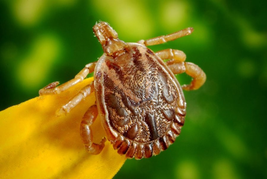
Ixodes scapularis (blacklegged or deer ticks)

The images below are of the Ixodes scapularis ticks, also known as blacklegged or deer ticks. From left to right, the male (M) with a dorsal scutum (also known as a shield on the hard-bodied tick) that covers the entire back on the male, the female (F) with only a portion of the back covered by the dorsal scutum, the nymph (N), and the larva (L).

Figure

Sourse: Procop GW, Church DL, Hall GS, et al. Koneman’s Color Atlas and Textbook of Diagnostic Microbiology. 7th edition. Philadelphia, PA: Wolters Kluwer Health, 2016.

Reported cases of babesiosis in the US1,2,22,23

Most cases of babesiosis in the US occur in seven states, five of which are located in the Northeast (MA, CT, RI, NY, and NJ) and two in the upper Midwest (MN and WI). The geographic range of babesiosis has expanded beyond these highly endemic areas and it is now reported all along the northeastern seaboard and inland, ranging from Maine to Maryland.

Sporadic cases of babesiosis have been reported in other areas of the US including the West Coast. Additionally, transfusion-associated cases of babesiosis can occur anywhere in the country. Congenital transmission of babesiosis has also been reported.

REFERENCES

1. Sanchez E, Vannier E, Wormser GP, Hu LT. Diagnosis, treatment, and prevention of Lyme disease, human granulocytic anaplasmosis, and babesiosis: a review. JAMA. 2016;315(16):1767–1777.

2. Vannier EG, Diuk-Wasser MA, Ben Mamoun C, Krause PJ. Babesiosis. Infect Dis Clin North Am. 2015;29(2):357–370.

3. Vannier E, Krause PJ. Human babesiosis. N Engl J Med. 2012;366(25):2397–2407.

4. Wormser GP, Dattwyler RJ, Shapiro ED, et al The clinical assessment, treatment, and prevention of Lyme disease, human granulocytic anaplasmosis, and babesiosis: clinical practice guidelines by the Infectious Diseases Society of America. Clin Infect Dis. 2006;43(9):1089–1134.

5. Centers for Disease Control and Prevention. Babesiosis surveillance—18 States, 2011. MMWR Morb Mortal Wkly Rep. 2012;61(27):505–509.

6. Levin AE, Krause PJ. Transfusion-transmitted babesiosis: is it time to screen the blood supply. Curr Opin Hematol. 2016;23(6):573–580.

7. Leiby DA. Babesiosis and blood transfusion: flying under the radar. Vox Sang. 2006;90(3):157–165.

8. Herwaldt BL, Linden JV, Bosserman E, Young C, Olkowska D, Wilson M. Transfusion-associated babesiosis in the United States: a description of cases. Ann Intern Med. 2011;155(8):509–519.

9. Scholtens RG, Braff EH, Healey GA, Gleason N. A case of babesiosis in man in the United States. Am J Trop Med Hyg. 1968;17(6):810–813.

10. Centers for Disease Control and Prevention. Notice to readers: final 2013 reports of nationally notifiable infectious diseases. MMWR Morb Mortal Wkly Rep. 2014;63(32):702–715.

11. Western KA, Benson GD, Gleason NN, Healy GR, Schultz MG. Babesiosis in a Massachusetts resident. N Engl J Med. 1970;283(16):854–856.

12. Diuk-Wasser MA, Vannier E, Krause PJ. Coinfection by Ixodes tick-borne pathogens: ecological, epidemiological, and clinical consequences. Trends Parasitol. 2016;32(1):30–42.

13. Herwaldt BL, McGovern PC, Gerwel MP, Easton RM, MacGregor RR. Endemic babesiosis in another eastern state: New Jersey. Emerg Infect Dis. 2003;9(2):184–188.

14. Thompson C, Spielman A, Krause PJ. Coinfecting deer-associated zoonoses: Lyme disease, babesiosis, and ehrlichiosis. Clin Infect Dis. 2001;33(5):676–685.

15. Paparone PW, Glenn WB. Lyme disease with concurrent ehrlichiosis. J Am Osteopath Assoc. 1994;94(7):568–570, 573, 577.

16. Hildebrandt A, Gray JS, Hunfeld KP. Human babesiosis in Europe: what clinicians need to know. Infection. 2013;41(6):1057–1072.

17. Wang G, Wormser GP, Zhuge J, et al Utilization of a real-time PCR assay for diagnosis of Babesia microti infection in clinical practice. Ticks Tick Borne Dis. 2015;6(3):376–382.

18. U.S. Food & Drug Administration. FDA approves first tests to screen for tickborne parasite in whole blood and plasma to protect the U.S. blood supply. 2018. http://www.fda.gov/newsevents/newsroom/pressannouncements/ucm599782.htm.

19. Facts and Comparisons. Drug. Facts and Comparisons 2013. St. Louis, MO: Wolters Kluwer Health; 2013.

21. Gelfand JA, Vannier EG. Clinical manifestations, diagnosis, treatment, and prevention of babesiosis. UptoDate. 2017. http://www.uptodate.com.

22. Joseph JT, Purtill K, Wong SJ, et al Vertical transmission of Babesia microti, United States. Emerg Infect Dis. 2012;18(8):1318–1321.

23. Centers for Disease Control and Prevention. Tickborne diseases of the United States. A reference manual for health care providers. 2017. http://www.cdc.gov/lyme/resources/TickborneDiseases.pdf.

____________________

**Comment**

According to Dr. Horowitz, an experienced LLMD (Lyme literate doctor), Babesia is a tenacious tick-borne infection that persists.  Experience has shown him patients often need 9-12 months of treatment, a far cry longer than what is suggested here.

For treatment options, please see:  https://madisonarealymesupportgroup.com/2016/01/16/babesia-treatment/

All patients of tick-borne infections need follow-up – years later from treatment.

While the research shows again and again the persistent symptoms of patients, the “powers that be” continue to treat this short-term and seemingly ignore the vast population out here struggling (and it’s far greater than the purported 5-10% of the patient population, I assure you).

The one drug, one disease paradigm also doesn’t work with most patients as we are often coinfected:  https://madisonarealymesupportgroup.com/2017/05/01/co-infection-of-ticks-the-rule-rather-than-the-exception/  This link shows that 45% of tested ticks were coinfected and carried up to 5 different pathogens. This directly translates to human infection and a survey substantiates this: https://madisonarealymesupportgroup.com/2014/11/14/studies-show-why-its-tough-to-treat-lyme-and-co/ The most common co-infections in the LDo study were Babesia (32%), Bartonella (28%), and Ehrlichia (15%) while a study by Dr. Janet Sperling in Canada found that the most common were Bartonella (36%), Babesia (19%), and Anaplasma (13%).

There is also the issue of tick bites igniting latent infections already within the human body such as Epstein Barr, numerous herpes viruses, and even Bartonella. Yet, patients are struggling with these – sometimes all at once.  Is it any wonder we are sicker than dogs?http://www.wildcondor.com/dr-horowitz-on-babesiosis.html Dr. Krause published in the New England Journal of Medicine that when a patient has Lyme and Babesia,Lyme is found three-times more frequently in the blood, proving Babesia suppresses the immune system. https://madisonarealymesupportgroup.com/2017/06/28/concurrent-babesiosis-and-lyme-in-patient/  Besides the fact it is a misnomer to think it novel that a patient has concurrent Lyme and Babesiosis, it is also a huge mistake to base treatment on geographical area as time and time again, entomologists are finding ticks in places they just shouldn’t be and ticks that shouldn’t be carrying pathogens, carrying them. Also, using logic, until every bird, fox, squirrel, lizard, deer, and every other rodent on the earth read the memo that they are not supposed to cross state and country boundaries, ticks are going to continue to defy the box “experts” put them into. And, there are other ways for pathogens to travel across state lines:https://doi.org/10.1111/tid.12741

Abstract
The potential for transmission of Babesia microti by blood transfusion is well recognized. Physicians may be unaware that products used for transfusion may be collected from geographically diverse regions. We describe a liver transplant recipient in South Carolina who likely acquired B. microti infection from a unit of blood collected in Minnesota.  Also, one must be careful of the “history of tick bite,” as well, as many never see the tick or subsequent bite, and fail to get a rash. A nymphal tick is nearly impossible to see. Lyme/MSIDS is a CLINICAL diagnosis.

So much research begging to be done.


https://dailyevergreen.com/38060/news/understanding-disease-transmission-in-ticks/

Understanding disease transmission in ticks

Study may help find ways to prevent future spread of pathogens

Understanding+how+immune+responses+affect+disease+transmission+could+be+the+first+step+toward+eventually+developing+a+preventative+treatment+for+tick-born+illnesses%2C+researchers+say.

Understanding how immune responses affect disease transmission could be the first step toward eventually developing a preventative treatment for tick-born illnesses, researchers say.
CAMERON SHEPPARD, Evergreen contributor
October 2, 2018


Research at WSU to understand how the mechanisms of tick immune systems operate could help uncover ways to affect how ticks carry and transmit diseases, many of which affect humans.

One type of tick, Ixodes scapularis, can transmit at least seven different diseases from feeding on the blood of a host, a number that is relatively high when compared to other bugs and insects that carry disease, said Dana Shaw, microbiologist and researcher at WSU.

Shaw said the I. scapularis’ high vector competency or ability to carry and transmit diseases, is what caught her attention. She wanted to study how the immune system of a tick operates to understand what mechanisms affect a tick’s ability to spread disease.

Shaw said much of what is known about the mechanisms and chemical pathways of arthropod immune systems comes from research done with Drosophila, a genus of fruit fly.

With the Drosophila, the biochemical pathway leading to an immune system response to certain gram-negative microbial infections is triggered by the presence of transmembrane peptidoglycan recognition proteins, or PGRPs.

In other words, the PGRPs are necessary to trigger an immune system response to these infections in the Drosophila.

Genome sequencing research done on the I. scapularis tick previous to Shaw’s study had indicated that transmembrane PGRPs and other downstream molecules were not present in I. scapularis ticks as they were in the Drosophila.

This led Shaw and colleagues to ask how the biochemical pathways that cause an immune response in the I. scapularis were being triggered without the presence of molecules similar to transmembrane PGRPs and other downstream molecules in the Drosophila.

Shaw’s research group is now hypothesizing that the immune response in ticks is triggered by a cellular reaction known as the unfolded protein response, or UPR.

After analyzing the transcriptional response of a sample of I. scapularis ticks, Shaw’s team found 14 genes that indicated the potential for a UPR activation in the event of an infection with the Anaplasma phagocytophilum pathogen.

In humans, it is known as known as granulocytic anaplasmosis, and can cause respiratory failure, organ failure or death in its late stages.

To test the hypothesis that UPR activation would trigger an immune response to infection, the researchers used tick cell lines that had been infected with A. phagocytophilum. Before infecting the tick cells, a sample set of cells underwent a process known as transcriptional knockdown.

Kristin Rosche, scientific assistant at WSU, said transcriptional knockdown blocks specific RNA from creating the proteins it would normally create. In this case, it was used to block the UPR genes in I. scapularis ticks.

After measuring and comparing the survivability rate of pathogens in the tick cells that had their UPR genes blocked to the tick cells that had not undergone transcriptional knockdown, it was evident that blocking the UPR did alter the infectious burden A. phagocytophilum had in the cells.

“Whether this is through an innate immune response or not is not yet known,” Shaw said. “We are still in early stages of the UPR project and we cannot draw any hard conclusions with our preliminary evidence.”

What this research may have uncovered is knowledge about a new immune signaling pathway that could affect a tick’s ability to carry and transmit pathogens.

Rosche said this research is significant because tick habitats are changing and expanding to new areas, and tick-borne diseases are difficult to diagnose and treat.

This research is a step toward developing preventatives for certain tick-borne illnesses, even if that possibility is far away.

“This research is necessary in order to have other people take the next steps,” Rosche said.

______________

**Comment**

  1. We are currently at 18 and counting diseases spread by ticks:  https://madisonarealymesupportgroup.com/2017/07/01/one-tick-bite-could-put-you-at-risk-for-at-least-6-different-diseases/
  2. The black legged tick is not the only transmitter of disease.
  3. While this new work presents some hopeful possibilities, let us never forget that the black legged tick is not the only way people can get infected and much, much more work needs to be done on vectors and transmission.  Willy Burgdorfer, the one who first “discovered” Borrelia, burgdorferi, the causative agent of LD, became infected while working in the lab when Bb infected rabbit urine got into his eye.  https://madisonarealymesupportgroup.com/2017/02/24/pcos-lyme-my-story/  Nobody talks about that inconvenient truth, or the fact there’s 33 years of data on congenital transmission:  https://madisonarealymesupportgroup.com/2018/06/19/33-years-of-documentation-of-maternal-child-transmission-of-lyme-disease-and-congenital-lyme-borreliosis-a-review/.  There is evidence to suggest sexual transmission of Lyme:  https://f1000research.com/articles/3-309/v3.  This was done in 2014 and no follow up studies have been done.

Why wouldn’t research on sexual transmission be done on the most common vector borne disease?  

This is the million dollar question that remains unanswered.


https://thevaccinereaction.org/2018/09/drug-companies-pay-fda-and-nih-to-fast-track-and-market-vaccines/

Drug Companies Pay FDA and NIH to Fast Track and Market Vaccines

Drug Companies Pay FDA and NIH to Fast Track and Market Vaccines


When it comes to cozy business relationships between government and industry, there is nothing like the lucrative one that Congress has encouraged federal health agencies to create with the drug and vaccine industry. One hand washes the other.

Have you ever wondered how some new drugs and vaccines vault to the front of the line of the FDA’s licensing process using fast track approvals? One way is through a federal law, the Food and Drug Administration Amendments Act passed by Congress in 2007, which allows a company developing a treatment for a neglected or rare pediatric disease to pay the FDA for a priority review voucher (PRV). Although FDA approval is not guaranteed, most of the time a PRV secures fast track approval in six rather than 10 months.1 2

According to the FDA, to earn a priority review designation, a pharmaceutical product must pose “significant improvements in the safety or effectiveness of the treatment, diagnosis, or prevention of serious conditions when compared to standard applications.” The company seeking approval must also provide “evidence of safety and effectiveness in a new subpopulation.”3

The PRV was created and included in the 2007 law to provide an incentive to companies developing non-profitable drugs for rare pediatric diseases, but has proved to be a windfall for companies producing vaccines.

Selling PRVs to Get Jump on Securing Market Share

Under the law, drug companies developing treatments for neglected and rare pediatric diseases may sell the PRVs they have purchased from the FDA to other companies, including vaccine manufacturers, for millions of dollars to fast track the licensure of completely different, profitable drugs and vaccines. When sold, the PRVs designed to help small companies fund their development of non-profitable disease treatments can give extreme advantages to multi-national corporations developing and selling other high priced drugs and vaccines.

“If you develop a new drug for malaria, your profitable cholesterol-lowering drug could go on the market a year earlier,” said Bill Gates at the World Economic Forum in Davos in 2008. Describing the 2007 law creating PRVs, Gates pointed out that, “This priority review could be worth hundreds of millions of dollars.”4 The Bill and Melinda Gates Foundation has invested hundreds of millions of dollars in the vaccine industry.5

Early PRV approval from the FDA gives drug companies several months of additional sales because the first licensed drug or vaccine in a category to reach the market often becomes the front-runner, leaving competitors in the dust. For example, Regeneron and Sanofi bought a PRV in the hopes that its cholesterol drug Praluent would beat Amgen’s Repatha to market.6 Gilead paid $125 million for a PRV and AbbVie paid $350 million, in addition to the  $2,706,000 paid to the FDA for the shortened review.7

As Gates pointed out, companies that purchase PRVs stand to make millions on pharmaceutical products that the FDA fast tracks to market. PRVs, then, appear to be primarily making money for drug companies rather than truly helping patients suffering with rare and neglected diseases.

Congress Gives Pharma Edge Over FDA Regulators

The FDA is charged with the legal duty to regulate the food and pharmaceutical industries to ensure that prescription drugs, vaccines and other biological products, medical devices, and certain types of foods are safe, labeled properly and effective before being released for use by the public.8 Reportedly, FDA officials objected to the priority review program, which was included in the 2007 law passed by Congress without soliciting input from FDA staff.9 According to an unnamed FDA source,

“FDA does not get a true seat at the table” during the legislative process so “well-meaning academics, advocates, and legislators ‘sold’ FDA to the highest bidder in setting up this program.”10

Critics of the PRV program point out that it does not really encourage drug development for rare diseases. Since medical reviewers in FDA cannot be easily moved from one review division to another in order to handle PRVs, it creates added workload strain to an overtaxed regulatory agency that is understaffed. The program also makes it easier for pharmaceutical products, for which there are existing treatments, such as for diabetes or cholesterol, to move to the front of the approval line at the expense of other, more important ones for which there are no treatments.

For example, Janssen used a PRV to accelerate the approval of Tremfya (guselkumab) to treat plaque psoriasis, a lucrative drug category competing with the best-selling psoriasis drug, Humira.11 Drug giants Gilead Sciences and Jazz Therapeutics have also bought PRVs.

Prescription Drug User Fee Act Paves Way for PRVs

Of course not all of the accelerated FDA reviews that big drug companies are enjoying involve priority review vouchers created under the 2007 law. In 1992, Congress passed the Prescription Drug User Fee Act (PDUFA) to accelerate FDA licensing approvals of new drugs and vaccines.12 It was the first law to allow pharmaceutical companies to pay the FDA to let them by-pass normal licensing procedures so they could fast track new products to market. The Act was reauthorized by Congress as PDUFA VI in the Food and Drug Administration Reauthorization Act of 2017 (PL 115-52).13

More than half of the FDA’s budget is now funded by the pharmaceutical industry through PDUFA fees.14 This raises serious questions about the integrity of the FDA licensing process when Congress has allowed drug companies to, in effect, bribe the FDA to lower licensing standards in order to grease the skids for certain drugs and vaccines to be fast tracked to licensure.15

Gardasil Vaccine’s Fast Track Licensing Under PDUFA

Recently, the FDA granted Priority Review to Merck’s new Supplemental Biologics License Application (sBLA) for Gardasil 9 vaccine under PDUFA. In a June 13, 2018 press release, Merck stated that the FDA has set a PDUFA, or target action, date of Oct. 6, 2018 for a decision about whether Merck will be granted “an expanded age indication for Gardasil 9 for use in women and men ages 27 to 45 to prevent certain cancers and diseases caused by the nine human papillomavirus (HPV) types covered by the vaccine.”16

Dr. Alain Luxembourg, a Merck official said, “Women and men ages 27 to 45 continue to be at risk for acquiring HPV, which can lead to cervical cancer and certain other HPV-related cancers and diseases. We look forward to working with the FDA on the review of this application for GARDASIL 9, which, if approved, would enable more people to have access to the vaccine.”

Serious reactions to Gardasil (and Cervarix, another HPV vaccine), including autoimmunity, brain dysfunction and infertility, have been reported in the U.S. and countries around the world and are documented in the medical literature.17 18 19 20 21 As of July 2018, there have been more than 57,000 HPV vaccine adverse events reported to the federal Vaccine Adverse Event Reporting System (VAERS) since 2006, including more than 15,000 emergency room visits, 5,600 hospitalizations and 358 deaths. Reported reactions include syncope (sudden loss of consciousness), Guillain Barre Syndrome (GBS), seizures, acute disseminated encephalomyelitis (ADEM), rheumatoid arthritis, lupus, thyroid disorders, deep vein thrombosis and blood clots, pancreatitis, postural orthostatic tachycardia syndrome (POTS), disabling fatigue, muscle and joint pain, memory loss and speech problems.22

In June 2006, the National Vaccine Information Center (NVIC) publicly criticized the FDA for fast tracking Gardasil to licensure before it had been fully evaluated for serious side effects and recommended for all 11-12 year old girls by the Centers for Disease Control (CDC).23 24 Merck’s pre-licensure clinical trials used an aluminum containing “placebo,” even though aluminum is an ingredient in Gardasil and can cause inflammation and nerve cell death.25

The next year, Congress passed the PRV legislation reinforcing and expanding the fast track licensing process.

Expanding Gardasil’s Market With Taxpayer Money

Merck is now the sole source manufacturer of HPV vaccine in the U.S.,26 although the well-known reactivity of HPV vaccine, together with its questionable effectiveness, has resulted in low vaccine uptake because of a reluctance by parents to give the vaccine to their children.27 28 There have been Gardasil vaccine injury lawsuits in Japan and France.29 30 In 2016, judges in India’s Supreme Court demanded answers after children died during a trial of Gardasil and Cervarix vaccines.31

In July 2018, the British Medical Journal published an indictment of a May 2018 Cochrane Collaboration clinical trial review of HPV vaccines that came to the conclusion that HPV vaccines,

“do not increase the risk of serious adverse events, miscarriage or pregnancy termination.”32>

A trio of well-credentialed epidemiologists authored the BMJ critique, detailing how the Cochrane group in charge of the review cherry picked 26 randomized clinical trials – all funded by vaccine manufacturers – to include in the review.33 Charging that the Cochrane review could not be considered “Trusted evidence” because it was influenced by reporting bias and biased trial designs, they pointed out that Cochrane used the biased review to publicly pronounce that HPV vaccine “causes no serious side effects,” even though the published review incompletely assessed serious and systemic HPV vaccine adverse events and failed to assess vaccine-related safety signals.34

The muddy record of HPV vaccine safety and parental resistance is clear and federal health officials are planning to use taxpayer money to launch a stepped up nationwide HPV vaccine promotion campaign in the U.S.35 At the same time, Merck is still determined to get its money’s worth by selling Gardasil in other countries, like Australia and China.36 37

The recent request to FDA to fast track an expanded use license for Gardasil is not Merck’s first attempt to enlarge the patient pool and market for its lucrative HPV vaccine. After a priority review in 2008, the FDA rejected the company’s application for Gardasil approval in females aged 27 to 45 years. But Merck is nothing if not persistent.38

A dose of Gardasil costs between $168 and $205 in the U.S.39 Like many drugs that enrich the drug industry due to high prices, much of Gardasil’s development was funded by the U.S. government and taxpayers, and the vaccine continues to receive taxpayer funding.40 41

In 2013, the NIH gave half a million dollars to the University of Texas SW Medical Center Dallas to try to

“identify an optimal and feasible self-persuasion intervention strategy to promote adolescent HPV vaccination in safety-net clinics”

also known as sell more vaccines.42 Nor was that the only marketing grant.

The University of Texas El Paso received $422,716 from the NIH to do similar free marketing and

“pilot test a future intervention to promote adoption of the HPV vaccine in the Latino community” while “considering cultural factors.”43

In 2013/2014, Yale University received $390,389 from the NIH to

“identify and describe barriers to HPV vaccination completion among lower income racial and ethnic minorities” and “generate ideas for future interventions that will be culturally relevant and have the greatest potential for impact.”44

In 2017 and 2018, NIH (National Cancer Institute) awarded Vanderbilt University Medical Center a total of $1,173,628 to fund a study project entitled

“Increasing HPV Vaccine Uptake in Community-Based Pediatric Practices” for the purpose of identifying “the optimal approach to implementing an evidence-based intervention for the uptake and completion of HPV vaccine among adolescents receiving care in the community, guided by implementation science theory.”

In plain language it means that the NIH grant is being given to a Vanderbilt researcher to develop strategies to sell more Gardasil vaccine. The problem, according to the grant is, “despite clear and indisputable value in cancer prevention, uptake and completion of the HPV vaccine series has lagged far behind the goal of 80%.”45

NIH Grants to Universities to Create Ways to Sell More Vaccines

The federal government helping the drug industry to market more vaccines is not limited to Gardasil and HPV vaccines. Another grant, this one to Emory University for $767,107 for fiscal year 2017, targets pregnant women and their children for vaccination using sophisticated sales and marketing techniques.

The Emory grant reads,

“Overall, the proportion of children not receiving all recommended vaccines or whose parents are consistently limiting visit-level vaccine administration is increasing… Additionally, despite evidence showing the effect of vaccinating pregnant women in reducing disease among infants too young to be fully vaccinated, maternal immunization rates remain low.”46

Grantees at Emory will explore how to sell more vaccines by using “vaccine champions, expanded reminder-recall systems,” “standardized talking points” and “interactive tablet computer (iPad) education application for pregnant women to view while waiting for care.”

Vaccine hesitancy and refusal is being addressed with a 5-year NIH grant for $1.7 million to Georgetown University researchers working with researchers from University of Georgia, Pennsylvania State and Emory University

“to identify areas of the country where vaccine refusal is on the rise.”

A Georgetown University press release announcing the NIH grant in November 2017, stated,

“With anti-vaccine activists growing in number and influence in recent years, public health professionals have become increasingly interested in identifying where and why people refuse vaccines and how this behavior drives the spread of vaccine-preventable disease.”47

Although the CDC tracks rates of vaccine refusal at the state level, the grant will be used to utilize datasets that can track vaccine refusal at the ZIP code level. Georgetown’s lead researcher on the grant commented,

“While there is previous work on what motivates individuals to engage in vaccine hesitancy, we don’t know much about the populations that tend to have higher rates of this behavior. But public health policy is made at the population-level. And our work will help us understand how to design and target effective population-level policies.”

NIH Grant to Study Safety of Childhood Vaccine Schedule

Finally, at least one NIH grant suggests that the federal agency is going to take a look at vaccine safety knowledge gaps associated with the childhood vaccine schedule, which vaccine safety advocates have spoken about for years. Those big gaps in vaccine safety research were highlighted by the Institute of Medicine in a 2013 report, Childhood Immunization Schedule and Safety: Stakeholder Concerns, Scientific Evidence and Future Studies.48 Acknowledging that

“a few of the existing studies show that there are cases in which the risk of adverse events depends on the vaccine schedule used,”

NIH has awarded a grant of $392,999 to Harvard Pilgrim Health Care, Inc. to evaluate the safety of the federally recommended childhood vaccine schedule and alternative schedules.

Researchers will evaluate

“the timing of individual vaccines; the timing between doses of the same vaccine; the interaction effect between vaccines and concurrent health conditions or pharmaceutical medications; the interaction effects of different vaccines given on the same day; the ordering of different vaccines; and the effect of cumulative summary metrics such as the total number of vaccines or the total amount of some vaccine ingredient.”49

The NIH funded project will also cover

“study designs for the comparative evaluation of the CDC recommended schedule, popular alternative schedules and completely unvaccinated children. Methods will be developed for both adverse events with an early onset, which are the easiest to study, and for adverse events with a late onset, including serious chronic conditions.”

So, while giving the green light to speedy vaccine approvals and aggressively marketing vaccines that yield big profits for drug companies, public health officials know there are outstanding questions about just how safe government recommended vaccines really are for infants and children being required by law to use them. It will be interesting to see if the design of the study funded by NIH and conducted by Harvard Pilgrim Health Care, Inc. will truly qualify as good science the public can trust or turn out to be just another transparent sales pitch that wastes the taxpayer’s money.

References:

1 FDA. Food and Drug Administration Amendments Act (FDAAA) of 2007. DHHS Mar. 29, 2018.
2 Ridley DB, Regnier SA. The Commercial Market for Priority Review Vouchers. Health Affairs 2016; 35(5). (Also see Priority Review Vouchers).
3 FDA. Priority Review. Jan. 4, 2018.
4 Polity. Davos: Gates: Speech on creative capitalism. Jan. 25, 2008.
5 Cáceres M. Gates Foundation Invests in for-Profit Ventures. The Vaccine Reaction Oct.12, 2017.
6 McCully M. What happened to the value of priority review vouchers (PRV)?  Locust Walk Mar. 2, 2017.
7 Pagliarulo N. Gilead snaps up Sarepta’s priority review voucher for $125M. Biopharmadive Feb. 21, 2017.
8 FDA. What does the FDA do? July 24, 2018.
9 Silverman E. FDA wants to nix voucher program for rare pediatric disease drugs. Pharmalot/STAT Mar. 3, 2016
10 Ibid.
11 Gaffney A, Mezher M, Brennan Z. Regulatory Explainer: Everything You Need to Know About FDA’s Priority Review Vouchers. Regulatory Focus (RAPS) July 23, 2018.
12 FDA. Prescription Drug User Fee Act (PDUFA). July 23, 2018.
13 Dabrowski A, Thaul S. Prescription Drug User Fee Act (PDUFA): 2017 Reauthorization as PDUFA VI. Congressional Research Service Mar. 16, 2018.
14 Ramsey L, Friedman LF. The government agency in charge of approving drugs gets a surprising amount of money from the companies that make them. Business Insider Aug. 17, 2016.
15 Llamas M. Misplaced Trust: Why FDA Approval Doesn’t Guarantee Drug Safety. Drug Watch July 9, 2018.
16 Merck. FDA grants priority review to Merck’s supplemental biologics license application (SBLA) for GARDASIL®9 in women and men ages 27 to 45 for the prevention of certain HPV-related cancers and diseases. Businesswire June 13, 2018.
17 MedAlerts. HPV4 and HPV9 (Gardasil) Vaccine Adverse Event Reports to VAERS as of June 14, 2018.
18 Palmieri B, Poddighe D et al. Severe somatoform and dysautonomic syndromes after HPV vaccination: case series and review of literature. Immunol Res 2017; 65(1): 106-116.
19 Chandler RE, Juhlin K et al. Current Safety Concerns with Human Papillomavirus Vaccine: A Cluster Analysis of Reports in VigiBase. Drug Safety 2017; 40:81-90.
20 Schofield JR, Hendrickson JE. Autoimmunity, Autonomic Neuropathy, and the HPV Vaccination: A Vulnerable Subpopulation. Clin Pediatr 2018; 57(5): 603-606.
21 Colafrancesco S, Pericone Ce et al. Human Papilloma Virus Vaccine and Primary Ovarian Failure: Another Facet of the Autoimmune/Inflammatory Syndrome Induced by Adjuvants. Am J Reprod Immunol 2013; 70(4): 309-316.
22 MedAlerts. Search the VAERS Database: HPV2, HPV4, HPV9 vaccine adverse events. July 14 2018.
23 National Vaccine Information Center. Merck’s GARDASIL Not Proven Safe for Little Girls NVIC. NVIC Press Release June 24, 2006.
24 Debold V, Downey C, Fisher BL. Human Papilloma Virus Vaccine Safety: Analysis of Vaccine Adverse Events Reporting System Reports (Part 3). National Vaccine Information Center Aug. 14, 2007.
25 Mold M, Shardlow E, Exley C. Insight into the cellular fate and toxicity of aluminum adjuvants used in clinically approved human vaccinations. Scientific Reports 2016; 6(31578).
26 Sagonowsky E. GSK exits US market with its HPV vaccine Cervarix. Fierce Pharma Oct. 21, 2016.
27 The Pharma Letter. US HPV Vaccine Uptake Low Compared to Other Adolescent Vaccines. Feb. 13, 2018.
28 Newman PA, Logie CH et al. Parent’s uptake of human papillomavirus vaccines for their children: a systematic review and meta-analysis of observational studies. BMJ Open 2018; 8.
29 Reuters. Sanofi sued in France over Gardasil vaccine. Nov. 24, 2013.
30 Aoki M. Suit opens in Tokyo court over cervical cancer vaccine side effects. The Japan Times Feb. 13, 2017.
31 Rosenberg M. Where is the vaccine safety grey area? Epoch Times Jan. 27, 2017.
32 Arbyn M, Xu L et al. Prophylactic vaccination against human papillomavirus to prevent cervical cancer and its precursors. Cochrane Library Database of Systematic Reviews May 9, 2018.
33 Jorgensen L, Gotzsche P, Jefferson T. The Cochrane HPV vaccine review was incomplete and ignored important evidence of bias. BMJ Evidence-Based Medicine July 27, 2018;
34 Cochrane. Media coverage of new Cochrane Review on HPV vaccine for cervical cancer prevention in girls and women. May 9, 2018.
35 National Vaccine Advisory Committee (NVAC). Strengthening the Effectiveness of National, State and Local Efforts to Improve HPV Vaccination Coverage in the United States: Recommendations from the National Vaccine Advisory Committee. Public Health Reports Aug. 9, 2018.
36 Gartland A. Australia rolls out Gardasil 9 amid reports that HPV vaccination is causing death, injury, and infertility. ChangingTimes Oct. 12, 2017.
37 Sagonowsky E. Boosted by China Launch, Merck’s Gardasil Turns in Big First Quarter Performance. Fierce Pharma May 8, 2018.
38 Liu A.|10 years after a rejection, Merck returns to the FDA with a Gardasil 9 age expansion bid. FiercePharma June 19, 2018.
39 CDC. Current CDC Vaccine Price List. July 2, 2018.
40 Padmanabhan S, Armin T, Sampat B et al. Intellectual Property, Technology Transfer and Developing Country Manufacture of Low-cost HPV vaccines – A Case Study of India. Nature Biotechnology 2010; 28 (7): 671–678.
41 Joshi, PP, Roberts, L. Hann, D. NIH’s Role in Developing an HPV Vaccine:  A Retrospective Analysis. Portfolio Analysis Poster Meeting. NIH Division of Program Coordination, Planning and Strategic Initiatives (DPCPSI) July 2014.
42 National Institutes of Health. NIH Grant to JA Tiro and AS Baldwin, University of Texas, SW Medical Center. Developing a self-persuasion intervention promoting adolescent HPV vaccination.
43 National Institutes of Health. NIH Grant to J Lechuga, University of Texas. Mother-daughter joint decision making to obtain the HPV vaccine.
44 National Institutes of Health. NIH Grant to LM Nicolai, Yale University. Disparities in HPV vaccine completion: Identifying and quantifying the barriers.
45 National Institutes of Health. NIH to PC Hull, Vanderbilt University Medical Center. Increasing HPV vaccine uptake in community-based pediatric practices. Project Information History (2017-2018). NIH Research Portfolio Online Reporting Tools (RePORT).
46 National Institutes of Health. NIH Grant to SB Omer and DA Salmon, Emory University: A Comprehensive Pre-Natal Intervention to Increase Vaccine Coverage.
47 Georgetown University. NIH Study to Determine If Childhood Disease Rates Higher in Vaccine Refusal Areas. Press Release Nov. 9, 2017.
48 Institute of Medicine. Childhood Immunization Schedule and Safety: Stakeholder Concerns, Scientific Evidence and Future Studies. Jan. 16, 2013.
49 National Institutes of Health. NIH Grant to M Kulldorff, Harvard Pilgrim Healthcare, Inc. Methods for Evaluation of Vaccination Schedules.

https://thevaccinereaction.org/2018/10/vaccine-for-acne-targets-bacteria-in-the-skins-microbiome/

Vaccine for Acne Targets Bacteria in the Skin’s Microbiome

Vaccine for Acne Targets Bacteria in the Skin’s Microbiome


Story Highlights

  • Current acne treatments such as antibiotics, birth control pills, etc., have largely proven to be ineffective.
  • A proposed vaccine to treat acne targets toxins secreted by a specific bacteria, P.acnes, which lives on the skin and is a part of the skin’s natural mircobiome.
  • Concerns have been raised regarding the vaccine because of its potential to disrupt the equilibrium of the skin’s microbiome.

A new study published in the Journal of Investigative Dermatology reports that a group of scientists at the University of California, San Diego are in the process of developing a vaccine to treat acne. According to the American Academy of Dermatology (AAD), acne is a common, non-life threatening skin condition affecting up to 50 million people annually.1

Acne is generally believed to begin during puberty but can occur at any stage of life. Late onset of acne is becoming increasingly common in women in their 30s, 40s and 50. Treating acne is a $3 billion industry in the United States, which includes everything from topical skin creams, cosmetic products to pharmaceutical drugs; however, current treatments have proved to be ineffective. Researchers are now saying that a new vaccine for acne appears to be promising for the future.2

Vaccine Targets Toxins Secreted from a Bacteria that is A Part of the Skin’s Natural Microbiome

It has long been believed that bacteria play a big role in triggering acne; therefore, antibiotics have always been the go-to treatment for acne. Nevertheless, many people who have used antibiotics for treating acne have found it to be ineffective, which essentially means that killing bacteria is not the answer.3

Past research has discovered that a bacterium known as Propionibacterium acnes (P.acnes) resides in the pores of the skin and releases a toxin known as Christie-Atkins-Munch-Peterson (CAMP) factor, that plays a role in the development of acne.4 Instead of targeting pathogens, which most vaccines are designed to do, the new acne vaccine would be the first of its kind to target this specific bacteria (P.acnes) that is already present in abundance on the human skin.4

Given that the CAMP factor is believed to be primarily responsible for the inflammation in acne lesions, researchers used mice models and ex vivo human skin cells to test whether they could inhibit inflammation by using antibodies to neutralize the CAMP factor.4 The findings suggest that using antibodies against the CAMP factor did in fact reduce the inflammatory response.4 Whether these same results can be replicated in human subjects is yet to be determined in future clinical trials.

Concerns Regarding the Effects of the Vaccine on the Skin’s Microbiome

Emmanuel Contassot, PhD at The University of Zurich in Switzerland who also authored an editorial accompanying the study, cautions that while such vaccines could be more effective than current acne treatments, such a vaccine could also be problematic to the skin’s microbiome. He states that,

“acne immunotherapies that target P. acnes-derived factors have to be cautiously designed to avoid unwanted disturbance of the microbiome that guarantees skin homeostasis [self-regulation].”5

He goes on to further explain that,

“Targeting P.acnes with a vaccine would be more specific and less toxic than chemical therapies but not all P. acnes bacteria is bad. It’s made up of different strains, and while some cause acne, others are beneficial. If it targets the wrong strains, the vaccine “might worsen patients’ condition by disturbing skin integrity.”3

A Band-Aid Solution to Acne

For such a common skin condition, acne is highly misunderstood and mistreated by health care professionals. Over the last few years, emerging evidence points to environmental factors such as diet, stress, etc., as the root cause of acne and not bacteria and genetics.6 Acne is a symptom of an underlying condition in the body that can easily be treated naturally.6 Just like many other drugs and vaccines on the market, this proposed vaccine is merely a band-aid solution to a problem that has been observed to be caused by factors that can be corrected without the use of a vaccine and pharmaceutical drugs, thus leading to better health.

This vaccine, yet again, is reflective of a medical model that continues to take on a reactive approach to health care by aggressively using vaccines and pharmaceutical drugs to suppress symptoms without addressing the underlying cause.

References:

1 American Academy of Dermatology. Skin Conditions by the Numbers. Aad.org.
2 Wang et al. The Anti-Inflammatory Activities of Propionibacterium acnesCAMP Factor-Targeted Acne Vaccines. Journal of Investigative Dermatology 2018.
3 Park A. Why Does Acne Still Exist? The Atlantic June 5, 2013.
4 On the Horizon: A New Acne Vaccine. Science Daily Aug. 29, 2018.
5 Cohut M. A New Vaccine Could Wipe Out Acne. Medical News Today Aug. 31, 2018.
6 Mercola J. The Root Cause of Your Acne Your Doctor Will Never Tell You About. Mercola.com May 31, 2011.


https://www.ncbi.nlm.nih.gov/m/pubmed/30240834/

Genome-wide analysis of Borrelia turcica and ‘Candidatus Borrelia tachyglossi’ shows relapsing fever-like genomes with unique genomic links to Lyme disease Borrelia.

Gofton AW, et al. Infect Genet Evol. 2018.

Abstract

Borrelia are tick-borne bacteria that in humans are the aetiological agents of Lyme disease and relapsing fever. Here we present the first genomes of B. turcica and B. tachyglossi, members of a recently described and rapidly expanding Borrelia clade associated with reptile (B. turcica) or echidna (B. tachyglossi) hosts, transmitted by hard ticks, and of unknown pathogenicity. Borrelia tachyglossi and B. turcica genomes are similar to those of relapsing fever Borrelia species, containing a linear ~ 900 kb chromosome, a single long (> 70 kb) linear plasmid, and numerous short (< 40 kb) linear and circular plasmids, as well as a suite of housekeeping and macronutrient biosynthesis genes which are not found in Lyme disease Borrelia. Additionally, both B. tachyglossi and B. turcica contain paralogous vsp and vlp proteins homologous to those used in the multiphasic antigen-switching system used by relapsing fever Borrelia to evade vertebrate immune responses, although their number was greatly reduced compared to human-infectious species. However, B. tachyglossi and B. turcica chromosomes also contain numerous genes orthologous to Lyme disease Borrelia-specific genes, demonstrating a unique evolutionary, and potentially phenotypic link between these groups. Borrelia tachyglossi and B. turcica genomes also have unique genetic features, including degraded and deleted tRNA modification genes, and an expanded range of macronutrient salvage and biosynthesis genes compared to relapsing fever and Lyme disease Borrelia. These genomes and genomic comparisons provide an insight into the biology and evolutionary origin of these Borrelia, and provide a valuable resource for future work.

_______________

**Comment**

We don’t hear much about the importance of reptiles but we should.  According to independent Canadian tick researcher, too much emphasis has been placed on the white footed mouse and not enough on reptiles, particularly in the West & South.  https://madisonarealymesupportgroup.com/2018/08/13/study-shows-lyme-not-propelled-by-climate-change/   Scott has shown that there are established populations of deer ticks in Manitoba as well as in insular, hyper-endemic Corkscrew Island, yet both are devoid of white-footed mice. He points out that there are numerous reservoir hosts that must be considered including other mammals, birds, and reptiles.

For decades we’ve been told it’s the mice. Yet a real problem are reptiles like skinks and lizards: https://madisonarealymesupportgroup.com/2017/12/03/biologists-at-sf-state-dig-into-ticks-and-ld/, https://madisonarealymesupportgroup.com/2018/06/25/the-confounding-geography-of-lyme-disease-in-the-u-s/

This current abstract brings up Echidna – or spiny ant-eaters, yet another potential vector/reservoir which are egg-laying mammals.

Digging a bit further, I discovered that varieties of uniquely Australian borrelia have been identified and are in fact related to Relapsing fever and Reptilian fever groups found in the paralysis tick, a common tick that bites people.  While the “powers that be” deny Lyme in Australia, this could be a way Australians are contracting a “Lyme-like” illness.  http://karlmcmanus.org.au/borrelia-echidna-ticks/  (Abstract found in link)

The research possibilities are endless, but one things’s for certain, Australians are getting sick and there’s a reason why.  To announce that Lyme doesn’t exist there is very short-sighted.  Whether it’s Lyme caused by Bb or a “Lyme-like” illness caused by another organism, WE NEED TO KNOW, because in the sidelines are thousands of very sick people who deserve answers.

https://madisonarealymesupportgroup.com/2018/10/03/aussie-widow-of-lyme-disease-victim-to-sue-nsw-health/  A SYDNEY woman will launch a class action against NSW Health after autopsy results showed her husband had been riddled with a disease the Health Department says does not exist in Australia….The autopsy indicated he had bacteria from Lyme disease in his liver, heart, kidney and lungs.

Hmmmmm…. now how in the world did that happen?!!

Patients need answers that will directly affect their immediate health, not a bunch of climate data that is for all intents and purposes is worthless to patient health outcome.