Archive for the ‘Viruses’ Category

Schizophrenia Linked With Abnormal Immune Response to Epstein-Barr Virus

https://www.hopkinsmedicine.org/news/newsroom/news-releases/schizophrenia-linked-with-abnormal-immune-response-to-epstein-barr-virus

Schizophrenia Linked with Abnormal Immune Response to Epstein-Barr Virus

01/09/2019
New research from Johns Hopkins Medicine and Sheppard Pratt Health System shows that people in the study with schizophrenia also have higher levels of antibodies against the Epstein-Barr virus (EBV), a herpes virus that causes infectious mononucleosis, so-called mono.


Researchers proposed two explanations for the association of heightened immune responses in patients with schizophrenia and EBV infection: schizophrenia might alter the immune systems of these patients and make them more susceptible to EBV, or EBV infection might increase the risk of schizophrenia.

The article was published online Nov. 20 in Schizophrenia Bulletin.

“We are interested in the role of infectious agents such as Epstein-Barr virus in schizophrenia and other serious psychiatric disorders, so we did this study to look at the associations,” said Robert Yolken, M.D., the Theodore and Vada Stanley Distinguished Professor of Neurovirology in pediatrics at Johns Hopkins Children’s Center and senior author of the study. Yolken cautioned that the study wasn’t designed to determine cause and effect.

Schizophrenia is a mental disorder where patients have distorted thinking, perception, emotions, language, sense of self and behavior. According to the World Health Organization, schizophrenia affects more than 21 million people worldwide.

While schizophrenia has some genetic associations, genes that have been found to date explain only a portion of the disease risk. Environmental exposures, including to some infectious agents, have also been identified in previous studies as increasing the risk for schizophrenia.

EBV initially causes fever and swollen lymph nodes, and is commonly transmitted through oral contact such as kissing. In severe cases, it can spread to the central nervous system and cause persistent infection. Researchers wanted to see the relationship between this EBV infection and schizophrenia.

The researchers conducted a study among 743 people—432 with a schizophrenia diagnosis and 311 without a history of a psychiatric disorder to serve as a control group. Around 55 percent of the participants were men.

The researchers first measured levels of antibodies against components of EBV by comparing antibody levels in healthy people with those of people who have schizophrenia. They looked at the odds of having these antibodies in the 50th, 75th and 90th percentiles and found that people with schizophrenia were 1.7 to 2.3 times more likely to have increased levels of some EBV antibodies compared with people without schizophrenia.

Then they measured the antibodies to other related viruses such as varicella/chicken pox or herpes simplex type 1/cold sore virus, and didn’t find an increase of antibodies against these viruses in people with schizophrenia. These findings suggest that only EBV was associated with increased risk of schizophrenia.

After that, the researchers sequenced a portion of the participants’ DNA to determine their genetic risk for schizophrenia. Results from the analysis showed that people who had both evidence of increased genetic risk for schizophrenia and increased antibody levels to EBV had a more than eight times higher chance of being in the schizophrenia group as compared with controls. Approximately 10 percent of the individuals with schizophrenia had increased levels of both antibodies and genetic risk as compared with slightly more than 1 percent of the controls.

“We found that individuals with schizophrenia had an unusual response to Epstein-Barr virus,” said Yolken. “This indicated that the prevention and treatment of Epstein-Barr virus might represent an approach for the prevention and treatment of serious psychiatric disorders such as schizophrenia.”

Currently, there are no treatments available for EBV approved by the Food and Drug Administration, but a number of compounds that may prevent or treat replication of the virus are under investigation. The researchers considered the development of these approaches a high priority so that people with schizophrenia or other disorders associated with susceptibility to EBV could use them. In the meantime, researchers recommend preventing EBV transmission through good hygienic practices such as hand-washing and avoiding oral contact, such as kissing, with infected people. 

The study is supported by the National Institute of Mental Health (MH-94268) and by the Stanley Medical Research Institute (07-1690).

Other authors on the study included Lorraine Jones-Brando of Johns Hopkins; Faith Dickerson, Cassie Stallings, Andrea Origoni, Emily Katsafanas, Kevin Sweeney and Sunil Khushalani from the Stanley Research Program at Sheppard Pratt; Glen Ford from VanPelt Biosciences; Giulio Genovese and Colm O’Dushlaine from Broad Institute of MIT and Harvard.

This press release is modified from a release prepared by Sheppard Pratt Health System.

_________________

**Comment**

EBV & other viruses can be key players with Lyme/MSIDS:  https://madisonarealymesupportgroup.com/2017/04/11/diagnosed-with-ebv-had-lyme/

https://madisonarealymesupportgroup.com/2018/04/25/ebv-protein-can-turn-on-genes-for-autoimmune-diseases/

https://madisonarealymesupportgroup.com/2017/11/04/24514/  (EBV – A Key player in Chronic Illness & Tips to Treat Reactivated EBV)

https://madisonarealymesupportgroup.com/2018/09/30/he-got-schizophrenia-he-got-cancer-and-then-he-got-cured/

A decade ago, Dr. Miyaoka accidentally discovered one. He treated two schizophrenia patients who were both institutionalized, and practically catatonic, with minocycline, an old antibiotic usually used for acne. Both completely normalized on the antibiotic.When Dr. Miyaoka stopped it, their psychosis returned. So he prescribed the patients a low dose on a continuing basis and discharged them.

Minocycline has since been studied by others. Larger trials suggest that it’s an effective add-on treatment for schizophrenia. Some have argued that it works because it tamps down inflammation in the brain. But it’s also possible that it affects the microbiome — the community of microbes in the human body — and thus changes how the immune system works.

https://madisonarealymesupportgroup.com/2018/09/20/schizophrenia-breakthrough-identifies-importance-of-immune-cells/

https://madisonarealymesupportgroup.com/2017/10/03/treat-the-infection-psychiatric-symptoms-get-better/  LLMD, Dr. Horowtiz, goes on record stating that antibiotics are effective in Schizophrenia. With irony he points out that the authors attribute the reason minocycline helped these patients is due to its ability to affect glutamate pathways in the CNS, blocking nitric oxide-induced neurotoxicity, and inflammation in the brain. He reminds them that minocycline is a tetracycline antibiotic that very well may be treating an infection. He also emphatically states that he has had several schizophrenic patients test positive for Bb, the agent of Lyme Disease. After taking doxycycline they improved significantly and with the help of their psychiatrist, were able to reduce and in some cases eliminate all of their antipsychotic medication. It is important to note that patients remained stable on antibiotics but their symptoms returned if they stopped treatment.

https://madisonarealymesupportgroup.com/2017/06/04/minocycline-for-ms-and-much-more/  BTW:  Mino was one of the most effective meds for my neuro issues and severe occipital headaches.  It crosses the blood brain barrier.

https://madisonarealymesupportgroup.com/2017/01/17/lymemsids-and-psychiatric-illness/  In this presentation, Dr. Markes questions if psychiatric disorders are inflammatory diseases. She lists: Autism, Alzheimer’s, Schizophrenia, Bipolar, PTSD, Depression, Stress, Sleep Deprivation, Self-harm, and Suicide Attempts. She also describes a study in England observing children for over a decade in which children with a high IL-8 at age 8 have an 81% change of developing depression by age 18 and a 2-fold chance of becoming psychotic.  


Multistate Infestation with the Exotic Disease Vector Tick Haemaphysalis Longhornis – U.S., Aug. 2017- Sept. 2018

https://www.cdc.gov/mmwr/volumes/67/wr/mm6747a3.htm

Multistate Infestation with the Exotic Disease–Vector Tick Haemaphysalis longicornis — United States, August 2017–September 2018

C. Ben Beard, PhD1; James Occi, MA, MS2; Denise L. Bonilla, MS3; Andrea M. Egizi, PhD4; Dina M. Fonseca, PhD2; James W. Mertins, PhD3; Bryon P. Backenson, MS5; Waheed I. Bajwa, PhD6; Alexis M. Barbarin, PhD7; Matthew A. Bertone, PhD8; Justin Brown, DVM, PhD9; Neeta P. Connally, PhD10; Nancy D. Connell, PhD11; Rebecca J. Eisen, PhD1; Richard C. Falco, PhD5; Angela M. James, PhD3; Rayda K. Krell, PhD10; Kevin Lahmers, DVM, PhD12; Nicole Lewis, DVM13; Susan E. Little, DVM, PhD14; Michael Neault, DVM15; Adalberto A. Pérez de León, DVM, PhD16; Adam R. Randall, PhD17; Mark G. Ruder, DVM, PhD18; Meriam N. Saleh, PhD14; Brittany L. Schappach10; Betsy A. Schroeder, DVM19; Leslie L. Seraphin, DVM3; Morgan Wehtje, PhD3; Gary P. Wormser, MD20; Michael J. Yabsley, PhD21; William Halperin, MD, DrPH22 (View author affiliations)

Summary

What is already known about this topic?

Haemaphysalis longicornis is a tick indigenous to Asia, where it is an important vector of human and animal disease agents, which can result in human hemorrhagic fever and substantive reduction in dairy production.

What is added by this report?

During 2017–2018, H. longicornis has been detected in Arkansas, Connecticut, Maryland, New Jersey, New York, North Carolina, Pennsylvania, Virginia, and West Virginia on various species of domestic animals and wildlife, and from two humans.

What are the implications for public health practice?

The presence of H. longicornis in the United States represents a new and emerging disease threat. Characterization of the tick’s biology and ecology are needed, and surveillance efforts should include testing for potential indigenous and exotic pathogens.

Haemaphysalis longicornis is a tick indigenous to eastern Asia and an important vector of human and animal disease agents, resulting in such outcomes as human hemorrhagic fever and reduction of production in dairy cattle by 25%. H. longicornis was discovered on a sheep in New Jersey in August 2017 (1). This was the first detection in the United States outside of quarantine. In the spring of 2018, the tick was again detected at the index site, and later, in other counties in New Jersey, in seven other states in the eastern United States, and in Arkansas. The hosts included six species of domestic animals, six species of wildlife, and humans. To forestall adverse consequences in humans, pets, livestock, and wildlife, several critical actions are indicated, including expanded surveillance to determine the evolving distribution of H. longicornis, detection of pathogens that H. longicornis currently harbors, determination of the capacity of H. longicornis to serve as a vector for a range of potential pathogens, and evaluation of effective agents and methods for the control of H. longicornis.

H. longicornis is native to eastern China, Japan, the Russian Far East, and Korea. It is an introduced, and now established, exotic species in Australia, New Zealand, and several island nations in the western Pacific Region. Where this tick exists, it is an important vector of human and animal disease agents. In China and Japan, it transmits the severe fever with thrombocytopenia syndrome virus (SFTSV), which causes a human hemorrhagic fever (2), and Rickettsia japonica, which causes Japanese spotted fever (3). Studies in Asia identified ticks infected with various species of Anaplasma, Babesia, Borrelia, Ehrlichia, and Rickettsia, and all of these pathogen groups circulate zoonotically in the United States (4,5). In addition, parthenogenetic reproduction, a biologic characteristic of this species, allows a single introduced female tick to generate progeny without mating, thus resulting in massive host infestations. In some regions of New Zealand and Australia, this tick can reduce production in dairy cattle by 25% (6). Before 2017, H. longicornis ticks were intercepted at U.S. ports of entry at least 15 times on imported animals and materials (James W. Mertins, U.S. Department of Agriculture [USDA], personal communication).

The USDA Animal and Plant Inspection Service coordinated cooperative efforts through telephone conference calls with various local, state, and federal agricultural and public health agencies. Through these efforts, enhanced vector and animal surveillance were implemented to detect additional tick infestations. Suspect archival specimens that were available among previously collected ticks were also examined. Ticks were identified definitively by morphology at the USDA National Veterinary Services Laboratories or by DNA sequence analysis (molecular barcoding) at Rutgers University Center for Vector Biology, Monmouth County (New Jersey) Mosquito Control Division; College of Veterinary Medicine, University of Georgia; and Center for Veterinary Health Sciences, Oklahoma State University. By definition, a “report” is any new morphologic or molecular identification of H. longicornis ticks with a new county or host species from that county, identified from August 2017 through September 2018. Subsequent repeat collections are not reported here.

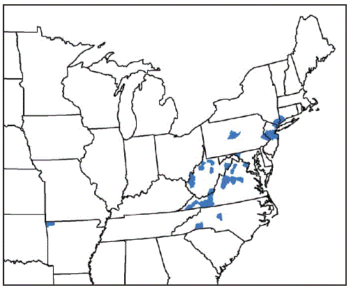
From August 2017 through September 2018, vector and animal surveillance efforts resulted in 53 reports of H. longicornis in the United States, including 38 (72%) from animal species (23 [61%] from domestic animals, 13 [34%] from wildlife, and two [5%] from humans), and 15 (28%) from environmental sampling of grass or other vegetation using cloth drags or flags* or carbon dioxide–baited tick traps.† With the exception of one report from Arkansas, the remaining reports of positively identified ticks are from eight eastern states: New Jersey (16; 30%), Virginia (15; 28%), West Virginia (11; 21%), New York (three; 6%), North Carolina (three; 6%), Pennsylvania (two; 4%), Connecticut (one; 2%), and Maryland (one; 2%) (Figure). Among the 546 counties or county equivalents in the nine states, ticks were reported from 45 (8%) counties (1.4% of all 3,109 U.S. counties and county equivalents) (Table 1). Excluding 15 reports of positive environmental sampling using flagging, dragging, or carbon dioxide traps, the remaining 38 reports reflect collection of ticks from infested host species (Table 2). Surveillance efforts did not include testing the ticks or hosts for pathogens. No cases of illness in humans or other species were reported. Concurrent reexamination of archived historical samples showed that invasion occurred years earlier. Most importantly, ticks collected from a deer in West Virginia in 2010 and a dog in New Jersey in 2013 were retrospectively identified as H. longicornis.

Discussion

Cooperative efforts among federal, state, and local experts from agricultural, public health, and academic institutions during the last year have documented that a tick indigenous to Asia is currently resident in several U.S. states. The public health and agricultural impacts of the multistate introduction and subsequent domestic establishment of H. longicornis are not known. At present, there is no evidence that H. longicornis has transmitted pathogens to humans, domestic animals, or wildlife in the United States. This species, however, is a potential vector of a number of important agents of human and animal diseases in the United States, including Rickettsia, Borrelia, Ehrlichia, Anaplasma, Theileria, and several important viral agents such as Heartland and Powassan viruses. Consequently, increased tick surveillance is warranted, using standardized animal and environmental sampling methods.

The findings in this report are subject to at least two limitations. First, the findings are limited by the variable surveillance methods used to identify the geographic and host distribution of H. longicornis. These methods included both passive and active surveillance. Conclusions about the geographic and host distribution might reflect the biases in the collection and submission of samples to states and USDA and the paucity of available information. Second, the data in this report reflect the collection of specimens that were positively identified by morphology or molecular barcoding. These represent sentinels that H. longicornis is present in different U.S. states and regions, and not a comprehensive assessment of the distribution of H. longicornis in the United States. The absence of positive samples from many states and counties might reflect the absence of infestation, absence of sampling, or failure to recover the tick. Even in states where H. longicornis has been found, the available data do not describe the actual extent or intensity of infestation.

The biology and ecology of H. longicornis as an exotic species in the United States should be characterized in terms of its vector competence (ability to transmit a pathogen) and vectorial capacity (feeding habits, host preference, climatic sensitivity, population density, and other factors that can affect the risk for pathogen transmission to humans) for tickborne pathogens known to be present in the United States (5). Surveillance for H. longicornis should include adequate sampling of companion animals, commercial animals, wildlife, and the environment. Where H. longicornis is detected, there should be testing for a range of indigenous and exotic viral, bacterial, and protozoan tickborne pathogens potentially transmitted by H. longicornis. Given the similarity between SFTSV and Heartland virus, a tickborne phlebovirus (https://www.cdc.gov/heartland-virus/index.html), further evaluation of the potential role of H. longicornis in transmission of this disease agent among animal reservoirs and possibly to humans is warranted. A broad range of interventions should be evaluated, including insecticide and acaricide sensitivity testing. Many state and federal agencies are developing and disseminating information for stakeholders, including development of hotlines, and some states are identifying ticks submitted by the public. The recently documented occurrence of H. longicornis in the United States presents an opportunity for collaboration among governmental, agricultural, public health agencies and partners in academic public health, veterinary sciences, and agricultural sciences to prevent diseases of potential national importance before onset in humans and other animal species.

Top

Acknowledgments

Wes Watson, Andrew D. Haddow, Naomi Drexler, Gleeson Murphy, Harry Savage, Howard Ginsberg, Kim Cervantes, field and laboratory personnel.

Corresponding author: C. Ben Beard, cbeard@cdc.gov, 970-221-6418.


* Drags consist of white cloth (usually 1 m2) that have a wooden leading frame and are dragged by a cord through grass or a leafy forest floor. Flags are similar but are used to brush uneven surfaces such as small bushes in wooded areas. Drags and flags are used to sample the environment for ticks trying to locate a host.

† Carbon dioxide traps consist of dry ice–filled small boxes with holes that allow the CO2 to escape which are placed on a white cloth or mat in a grassy area or forest floor. Ticks, attracted by the CO2, crawl on to the cloth or mat surface, which is inspected for ticks after a period of time.

Top

References

  1. Rainey T, Occi JL, Robbins RG, Egizi A. Discovery of Haemaphysalis longicornis (Ixodida: Ixodidae) parasitizing a sheep in New Jersey, United States. J Med Entomol 2018;55:757–9. CrossRef PubMed
  2. Luo L-M, Zhao L, Wen H-L, et al. Haemaphysalis longicornis ticks as reservoir and vector of severe fever with thrombocytopenia syndrome virus in China. Emerg Infect Dis 2015;21:1770–6. CrossRef PubMed
  3. Mahara F. Japanese spotted fever: report of 31 cases and review of the literature. Emerg Infect Dis 1997;3:105–11. CrossRef PubMed
  4. Kang J-G, Ko S, Smith WB, Kim H-C, Lee I-Y, Chae J-S. Prevalence of Anaplasma, Bartonella and Borrelia species in Haemaphysalis longicornis collected from goats in North Korea. J Vet Sci 2016;17:207–16. CrossRef PubMed
  5. Rosenberg R, Lindsey NP, Fischer M, et al. Vital signs: trends in reported vectorborne disease cases—United States and territories, 2004–2016. MMWR Morb Mortal Wkly Rep 2018;67:496–501. CrossRef PubMed
  6. Heath A. Biology, ecology and distribution of the tick, Haemaphysalis longicornis Neumann (Acari: Ixodidae) in New Zealand. N Z Vet J 2016;64:10–20. CrossRef PubMed
Return to your place in the textFIGURE. Counties and county equivalents* where Haemaphysalis longicornis has been reported (N = 45) — United States, August 2017–September 2018

The figure is a map showing the counties and county equivalents where Haemaphysalis longicornis has been reported (N = 45), in the United States, during August 2017–September 2018.* Benton County, Arkansas; Fairfield County, Connecticut; Washington County, Maryland; Bergen, Hunterdon, Mercer, Middlesex, Monmouth, Somerset, and Union Counties, New Jersey; Davidson, Polk, and Rutherford Counties, North Carolina; Richmond, Rockland, and Westchester Counties, New York; Bucks and Centre Counties, Pennsylvania; Albemarle, Augusta, Carroll, Fairfax, Giles, Grayson, Louisa, Page, Pulaski, Rockbridge, Russell, Scott, Smyth, Staunton City, Warren, and Wythe Counties, Virginia; Cabell, Hardy, Lincoln, Mason, Marion, Monroe, Putnam, Ritchie, Taylor, Tyler, Upshur Counties, West Virginia.

Top

TABLE 1. Percentage of Haemaphysalis longicornis–infested counties or county equivalents in infested states — nine states, August 2017–September 2018Return to your place in the text
State No. of counties* per state No. (%) of counties* with H. longicornis on host or in environment
Arkansas 75 1 (1)
Connecticut 8 1 (13)
Maryland 24 1 (4)
New Jersey 21 7 (33)
New York 62 3 (5)
North Carolina 100 3 (3)
Pennsylvania 67 2 (3)
Virginia 134 16 (12)
West Virginia 55 11 (20)
Total 546 45 (8)

* Counties or county equivalents

TABLE 2. Distribution of Haemaphysalis longicornis, by host and species — nine states, August 2017–September 2018Return to your place in the text
Host category, no. (% of total)/Species No. (% of host category)
Domestic animal, 23 (61)
Cat 1 (4)
Cow 4 (17)
Dog 12 (52)
Goat 2 (9)
Horse 2 (9)
Sheep 2 (9)
Total 23 (100)
Wildlife, 13 (34)
Coyote 1 (8)
White-tailed deer 7 (54)
Gray fox 1 (8)
Groundhog 1 (8)
Virginia opossum 2 (15)
Raccoon 1 (8)
Total 13 (100)
Human, 2 (5) 2 (100)
Total 38 (100)

Beard CB, Occi J, Bonilla DL, et al. Multistate Infestation with the Exotic Disease–Vector Tick Haemaphysalis longicornis — United States, August 2017–September 2018. MMWR Morb Mortal Wkly Rep 2018;67:1310–1313. DOI: http://dx.doi.org/10.15585/mmwr.mm6747a3.

____________________

**Comment**

In the section discussing the species and the other pathogens it’s been known to transmit, Theileria was mentioned. Theileria is a malarial-like pathogen similar to Babesia:

https://en.wikipedia.org/wiki/Theileria_microti

Babesia IS also spread by ticks and is a frequent coinfection with Lyme.

An important difference from malaria is that T. microti does not infect liver cells. Additionally, the piroplasm is spread by tick bites (Ixodes scapularis, the same tick that spreads Lyme disease), while the malaria protozoans are spread via mosquito. Finally, under the microscope, the merozoite form of the T. microti life cycle in red blood cells forms a cross-shaped structure, often referred to as a “Maltese cross“, whereas malaria forms more of a diamond ring structure in red blood cells.[3]

Much is yet to be discovered about the Asian tick that clones itself and can drain cattle of its blood.  For more:  https://madisonarealymesupportgroup.com/2018/09/12/three-surprising-things-i-learned-about-asian-longhorned-ticks-the-tick-guy-tom-mather/

One of the biggest discoveries by Mather was how the ticks line up on stalks of grass resembling grains of wheat.  When anything touches this, it’s like a tick cluster bomb and ticks go everywhere.  Not just one or two, mind you, but hundreds at one time.  See link for pictures.

https://madisonarealymesupportgroup.com/2018/03/01/asian-ticks-mysteriously-turn-up-in-new-jersey/

https://madisonarealymesupportgroup.com/2018/10/03/1st-person-bitten-by-east-asian-longhorned-tick/

https://madisonarealymesupportgroup.com/2018/11/05/hawk-found-carrying-asian-long-horned-tick-the-one-that-drains-cattle-of-all-their-blood/


Transverse Myelitis & Guillain-Barre Associated with Bartonella

https://www.ncbi.nlm.nih.gov/pmc/articles/PMC6106413/

Transverse Myelitis and Guillain-Barré Syndrome Associated with Cat-Scratch Disease, Texas, USA, 2011

Emerg Infect Dis. 2018 Sep; 24(9): 1754–1755.

Abstract

We describe a case of coexisting transverse myelitis and Guillain-Barré syndrome (GBS) related to infection with Bartonella henselae proteobacterium and review similar serology-proven cases. B. henselae infection might be emerging as a cause of myelitis and Guillain-Barré syndrome and should be considered as an etiologic factor in patients with such clinical presentations.

__________________

**Comment**

This case study is of a previously healthy 10 year old girl who presented with lower extremity weakness, abdominal pain, vomiting, urinary retention, lymphadenopathy, headache, neck pain, lower back pain, burning in wrists, knees, ankles, and feet, and increases sensitivity to pain in her legs.

She had evidence of myelitis, brain lesions, and peripheral nerve involvement.

They administered rifampin and doxycycline for a possible CSD (Cat Scratch Disease) diagnosis but had to discontinue rifampin due to rising liver enzyme levels.  She was then given IV immunoglobulin for possible GBS (Guillain Barre Syndrome) and showed tremendous improvement with resolution of urinary retention and decrease in pain & weakness.  Four months later she had only residual sensory deficits.

According to the study, by 1971, 40 cases of Bartonella with neurological complications occurred with 90% involving encephalitis with a few myelopathy.  Other CSD associated myelitis cases and other GBS-associated Bartonella cases have been reported.

Key quote:

Studies of the efficacy of treatments for CSD-associated neurologic manifestations are lacking, and thus, the optimal regimen and duration of therapy are unknown. However, we suggest that clinicians consider CSD early in disease courses involving neurologic complications; the possibility of GBS, myelitis, or both in the setting of possible CSD should prompt clinicians to initiate antimicrobial treatment early and consider steroid or intravenous immunoglobulin therapy to prevent progression of disease.

For Lyme/MSIDS patients with these presentations, steroids would be counter-indicated due to the immune suppression which would enhance pathogen involvement:  http://www.lymenet.de/literatur/steroids.htm

“It is interesting to note that in dogs who had Lyme disease, injections of dexamethasone, a corticosteroid, enabled Borrelia burgdorferi to be cultured from blood drawn on the following day.”

This was done by Dr. Elizabeth Burgess at the University of Wisconsin. This suggests that the steroid suppresses a mechanism for keeping the bacteria out of the circulatory system, since ordinarily it is difficult to grow the Lyme organism from the blood. Entrance of the bacteria into the bloodstream can allow seeding of other organs.

“In conclusion, the decision to use the steroids in a Lyme patient must be given considerable thought and the possible benefits must be weighed against the risks. I would not use steroids unless the patient was also on antibiotics.“

Please keep in mind the difference between catabolic (break down) steroids and anabolic (build up) steroids.


Vaccines Likely Infected With Retroviruses & Linked to Chronic Disease

https://articles.mercola.com/sites/articles/archive/2018/12/09/retrovirus.aspx?

Plague: One Scientist’s Intrepid Search for the Truth About Human Retroviruses and Chronic Disease

 Approx 3 Min

Written by Dr. Joseph Mercola

Story at-a-glance

  • A retrovirus is a virus that contains RNA encoded genes rather than DNA. Using reverse transcriptase, the retrovirus is able to transform the single-stranded RNA into a double-stranded DNA
  • When the retrovirus infects a host, it integrates its DNA into the DNA of the host cell, which allows the retrovirus to replicate itself and spread through the host
  • One example of a transmissible retrovirus is the HIV virus, which can cascade into the clinical symptoms of acquired immunodeficiency syndrome (AIDS)
  • A retrovirus family known as xenotropic murine leukemia virus-related viruses (XMRV) may play a causal role in chronic fatigue syndrome, chronic myalgic encephalopathy (ME) and other diseases, including autism
  • Some retroviruses, including XMRV (but not HIV as far as we know), infect your germ cells, which means they are transmitted to your offspring

Judy Mikovits, Ph.D., a virologist, researcher and founding research director of the Whittemore Peterson Institute — which researches and treats chronic fatigue syndrome (CFS) in Reno, Nevada — got embroiled in controversy when, in 2009, she was the senior author on a paper which reported that a retrovirus known as xenotropic murine leukemia virus-related virus (XMRV) may play a causal role in CFS and other diseases, including autism.

Her book, “Plague: One Scientist’s Intrepid Search for the Truth About Human Retroviruses and Chronic Fatigue Syndrome (ME/CFS), Autism and Other Diseases,” details her research and personal trials that arose as a consequence of her work.

“Kent Heckenlively essentially wrote it,” Mikovits says, “because I write like a scientist. We wrote it using the genre of flashback. He taped hours and hours of me telling the story as he asked me questions — because he’s trained as an attorney — and then he turned that into this suspense-thriller. Interestingly enough, it almost has to read like fiction because of the lawyers it took to … make sure we weren’t sued.”

What Are Retroviruses?

Before we go further, let’s review what a retrovirus is. A retrovirus is a ribonucleic acid (RNA) virus — in other words, a virus that contains RNA encoded genes rather than deoxyribonucleic acid (DNA). Using reverse transcriptase, the retrovirus is able to transform the single-stranded RNA into a double-stranded DNA.

When the retrovirus infects a host, it integrates its DNA into the DNA of the host cell, which allows the retrovirus to replicate itself and spread through the host. As more and more cells are infected, you become increasingly sicker. Mikovits explains:

“Humans have a DNA genome. Our blueprint is DNA. Retroviruses have an RNA genome, but they also are unique in the RNA family of viruses, where their RNA genome is reverse-transcribed. That is, written backwards by an enzyme unique to retroviruses called reverse transcriptase. That enzyme writes the RNA into DNA.

Then they have another enzyme called integrase. Integrase is like a pair of scissors that cuts open your DNA and then inserts the retrovirus, which is only about 8,000 base pairs, a very, very, very small virus, 50 to 100 nanometers on an electron micrograph. That piece of DNA — called a provirus — is now in the DNA of your cells forever. Every time your cells replicate, you make more viruses.”

Now, this DNA insertion has been ongoing throughout human history. According to Mikovits, about 10 percent of the human genome is retroviral in origin. These are called human endogenous retroviruses. These, however, differ in that they’ve been crippled in part by our DNA methylation machinery (which modulates genes expression and the human immune system — so that they can no longer make complete viruses and therefore cannot infect others.

However, when you’re infected with a retrovirus such as human T-lymphotropic virus (HTLV-1), HIV HBRV or Borellia as in chronic Lyme disease and develop DNA methylation and immune dysfunction, these endogenous retroviruses begin to be expressed, and this is yet another really important finding.

HIV — One Example of a Transmissible Retrovirus

One example of a transmissible retrovirus is the HIV virus, which can cascade into the clinical symptoms of acquired immunodeficiency syndrome (AIDS). HIV was discovered in 1982, and as mentioned above, was part of Mikovits’ early research work. Her book includes the history of that important discovery.

When Mikovits first began studying retroviruses, HIV/AIDS was completely unknown, but they suspected a retrovirus was at play because of how retroviruses affect the human immune system and lead to acquired immune deficiencies and cancers.

“You don’t just one day get this virus and you’re sick. In fact, we now know millions of people have HIV and will never develop AIDS. We talk about that in the book, because the book ultimately is one of hope that we fix HIV.

I can honestly tell you in 1999, when I was running the lab of antiviral drug mechanisms, I did not ever expect we would solve that problem. Now, AIDS patients on antiretroviral therapy are probably healthier and develop fewer cancers … than most of the rest of society.”

Some retroviruses, including XMRV (but not HIV), also infect your germ cells, which means they not only cause continuous infection in your body but also transfer to your offspring.

“XMRV, the xenotropic murine (mouse) leukemia retrovirus, is the mouse-related retroviruses that cause cancer and lots of neurological diseases. Those affect the stem cells, the egg, the sperm — every cell in your body. That was one of the big ‘Oh, my Gods,’ about our discovery,” Mikovits says.

When it comes to treatment, the key is to keep the virus silent, because when they’re not, each time your cells divide you’re making more retroviruses. For this, antiretroviral treatments are used, some of which will be discussed later in this article.

From AIDS to ME/CFS

After 9/11, Mikovits started working with a woman whose daughter was severely ill with chronic fatigue syndrome. “Basically, that was the first time I ever saw the disease called ME/CFS,” she says.

“This person was looking at a herpes virus known as human herpesvirus 6 (HHV-6). This is a virus prominent in people with Kaposi sarcoma, [which] became associated with HIV and AIDS. Dr. Patrick Moore and Dr. Yuan Chang [discovered] that Kaposi sarcoma was actually caused by a herpes virus — then known as Kaposi sarcoma herpes virus; now, it’s HHV-8.

Because the immune system is crippled, you wake up the sleeping herpes viruses. People with autism, ME/CFS and cancers have a lot of chronic active infections, so we often see the Epstein-Barr virus (EBV) associated with outbreaks of ME/CFS …

This woman introduced me to Dr. Dan Peterson and Annette Whittemore in Incline Village, Nevada, where he had been studying outbreaks of ME/CFS for probably 25 years. He said he had a bank of samples. We went up there. I met all the patients.

I interviewed them in great length and developed a hypothesis, which had actually been shown before by Elaine Defreitas, Ph.D., another scientist many years earlier …

Defreitas had isolated retroviruses from patients with ME/CFS. A doctor … named Sidney Grossberg had also isolated retroviruses from at least one patient with ME/CFS. So, the retroviral hypothesis wasn’t new. Everything about it fit …

One of the most severely injured patients at that time was Whittemore’s daughter, Andrea. That summer (2006), I went up there … and started studying it … I used the systems biology approach, because there’s a lot of heterogeneity.

We know AIDS patients who have HIV and will never get AIDS … I interviewed patients in Peterson’s office all summer and took blood, urine, saliva and all kinds of samples to isolate that virus, which is what you need to do to show it’s associated with a disease.”

The Discovery of Infectious Retroviruses

Eventually, she brought together several of her former and current colleagues who were world experts in HIV sequencing to look at ME/CFS. Among them was the world’s leading electron microscopist, Kunio Nagashima, who has done the electron micrographs of every family of human retroviruses discovered: the human beta retrovirus, human delta virus, lenti-virus (such as HIV) and gamma retroviruses.

Working in collaboration with the Cleveland Clinic, Mikovits and her team isolated the virus and spent the better part of 2008 and 2009 putting a paper together, proving the XMRV retrovirus was infectious and transmissible and not just another crippled human endogenous retrovirus.

“To our horror, we learned these [retroviruses] could be aerosolized. This was in 2011 … That was really the first nail in my coffin. Pun intended, because the national academy member, John Coffin, Ph.D. — who had told Frank Ruscetti, ‘There is no such thing as human retroviruses. Don’t study them’ — then made a fortune out of HIV and did everything he could to destroy me and the patients,” Mikovits says.

“Prior to publication in 2009, we wrote a patent on the detection of these retroviruses, these pieces and parts as contaminants of the cell cultures, of the cell lines from which we make vaccines. After they destroyed my reputation and career and forced the retraction of our paper from [the journal] Science, Coffin turned around and wrote a patent on the detection of these viruses in contaminating cell linings and contaminating biologicals in our labs.”

This PDF includes emails, letters and supporting documentation showing how the retraction of Mikovits’ Science paper was forced, after which Coffin filed his own patent for a detection method of the contaminants in cell lines used for vaccines and other biologicals. There’s also documentation detailing the scientific fraud Mikovits asserts in this interview.

Infectious Retroviruses May Contaminate Blood Supply and Vaccines

In her book, she also details how infectious retroviruses are still likely infecting many biological solutions used clinically today, such as vaccines and other therapies. To say that this is a concern would be an understatement. Children’s Health Defense discusses this, and more, in “Looking Back, Looking Forward: Cancer and Vaccines.”1 Mikovits explains:

“That was really at the heart of the big ‘Oh, my God.’ The worst I learned in this whole experience is how corrupt scientific journals are. In fact, Ruscetti now calls Science, that prestigious journal, ‘The National Inquirer,’ because they literally engineered the whole thing to destroy MEC/FS patients and any association this virus [XMRV] had with these diseases …

All of the studies showed that the control population was between 3.75 and 6.8 percent infected. When you do a study and there’s evidence of infection in 6 percent of the human population, that’s 25 million Americans. To put that in context, at the height of HIV/AIDS in 1995, it was 1 million Americans. It would crush our health care system if they had to pay for what they caused.”

The result of Mikovits’ findings was nothing short of personal devastation. Not only was her paper retracted by Science, she was even arrested for “stealing” her own lab notes. Charges were ultimately dropped, but the damage to her reputation was a done deal.

“Basically, our paper came out on October 8, 2009. It was literally like ‘the shot heard around the world.’ I was on the road every single day. Everywhere I went doctors were like, ‘She’s got it. She’s got it. She’s got it,’ and not just with MEC/FS but also with cancer, leukemia, lymphoma, with prostate cancer.

When you start looking at the inflammatory events in the acquired immune deficiencies, with autoimmune disease, with Lou Gehrig’s disease, the problem became this [retro]virus. Well, there’s no single virus. There’s no HIV. There’s a whole family of HIVs. There’s an HIV 1. There’s an HIV 2. There’s a strain A, B, C and D.

Why do we do influenza vaccines for this strain de jour or every year? [Because] there are strains of viruses. There are families of viruses … The second that we published this paper, we started working to get a diagnostic test for the blood supply to show it wasn’t contaminated, which, in fact, it was.

Later that year, the last talk I ever gave was on a science paper that came out September 22, 2011 … That talk was basically a debate for the evidence that there are human retroviruses of the XMRV family that aren’t VP62 (the infectious molecular clone, not the natural isolates of our paper).

We could show in the original paper that there was evidence of murine leukemia viruses, gamma retroviruses that were infectious and transmissible, just as we had said.

Coffin was on the other end of that debate. He said it was all a recombination event. He published a paper in 2013 saying, ‘When we worked with mouse cells, they expressed a lot of pieces and parts of retroviruses. This just happened to happen in the laboratory.’

[Hence, he claimed] that’s what we had isolated. [Coffin claimed] that what we were looking at were just contaminants in the laboratory. ‘It’s all a lab contaminant,’ [Coffin said], ‘You can all go home. You’re safe.'”

Massive Public Health Concerns Swept Under the Rug

As one might expect, Mikovits’ research caused massive concern in the professional community, because here was a newly identified, infectious and transmissible retrovirus that no one was screening for, and it was potentially contaminating 10 percent of the human blood supply. But rather than face the problem head on, it was rapidly swept under the proverbial rug.

“My mom was watching Good Morning America one morning. Across the bottom of the ticker tape said, ‘XMRV all a hoax’ … It was horrible. We started to realize our fake news and fake science.”

Today, the blood supply is unlikely to be contaminated, thanks to a decontamination procedure developed by a California-based company called Cerus and which Mikovits proved to inactivate XMRV, rendering it noninfectious.

Other biologicals, including vaccines, however, may not be routinely decontaminated using this process, in large part because they’re not required to do so, and drug companies are not liable for vaccine-induced harm. What’s more, decontaminating the vaccine may render it ineffective.

“It won’t work. It will no longer be a vaccine … The Cerus method cleans up Ebola. It cleans up Zika. It cleans up essentially any RNA viruses, including HIV and all three human retroviruses. The Cerus system is extremely valuable to cleaning up the blood supply.

But they cannot clean up the vaccines for another reason. If they do, they prove Andy Wakefield right. They prove me right. They prove they’ve got 25 million Americans, who they have to support for the rest of their lives and pay damages [to] …”

The Price of Making an Unpopular Scientific Discovery

On a personal level, Mikovits has taken an enormous personal hit. September 29, 2011, she was fired from the Whittemore Peterson Institute for insolence and insubordination, and was driven into bankruptcy after being falsely arrested for stealing her own lab notes. (She never was and to this day is not in possession of her notebooks or any of the two offices full of her work done in her entire career.)

She explains her firing saying that Whittemore had been selling a diagnostic test and the director of their for-profit commercial laboratory was using federal grant funds to do that work (with full knowledge and under the direction of Annette and Harvey Whittemore), which is misappropriation of federal funds. Mikovits became aware of this in August that year, and wrote him off the grant.

“The Whittemores basically fired me immediately in an attempt … to get this scientist, Vince Lombardi, Ph.D. … to recreate the work while I was out of town and say I was a lunatic — that he’d been doing the work all along, and he hadn’t misappropriated any of the funds.

They fired me on September 29 and immediately locked down the entire university to me or my staff … The insolence and insubordination was I had refused a direct order to misappropriate federal funds, basically. I wasn’t ever going to do that. The insolence I’m trying to learn not to do, because it probably would have gone a lot better for me if I didn’t say ‘F-you,’ at the same time …

It was September 22, 2011, when I gave my last talk. They had three weeks to get a Science paper out there that would destroy my reputation in the ME/CFS community … Ruscetti had to sign that paper, or he and Sandy Ruscetti would be fired … [and] lose their entire retirement, which is 75 years.

That was one of the few times I sobbed. I was sitting in my bed screaming …It was 6 o’clock in the morning. They were on the East Coast and they needed to get this paper published fast by Science.

I called the Ruscettis and said, “Frank, they agreed to change the language. They agreed to change the title. They agreed it wasn’t an association study … [they say] we didn’t have a diagnostic test. Either way, the Whittemores are going to kill me because they’re selling the diagnostic test.’

So Frank [Ruscetti] signed the paper. They didn’t change the wording. [What they did] is pure fraud. Here, the head of the National Heart, Lung, and Blood Institute published pure fraud in the journal Science, just as two years later, Ian Lipkin published pure fraud. It is fake news. It is so corrupt, everything about it.

It’s not [the researchers]. It’s the top of the line. It’s Dr. Tony Fauci. We’re only allowed to make incremental advances. When you make a discovery of this nature, it changes all of everything. This is misogyny … This is a bunch of little boys … fighting over who gets credit, while the world dies, while you kill an entire continent.

That’s why I do shows like this. Because we’re going to teach doctors. When doctors understand the science — and they’re coming around a lot — because the science is there. Nothing about our paper, except the sequence of the virus, has ever been wrong. We knew that in the beginning.”

Individuals Infected With Retroviruses Should Avoid Vaccinations

According to Mikovits, retroviruses such as XMRV affect entire families, as it can be transmitted to your offspring. Many of these families also have children with autism, which Mikovits believes may be connected to the retrovirus. The question is, what can you do if you’re infected? For starters, Mikovits recommends avoiding vaccinations.

“Until 2011, not inconsequentially, we didn’t vaccinate AIDS patients the same way. It’s in the book. You don’t vaccinate the immune-compromised … By definition, you have an immune system that doesn’t work. Why would you vaccinate them? Why would you vaccinate somebody under 3 years old, who has an immune and detox systems that don’t work?

This was the key of the RNaseL story (a genetic susceptibility not to degrade RNA viruses), of the Thompson fraudulent paper [Editor’s note: This refers to William Thompson, Ph.D., a former senior scientist at the CDC’s National Center for Immunizations and Respiratory Diseases, who confessed he conspired to cover up links found between the MMR vaccine and autism].

All they had to do was wait for black boys to be 3 years old, and they would have been able to degrade the RNA virus. That’s criminal. That’s beyond comprehension …

The pearl of wisdom is this DNA methylation. Keep the violent virus silent … DNA methylation has to silence them. You can’t inject them in a vaccine. We’re injecting millions of pieces in parts of retroviruses in every vaccine, by definition (and admission).

I am working on an ongoing cancer lawsuit that says vaccines cause childhood cancer, a lymphoma. By these same mechanisms, you’ve destroyed the DNA methylation machinery’s ability [to silence the virus]. You’ve simply overwhelmed the substrate. You’ve overwhelmed the ability to methylate.

Every time those viruses integrate, you have a better chance at insertional mutagenesis. Don’t expose anybody to human (or animal) retroviruses. Use antiretroviral therapy, which are natural products … There are lots of natural products. We published on them. Those are actually therapy for these kids.

[A 100-year-old drug called Suramin] was one of the first antiretroviral therapies for HIV … [It] worked best against the murine leukemia virus-related viruses, against the mouse retroviruses, the gamma retroviruses …

[Dr. Robert] Naviaux [professor of medicine, pediatrics and pathology at University of California San Diego School of Medicine] did a small clinical trial.2 These kids got their life back.3 They started talking again. What did Bayer do? They stopped the trial and took the drug away from everyone. Now, you can’t get it …

We could help millions of people get over [autism]. But when you show cure, you know cause. That’s it. I would be right … Millions of people would get their lives back, and it’s all about money.”

XMRV Is a Significant Threat

As mentioned, there are several different retroviruses, which are part of four viral families (delta, lenti, beta and gamma). Aside from HIV and XMRV, there’s the human T-cell leukemia lymphoma virus (HTLV-1) family. There are five or six HTLV viruses, but HTLV-1 is the only one known to cause severe disease.

Human beta retrovirus is another virus associated with primary biliary cirrhosis. Many patients with MEC/FS also have family members with primary biliary cirrhosis. As for which one might be the most significant threat, Mikovits believes XMRV is among the most pressing, because while HIV is well-contained at present, XMRV is not, and it appears to play a significant role in diseases of methylation.

Disturbingly, they’re now using murine leukemia viruses as vectors for gene therapy and a novel cancer therapy called chimeric antigen receptor (CAR) T-cell therapy. In other words, they’re causing cancer and other retroviral illnesses.

“The same thing with Gardasil … We’re causing these diseases and we know it because we’re using these [retroviruses] as vectors. We don’t need infectious viruses. That’s one thing that’s really important to know. You don’t need infectious viruses if you’re injecting the provirus, or the pieces and parts. You inject it, past your immunity, past your gut, past RNA cell, past everything. You bypass the immune system. They don’t need to be infectious.

All you need is an envelope to cause that prostate cancer. That’s a paper that was published 2013. In most of our studies, all we detected was the envelope. The envelope alone causes vasculitis … Another strain of XMRV gamma retrovirus from mice was identified by Gary Owens … associated with cardiovascular disease. This is just a nightmare that we’ve unleashed in our environment.”

Retroviruses and ME/CFS

According to Mikovits, 6 to 8 percent of the general population are infected with infectious and transmissible XMRV-retroviruses, and in the chronic fatigue population, that prevalence shoots up to about 30 to 40 percent. As with HIV, antiretroviral therapies can be very helpful in the treatment of ME/CFS, including low-dose naltrexone.

“You have to silence the other pathogens, so taking care of mycoplasma, taking care of mold, absolutely supporting the gut microbiome [will help],“ Mikovits says. “We learned with AIDS and cancer patients that if they don’t have the diversity in the microbiome, just like in autism, just like in MEC/FS, it’s because the retrovirus is causing leaky gut …

The nonspecific inflammation [is] the retroviruses. If you keep the gut healthy, you can heal. The primary is the diversity in the microbiome, or you can’t respond to the drugs. There’s a lot of hope. That’s what we end the show with. There are therapies. We could fix this tomorrow. That’s why I do it.”

To learn more, be sure to pick up a copy of “Plague: One Scientist’s Intrepid Search for the Truth About Human Retroviruses and Chronic Fatigue Syndrome (ME/CFS), Autism and Other Diseases,” which reads more like a fictional thriller than a nonfictional book about the science of disease.

________________

For More:  https://madisonarealymesupportgroup.com/2017/10/15/vaccines-and-retroviruses-a-whistleblower-reveals-what-the-government-is-hiding/

https://madisonarealymesupportgroup.com/2018/03/01/vaccines-could-contribute-to-disease-epidemics-due-to-retrovirus-contamination/

https://madisonarealymesupportgroup.com/2018/06/23/the-role-of-retroviruses-in-chronic-illness-a-clinicians-perspective/

They should make Mikovits’s book into a movie.  It’s beyond belief.


Acute Flaccid Paralysis is (Most Often) Guillain Barre Syndrome

https://jameslyonsweiler.com/2018/12/01/acute-flaccid-paralysis-is-most-often-guillain-barre-syndrome/

ACUTE FLACCID PARALYSIS IS (MOST OFTEN) GUILLAIN BARRE SYNDROME

by jameslyonsweiler

The US press has been pushing a view of acute flaccid paralysis as a mysterious condition of unknown etiology (unknown cause). Checking the scientific literature, however, tells us that AFP is most often Guillain Barre Syndrome, a condition that appears on the National Vaccine Injury Compensation Program as a “Table Condition” – i.e., one that the US HHS has no defense against when parents file in the NVICP for compensation for GBS as a vaccine injury in their children.

Here are some quotes from the abstracts of a collection of studies on AFP:

“Within the last few years, an enterovirus D68 outbreak has been associated with cases of acute flaccid paralysis in children, and emerging Zika virus infection has been concurrent with cases of acute flaccid paralysis due to Guillain-Barré syndrome, although cases of myelitis have also been reported.”

“Cases (of AFP) exhibited heterogeneous paralysis patterns from 1- to 4-limb involvement, but all definite cases had longitudinal spinal gray matter lesions on magnetic resonance imaging (median, 20 spinal segments). Cerebrospinal fluid pleocytosis was observed in 50 of 59 cases (85%), and 8 of 29 (28%) were positive for antiganglioside antibodies, as frequently observed in Guillain-Barré syndrome.”

“The syndrome of acute flaccid paralysis (AFP) is a common medical emergency in children. In the era of poliomyelitis eradication, the common causes of AFP include Guillain-Barré syndrome (GBS), transverse myelitis and traumatic neuritis.”

“One-hundred thirty-nine children aged <15 years were reported to the Center for Diseases Control with AFP. In 138 (99%) stool samples no poliovirus was isolated. None of the patients was diagnosed as having acute poliomyelitis or polio-compatible paralysis. Guillain-Barré syndrome was the most frequent final diagnosis (79 cases) followed by Transverse Myelitis (7 cases) and Encephalitis (6 cases).”

“The major clinical diagnoses associated with AFP were Guillain-Barré Syndrome (GBS, 40%) and encephalomyelitis/myelitis (13%).”

“Guillain-Barré syndrome represented more than half of the reported cases (of AFP) (N = 2611, 52.5%), followed by traumatic neuritis (N = 715, 14.4%), and other CNS infections (N = 292, 5.9%).”

“Guillain-Barré syndrome represented more than half of the reported cases (of AFP) (N = 2611, 52.5%), followed by traumatic neuritis (N = 715, 14.4%), and other CNS infections (N = 292, 5.9%).”

“Of these (cases of AFP), nineteen (45%) cases were classified as Guillain-Barré syndrome on both registries.”

“In 44.5% of cases (of AFP) the definite diagnosis was Guillain Barrè syndrome.”

“Guillain-Barre syndrome dominated among non-polio AFP (39.3% of cases); more rare were traumatic neuritis (27.9% of cases), transient monoparalysis (12.1%), myelitis (7.6%).”

“A neurological cause was identified in 67.5% of cases (of AFP), of which the most common was Guillain-Barre syndromee (42%), followed by transverse myelitis (15%)”

“The major clinical diagnosis associated with AFP were Guillain-Barre syndrome (30.2%), central nervous system infection (16.2%), transverse myelitis (10.6%) non-polio enterovirus infection (6.2%), and hypokalaemic paralysis (5.2%).”

“Among (AFP cases), Guillain-Barré syndrome (118 cases, 41.5% of all non-polio AFP cases), traumatic neuritis (63 cases, 22.2%), transient monoparesis of limb (35 cases, 12.3%), myelitis (26 cases, 9.2%) were registered most frequently.

“To describe the epidemiology and causes of acute flaccid paralysis (AFP) in Australian children, and the clinical features of the two most common causes of AFP, Guillain-Barré syndrome and transverse myelitis.”

“The most common causes of AFP were Guillain-Barré syndrome in 67 (47%) and transverse myelitis in 27 (19%)”

“Guillain-Barré syndrome was the commonest single cause of AFP.”

“…acute flaccid paralysis (AFP) attributed to a peripheral demyelinating process (Guillain-Barré Syndrome [GBS]), or to an anterior myelitis.”

Additional Information:

How to file your vaccine injury in VAERS

How to file a case in the National Vaccine Injury Compensation Program

AFP IS GBS ABSTRACTS <<< download the abstracts

________________

**Comment**

I’ve been sitting on this article until today when it seemed appropriate to repost due to this:  https://madisonarealymesupportgroup.com/2018/12/07/nevada-man-diagnosed-with-guillain-barre-syndrome-after-getting-flu-shot/

Various vaccines are definitely causing GBS.

https://madisonarealymesupportgroup.com/2018/10/19/rise-in-acute-flaccid-myelitis-cases-and-the-link-to-vaccinations/

https://madisonarealymesupportgroup.com/2016/11/07/connection-of-acute-flaccid-myelitis-and-vaccinations/ The connection between vaccination and paralysis has been known since the 40’s and 50’s and was written about in The Lancet by Stephen Mawdsley in an article titled, “Polio Provocation: Solving a Mystery With the Help of History.”

Mawdsley states:

“The application of epidemiological surveillance and statistical methods enabled researchers to trace the steady rise in polio incidence along with the expansion of immunization programs for diphtheria, pertussis, and tetanus. A report that emerged from Guy’s and Evelina Hospitals, London, in 1950, found that 17 cases of polio paralysis developed in the limb injected with pertussis or tetanus inoculations. Results published by Australian doctor Bertram McCloskey also showed a strong association between injections and polio paralysis. Meanwhile, in the USA, public health researchers in New York and Pennsylvania reached similar conclusions.Clinical evidence, derived from across three continents, had established a theory that required attention.”

So what happened to this theory that piercing the skin during injection drives the polio virus into deep tissue where it then enters the central nervous system where it ultimately leads to paralysis and even death?

Good question.

The theory was essentially proven in 1998 in an article titled, “Mechanism of Injury-Provoked Poliomyelitis,” in the Journal of Virology. Researchers state:

“Skeletal muscle injury is known to predispose its sufferers to neurological complications of concurrent poliovirus infections. This phenomenon, labeled ‘provocation poliomyelitis,’ continues to cause numerous cases of childhood paralysis due to the administration of unnecessary injections to children in areas where poliovirus is endemic. Recently, it has been reported that intramuscular injections may also increase the likelihood of vaccine-associated paralytic poliomyelitis in recipients of live attenuated poliovirus vaccines. We have studied this important risk factor for paralytic polio in an animal system for poliomyelitis and have determined the pathogenic mechanism linking intramuscular injections and provocation poliomyelitis.  Skeletal muscle injury induces retrograde axonal transport of poliovirus and thereby facilitates viral invasion of the central nervous system and the progression of spinal cord damage. The pathogenic mechanism of provocation poliomyelitis may differ from that of polio acquired in the absence of predisposing factors.”

It’s such a big deal that even the CDC has created a AFM Task Force:  https://thevaccinereaction.org/2018/11/cdc-creates-afm-task-force/  While the cause of AFM remains a mystery, Nancy Messonnier, MD, director of the CDC’s National Center for Immunization and Respiratory Diseases recently said that AFM “seems to be more of an autoimmune syndrome, as opposed to a direct result of a virus.” She has also compared AFM to what happens with the autoimmune neurological disorder known as Guillain-Barré Syndrome, in which the immune system attacks healthy nerve cells in the peripheral nervous system.7 8 

Why do I get the impression that the CDC will state that while the cause of AFM is a mystery, it’s DEFINITELY NOT VACCINES.

In early November the CDC blamed a virus:  https://thevaccinereaction.org/2018/11/under-siege-by-critics-cdc-moves-to-blame-a-virus-for-polio-like-cases-of-afm/

BTW:  in 1995, Swiss Scientists found reverse transcriptase in the live measles & mumps vaccine as well as some influenza vaccines.  https://thevaccinereaction.org/2018/11/virus-enzyme-found-in-mmr-vaccine/  Excerpt:

RT activity is associated with the presence of retroviruses, a class of viruses that can permanently alter the genes of the cells they infect. AIDS is a retrovirus. Scientists at Merck & Co., the US manufacturer of MMR vaccine, are reportedly trying to find out where the RT came from. One possibility is that the RT activity in MMR vaccine signals the presence of an avian leucosis virus (AVL), a retrovirus that infects some birds and can cause a leukemia-like illness.

Another possibility is that the chickens used to make the vaccine carry RT coding genes derived from retroviruses that infected the species tens of thousands of years ago.

Mind you, this was going on in 1995.  The CDC’s response:  Continue vaccinating in spite of chicken viruses being in vaccines!

And now, to prevent absenteeism, kids are getting the Flu Shot in the Classroom for convenience:  https://thevaccinereaction.org/2018/12/children-given-flu-shots-in-school-classrooms/  “Healthy Schools, LLC,” was founded by former Jacksonville Jaguars pro-football player Tony Boselli and   is getting paid to handle the entire vaccination program.  Boselli recently sold the entity to CareDox, Inc. of New York, where he remains as a partner and President.  CareDox IS a for-profit company with investors.  The program hopes to give the jab to 600,000 kids in 8 states for free due to reimbursement contracts with Medicaid & other insurance providers.  The really scary aspect of this program is there are no physical exams or histories taken.  There is no mention of monitoring for adverse reactions and/or reporting to VAERS (Vaccine Adverse Event Reporting System).  It appears everything about the program is handled digitally and there also remains the question of informed consent.

https://www.nvic.org/NVIC-Vaccine-News/December-2018/internet-police-protect-you-from-freedom-of-speech.aspx? Barbara Loe Fisher explains in an informative video that we may not be able to find information about vaccine science, policy and law due to the cancellation of net neutrality in 2017.  She states that the public-private business partnership between government and corporations/institutions has, “cleared the way for factual information about health to be censored as ‘fake news’ and quietly removed from the internet if it does not conform with public health policy and government recommendations for use of pharmaceutical and food products.”  Also within this link is the story of a nurse who was fired for refusing the Flu shot.

This will affect much more than vaccine information.

It will affect the juggernaut of Lyme/MSIDS as well as any other politically incorrect disease.