Archive for the ‘research’ Category

Prozac & Triclosan Can Directly Induce Antibiotic Resistance Studies Say

https://www.sciencealert.com/antidepressant-medication-fluoxetine-prozac-may-raise-antibiotic-resistance

One of The Most Widely Used Antidepressants Has Just Been Implicated in Breeding Antibiotic Resistance

MICHELLE STARR
10 SEP 2018


In recent years, bacteria have been growing increasingly resistant to antibiotics – our strongest weapon against them.

It’s generally thought that it’s the overuse of antibiotics driving this trend, but now it looks like non-antibiotic medications can play a significant role too.

Specifically, as researchers from Australia’s University of Queensland have just discovered, a drug called fluoxetine – a selective serotonin reuptake inhibitor (SSRI), and the key ingredient in antidepressants such as Prozac and Sarafem.

The researchers embarked on the study to demonstrate that it’s not just antibiotics that are triggering the rise of the superbug.

“Our previous study reported that triclosan, a common ingredient in toothpaste and hand wash can directly induce antibiotic resistance,” said engineer Jianhua Guo of the University’s Advanced Water Management Centre.

“We also wondered whether other non-antibiotic pharmaceuticals such as fluoxetine can directly induce antibiotic resistance.”

The test involved exposing the bacterium Escherichia coli (or E. coli) to different concentrations of fluoxetine, in body-temperature baths that were changed daily for 30 days.

After this, the mutated bacteria were transferred to agar plates that contained an antibiotic. The researchers found that these bacteria had a greatly increased resistance to antibiotics chloramphenicol, amoxicillin and tetracycline – up to 50 million-fold compared to the control.

The stronger the concentration of fluoxetine, the faster the bacteria mutated over time.

And isolated mutant bacteria with a resistance to one of those antibiotics was also found to show multiple resistances against fluoroquinolone, aminoglycoside, β-lactams, tetracycline and chloramphenicol, the researchers found.

According to the researchers, up to 11 percent of the fluoxetine in medication travels through the body unchanged, and enters the environment via the sewer system after the patient urinates it out.

“Fluoxetine is a very persistent and well-documented drug in the wider environment, where strong environmental levels can induce multi-drug resistance,” Guo said.

“This discovery provides strong evidence that fluoxetine directly causes multi-antibiotic resistance via genetic mutation.”

That doesn’t mean that everyone needs to stop taking it immediately; for many people, fluoxetine is a fantastic life-saving medication. However, the study does point to other areas that need to be looked into.

First, it was conducted in the lab. How the drug impacts the microbiome inside the human body is yet to be investigated.

Second, what other non-antibiotic drugs could be having a similar effect?

A 2016 study estimated that by 2050, antibiotic-resistant superbugs will be killing 10 million people per year. So it’s vitally important that we figure this out – and try to find a way to fix it.

“[Fluoxetine] has previously been an invisible factor in the spread of antibiotic resistance,” said researcher Min Jin, “but we should consider this a warning.”

The research has been published in the journal Environment International.
 

Bartonella & Langerhans’ Cell Histiocytosis (Cancer)

https://www.ncbi.nlm.nih.gov/pubmed/30556266

2018 Dec 17:e27573. doi: 10.1002/pbc.27573. [Epub ahead of print]

Disseminated Bartonella henselae disease mimicking Langerhans’ cell histiocytosis.

Abstract

Bartonella henselae, the causative agent of cat-scratch disease, has been recognized to be responsible for a broad range of clinical syndromes. We report the case of a patient with disseminated B. henselae infection mimicking Langerhans cell histiocytosis at presentation and its successful management with neurosurgery, prolonged antibacterial therapy, and observation.

________________

**Comment**

Langerhans’ cell histiocytosis is a supposed “rare” disorder that looks like cancer (some say it is cancer).  The above case study implicates Bartonella.

Bartonella is known to cause granulomas

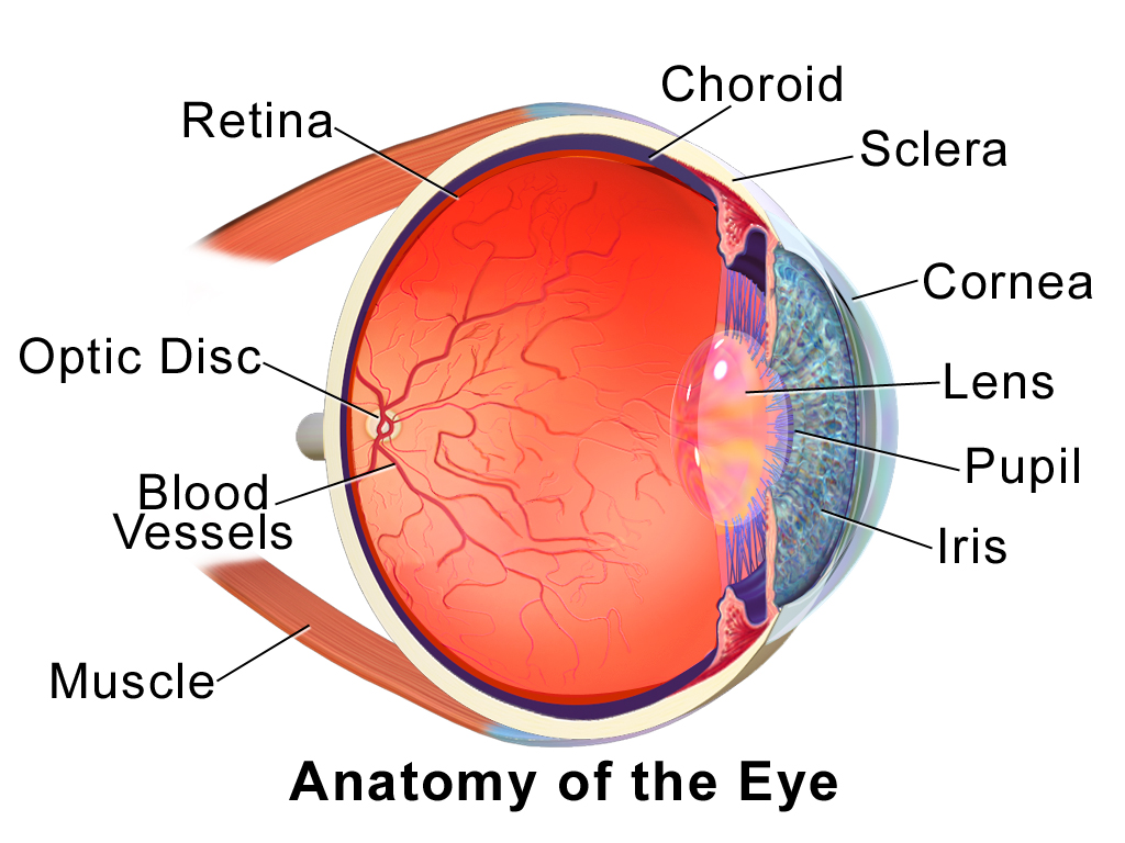
https://madisonarealymesupportgroup.com/2018/09/06/ocular-manifestations-of-bartonellosis/  Bartonella spp. infections continue to be a common cause uveitis with ophthalmic manifestations ranging from neuroretinits, vascular occlusions, to choroidal granulomas.

Blausen.com staff (2014). “Medical gallery of Blausen Medical 2014”. WikiJournal of Medicine 1 (2). DOI:10.15347/wjm/2014.010. ISSN 2002-4436. – Own work, CC BY 3.0, https://commons.wikimedia.org/w/index.php?curid=29025014

The choroid is the vascular layer of the eye, containing connective tissues, and lying between the retina and the sclera.

https://madisonarealymesupportgroup.com/2018/03/04/bartonella-erythema-nodosum-atypical-presentations/  The finding of bilateral enlarged axillary lymph nodes with necrosis and granulomas led to the diagnosis of Bartonella infection, an unusual cause of erythema nodosum.

Granulomas represents a chronic inflammatory response initiated by various infectious and noninfectious agents.  https://medical-dictionary.thefreedictionary.com/granuloma

Langerhans cell histiocytosis is a type of eosinophilic granuloma

https://ghr.nlm.nih.gov/condition/langerhans-cell-histiocytosis  Excerpt below:

Langerhans cell histiocytosis is a disorder in which excess immune system cells called Langerhans cells build up in the body. Langerhans cells, which help regulate the immune system, are normally found throughout the body, especially in the skin, lymph nodes, spleen, lungs, liver, and bone marrow. In Langerhans cell histiocytosis, excess immature Langerhans cells usually form tumors called granulomas. Many researchers now consider Langerhans cell histiocytosis to be a form of cancer, but this classification remains controversial.

In approximately 80 percent of affected individuals, one or more granulomas develop in the bones, causing pain and swelling. The granulomas, which usually occur in the skull or the long bones of the arms or legs, may cause the bone to fracture.

Granulomas also frequently occur in the skin, appearing as blisters, reddish bumps, or rashes which can be mild to severe. The pituitary gland may also be affected; this gland is located at the base of the brain and produces hormones that control many important body functions. Without hormone supplementation, affected individuals may experience delayed or absent puberty or an inability to have children (infertility). In addition, pituitary gland damage may result in the production of excessive amounts of urine (diabetes insipidus) and dysfunction of another gland called the thyroid. Thyroid dysfunction can affect the rate of chemical reactions in the body (metabolism), body temperature, skin and hair texture, and behavior.  In 15 to 20 percent of cases, Langerhans cell histiocytosis affects the lungs, liver, or blood-forming (hematopoietic) system; damage to these organs and tissues may be life-threatening.

Older names that were sometimes used for forms of Langerhans cell histiocytosis include eosinophilic granuloma, Hand-Schüller-Christian disease, and Letterer-Siwe disease.

More on Bartonella:  https://madisonarealymesupportgroup.com/2016/01/03/bartonella-treatment/

https://madisonarealymesupportgroup.com/2018/09/06/ocular-manifestations-of-bartonellosis/

https://madisonarealymesupportgroup.com/2017/01/04/endocarditis-consider-bartonella/

https://madisonarealymesupportgroup.com/2018/11/05/skull-infection-due-to-bartonella/

https://madisonarealymesupportgroup.com/2017/09/13/dr-fox-cat-scratch-fever-warning/

https://madisonarealymesupportgroup.com/2016/11/29/bartonella-seizures/

https://madisonarealymesupportgroup.com/2018/11/10/neurological-presentations-of-bartonella-henselae-infection/

https://madisonarealymesupportgroup.com/2018/11/05/skull-infection-due-to-bartonella/


Bartonella in Entire Canadian Family

https://www.ncbi.nlm.nih.gov/m/pubmed/30589638/

Bartonella spp. Bloodstream Infection in a Canadian Family

Breitschwerdt EB, et al. Vector Borne Zoonotic Dis. 2018.

Abstract

Historically, Bartonella spp. have been associated with febrile illness (Oroya fever, trench fever, and cat scratch disease), endocarditis (numerous Bartonella spp.), and vasoproliferative lesions (Bartonella bacilliformis, Bartonella quintana, Bartonella henselae, and Bartonella vinsonii subsp. berkhoffii), occurring most often but not exclusively in immunocompromised patients. Recently, bloodstream infections with various Bartonella spp. have been documented in nonimmunocompromised individuals in association with a spectrum of cardiovascular, neurologic, and rheumatologic symptoms. As documented in this family, symptoms for which the medical implications remain unclear can occur in multiple family members infected with one or more Bartonella spp. Serial serologic and molecular microbiological findings supported exposure to or infection with Bartonella spp. in all seven family members. Either antibiotics failed to eliminate bacteremic infection, resulted in partial resolution of symptoms, or potentially reinfection occurred during the 19-month study period. There is a substantial need for clinical research to clarify the extent to which Bartonella spp. bacteremia induces nonspecific cardiovascular, neurologic, or rheumatologic symptoms for ongoing improvement in the sensitivity and specificity of diagnostic testing, and clarification as to if, when, and how to treat patients with documented Bartonella spp. bacteremia.

_________________

More on Bartonella:  https://madisonarealymesupportgroup.com/2018/05/07/fox-news-bartonella-is-the-new-lyme-disease/

https://madisonarealymesupportgroup.com/2016/08/09/a-bartonella-story/

https://madisonarealymesupportgroup.com/2016/01/03/bartonella-treatment/

Various strains have been found in eye fluid, the heart (myocarditis and endocarditis), and cysts, and can infect by nearly anything puncturing the skin and exchanging bodily fluids – including needles. Evidence also suggests congenital transmission.

Symptoms are largely associated with where the blood flow is compromised. The reason many have pain in the soles of their feet is due to inflammation caused by microvascular trauma. It has been known to cause cysts around dental roots leading to chronic and hard to diagnose head and face pain as well as root canals. This microvascular trauma is also to blame for brain issues causing psychological issues such as anxiety, anger, and suicidal thoughts, since the small vessel disease affects executive function. A cog is literally caught in the wheel. As neurotransmitters become depleted due to overstimulation, depression rears its ugly head. A vicious cycle ensues.
Due to the cyclical nature of Bartonella and that it exists in very low amounts in human blood, blood tests are unreliable. It also has a long division time between 22-24 hours and requires a special growth environment. There is a Triple Draw through Galaxy which collects blood over 8 days to maximize the test, stating a 90% reduction in false negatives.


The History Of Lyme Disease – Dr. Burrascano

https://www.ilads.org/dr-burrascano-happy-holidays/?  Approx. 43 Min.

The History of Lyme Disease

Dr. Burrascano explains and debunks much of what is accepted and blindly promoted in main-stream medicine about Lyme.

For a summary of the video go here.  Go here for an article that presents a bird’s-eye view of the entire issue.

He also shares what he discovered worked with patients, including himself, often by trial and error.

Important takeaways:

  • Burrascano found that blood levels of various drugs in some patients was low or even undetectable.  In other words, the level of doxycycline in the blood for instance, was too low to kill anything, which helps explain why some fail antibiotic therapy.  (Scroll to 17:30 for this information.)  He also cringes because the CDC guidelines state all patients should receive 200mg of doxycycline, which sets some patients up for failure.  Doctors should do blood levels on patients to determine if it’s working.
  • He, as well as ILADS, recommends being symptom-free for 2-4 months before stopping treatment.  Then, wait until symptoms return and go back on treatment.  This is called Cycling.  He found that cycling 3-4 times typically worked for most patients.  Ironically, the 3rd cycle yielded the worst herx.  Pam Weintraub wrote about this in, “Cure Unknown:  Inside the Lyme Disease Epidemic,” way back in the 90’s, yet few doctors do this.
Please share this with family, friends, doctors, and researchers.

Widespread Inflammation in Brains of Those With Fibromyalgia

https://neurosciencenews.com/inflammation-fibromyalgia-9925/?

Widespread Inflammation in Brains of Those with Fibromyalgia

Summary: A new study reveals elevated glial activation in the brains of those with fibromyalgia.

Source: Mass General.

A study by Massachusetts General Hospital (MGH) researchers – collaborating with a team at the Karolinska Institutet in Sweden – has documented for the first time widespread inflammation in the brains of patients with the poorly understood condition called fibromyalgia. Their report has been published online in the journal Brain, Behavior and Immunity.

“We don’t have good treatment options for fibromyalgia, so identifying a potential treatment target could lead to the development of innovative, more effective therapies,” says Marco Loggia, PhD, of the MGH-based Martinos Center for Biomedical Imaging, co-senior author of the report. “And finding objective neurochemical changes in the brains of patients with fibromyalgia should help reduce the persistent stigma that many patients face, often being told their symptoms are imaginary and there’s nothing really wrong with them.”

Characterized by symptoms including chronic widespread pain, sleep problems, fatigue, and problems with thinking and memory, fibromyalgia affects around 4 million adults in the U.S., according to the Centers for Disease Control and Prevention. Previous research from the Karolinska group led by Eva Kosek, MD, PhD, co-senior author of the current study, suggested a potential role for neuroinflammation in the condition – including elevated levels of inflammatory proteins in the cerebrospinal fluid – but no previous study has directly visualized neuroinflammation in fibromyalgia patients.

A 2015 study by Loggia’s team used combined MR/PET scanning to document neuroinflammation – specifically activation of glial cells – in the brains of patients with chronic back pain. Hypothesizing that similar glial activation might be found in fibromyalgia patients as well, his team used the same PET radiopharmaceutical, which binds to the translocator protein (TSPO) that is overexpressed by activated glial cells, in their study enrolling 20 fibromyalgia patients and 14 control volunteers.

At the same time, Kosek’s team at Karolinska had enrolled a group of 11 patients and an equal number of control participants for a similar study with the TSPO-binding PET tracer. Since that radiopharmaceutical binds to two types of glial cells – microglia and astrocytes – they also imaged 11 patients, 6 who had the TSPO imaging and 5 others, and another 11 controls with a PET tracer that is thought to bind preferentially to astrocytes and not to microglia. At both centers, participants with fibromyalgia completed questionnaires to assess their symptoms. When the MGH team became aware of the similar investigation the Karolinska group had underway, the teams decided to combine their data into a single study.

a brain scan

The results from both centers found that glial activation in several regions of the brains of fibromyalgia patients was significantly greater than it was in control participants. Compared to the MGH team’s chronic back pain study, TSPO elevations were more widespread throughout the brain, which Loggia indicates corresponds to the more complex symptom patterns of fibromyalgia. TSPO levels in a structure called the cingulate gyrus – an area associated with emotional processing where neuroinflammation has been reported in patients with chronic fatigue syndrome – corresponded with patients reported levels of fatigue. The Karolinska team’s studies with the astrocyte-binding tracer found little difference between patients and controls, suggesting that microglia were primarily responsible for the increased neuro-inflammation in fibromyalgia patients.

“The activation of glial cells we observed in our studies releases inflammatory mediators that are thought to sensitize pain pathways and contribute to symptoms such as fatigue,” says Loggia, an assistant professor of Radiology at Harvard Medical School. “The ability to join forces with our colleagues at Karolinska was fantastic, because combining our data and seeing similar results at both sites gives confidence to the reliability of our results.”

Original Research: Open access research for “Brain glial activation in fibromyalgia – A multi-site positron emission tomography investigation” by Daniel S.Albrecht, Anton Forsberg, Angelica Sandström, Courtney Bergan, Diana Kadetoff, Ekaterina Protsenko, Jon Lamp, Yvonne C. Lee, Caroline Olgart Höglund, Ciprian Catana, Simon Cervenka, Oluwaseun Akeju, Mats Lekander, George Cohen, Christer Halldin, Norman Taylor, Minhae Kim, Jacob M. Hooker, Robert R. Edwards, Vitaly Napadowa, Eva Kosek, and Marco L.Loggia in Brain, Behavior and Immunity. Published September 14 2018.
doi:10.1016/j.bbi.2018.09.018

_________________
**Comment**
Many Lyme/MSIDS patients are initially diagnosed with fibromyalgia.
https://www.lymedisease.org/lyme-sci-fibro/  In this informative article you will read about how Rheumatologist, Dr. Miller, became a Lyme activist due to his daughter-in-law’s misdiagnosis of fibromyalgia.
He believes all patients who have been given a diagnosis of a neurodegenerative disease—including ALS, MS, lupus, and fibromyalgia—should be evaluated for Lyme disease.

Almost all of these diseases are accompanied by pain, fatigue, sleep issues, cognitive issues, headache, numbness and tingling.

And, according to a survey of over 4000 patients with Lyme disease,

“roughly 20% of those with chronic Lyme disease were initially misdiagnosed with a neurologic disease including MS, ALS, Parkinson’s and multiple systems atrophy.”

Dr. Miller’s 4-part Lyme series:  https://madisonarealymesupportgroup.com/2017/05/11/dr-al-miller-lyme-disease-series/

Interview with Dr. Miller:  https://madisonarealymesupportgroup.com/2017/10/13/dr-miller-a-new-perspective-on-lyme-disease/