Archive for the ‘Babesia’ Category

Updates and News From Russell Labs – Wisconsin

http://labs.russell.wisc.edu/wisconsin-ticks/

Updates

August, 2018: Nymphal deer ticks are less abundant but still active in Wisconsin right now. About 20-25% of nymphs are infected with the Lyme spirochete. Overall, 2018 has been normal in terms of tick numbers.

Live in Wisconsin and want your tick identified?


Take a picture of ticks on your phone and go here:  https://uwmadison.co1.qualtrics.com/jfe/form/SV_3s1wBopYCcW0lzT

Wisconsin ticks:  http://labs.russell.wisc.edu/wisconsin-ticks/

Go to link for pictures and information on each.  There are 4 ticks listed including the Lone Star Tick, which was until recently considered a Southern tick but is here as well.  Wisconsin had its first RMSF death, transmitted by the Lone Star Tick, recently:  https://madisonarealymesupportgroup.com/2018/07/10/first-rmsf-death-in-wisconsin/

There is also a tab titled “Tick-Borne Diseases.”  Go to link to read about them.  They give WI stats as well.  Please remember ALL the numbers are low as many go unreported:

  • Lyme (Bb or Bm)
  • Borrelia miyamotoi (relapsing fever)
  • Anasplasmosis
  • Ehrlichia muris eauclairensis (EML)
  • Babesiosis
  • Powassan virus/deertick virus
  • Ehrlichia chaffeensis
  • Rocky Mountain Spotted Fever

__________________

A few points stick out to me:

  1. Please take pictures of these ticks & send them in so we finally have an accurate record.  They are asking us for help so let’s give it.  It will only help us in the end.  Flood them with ticks!
  2. Baronella didn’t make the list, yet nearly everyone I work with has it.  WHY?  Because while Bart has been found in ticks, it hasn’t been proven conclusively they transmit.  Bart is a nasty, nasty bug and alone can kill you.  Coupled with Lyme it can make you want to die.
  3. For viruses, they only list Powassan when many more are on record including Heartland and Bourbon (unfortunately they aren’t mandatory to report).  They know Heartland is transmitted by the Lone Star tick but I couldn’t even find the tick supposedly responsible for Bourbon, although it’s a killer:  https://madisonarealymesupportgroup.com/2017/07/01/one-tick-bite-could-put-you-at-risk-for-at-least-6-different-diseases/
  4. The lack of data is glaring.  Seriously.  Glaring.  Zika makes front page news here and our mosquitoes can’t even carry it.  https://madisonarealymesupportgroup.com/2018/03/13/wed-nite-the-lab-talk-on-mosquitoes-ticks-disease/  There were only 46 cases of Zika in the U.S. in 2018 – ALL due to travelers returning from affected areas.The CDC “estimates” that there are 300,000 NEW Lyme Disease cases annually in the U.S.  Anyone see a disparity here between Zika and Lyme?  (Other tick-borne diseases aren’t even on the radar yet)


Increase of Infected Ticks Means Higher Risk of Tick Bites

https://www.lymedisease.org/lymesci-tick-bites/  (Please see comment at end of article)

LYMESCI: Increase of infected ticks means higher risk of tick bites

by Lonnie Marcum

More ticks in more places means more tick bites.
More tick bites resulting in more sick people.

That’s the result the Entomological Society of America (ESA) was trying to avoid, when in 2015, it published a “Position Statement on Tick-Borne Diseases.”

The article describes a multitude of factors that have created a near “perfect storm,” leading to more infected ticks in more places throughout the United States.

Along with other steps, the ESA recommended engaging the help of citizen-scientists. In 2016, the Bay Area Lyme Foundation (BALF) decided to do just that. The results of this groundbreaking nationwide project have recently been published—and the news is NOT GOOD!

Among many disturbing facts, the new study found blacklegged ticks —carriers of 7 of 18 US tick-borne diseases—in 83 counties where they had never previously been recorded.

More tick bites = more sick people

Ticks have undergone a population explosion over the past two decades, with Ixodes ticks, the primary source of Lyme disease, now found in nearly 50% more U.S. counties than in 1996.

“Since the late 1990s, the number of counties in the northeastern United States that are considered high-risk for Lyme disease has increased by more than 320%,” says Rebecca Eisen from the Division of Vector-Borne Diseases at the CDC. “The tick is now established in areas where it was absent 20 years ago,” she adds.

The reasons for the explosion of tick populations involves complex human and environmental factors and varies by geographic region. Experts feel the major contributing factors are:

  • Warming winter temperatures
  • Migratory bird patterns
  • Changes in landscape, land use and fragmented forests
  • Abundance of vertebrate hosts (mice, deer, squirrels, etc)
  • Reduction in natural predators (foxes, bobcats, etc.)
  • Invasive and non-native plant species
  • Accidental transport by humans (pets, livestock)

With the increase in ticks, we’ve seen a sharp rise in tick bites and tick-borne diseases, with reports of Lyme disease now coming from all 50 states, costing upwards of $75 billion per year.

What’s being done?

While the CDC lists Lyme disease as a nationally notifiable disease, the responsibility for reporting falls to each state’s health departments. The fact is, many states do not (or can’t) enforce these reporting requirements.

In addition to Lyme disease, the CDC lists six other tick-borne diseases as reportable—anaplasmosis, babesiosis, ehrlichiosis, spotted fever rickettsiosis (including Rocky Mountain spotted fever), and tularemia. Again, many states don’t put resources in tracking these illnesses.

The lack of accurate disease reporting leads to a reduction in public health awareness and medical education in areas where it’s needed. This then hinders a patient’s access to timely and accurate diagnosis and early treatment—which are absolutely critical to a good prognosis.

Citizen scientists collect ticks

BALF’s recently published tick study invited citizens from all over the US to send ticks to Northern Arizona University (NAU) for free testing, with the goal of mapping ticks and the diseases they carry.

Many Lyme advocacy groups helped spread the word. For instance, there were more than 16,000 website hits on LymeDisease.org’s announcement about the project.

Researchers had expected that maybe 2,400 ticks would be sent in. To their astonishment, they received over 16,000 ticks collected from 49 states and Puerto Rico. No ticks were received from Alaska.

As lead author Nate Nieto, PhD, associate professor in NAU’s Department of Biological Sciences, explains,

“This study offers a unique and valuable perspective because it looks at risk to humans that goes beyond the physician-reported infection rates and involves ticks that were found on or near people.”

This represents the first nationwide tick study with the goal of mapping the prevalence of disease-carrying ticks throughout the United States. During the period from January 2016 through August 2017, people could send ticks to NAU, free of charge, for testing of the most common tick-borne infections:

  • Borrelia burgdorferi, the cause of Lyme disease,
  • Borrelia miyamotoi, which causes tick-borne relapsing fever (TBRF), a Lyme-like illness,
  • Anaplasma phagocytophilum, the cause of granulocytic anaplasmosis, and
  • Babesia microti, the protozoan parasite that causes Babesiosis.
The Findings
  • Over 70% of submissions were the result of human tick bites.
  • Blacklegged ticks were found in 83 counties (in 24 states) where they had not previously been recorded.
  • All four pathogens tested for (Anaplasma, Babesia, Borrelia burgdorferi and Borrelia miyamotoi) were found in all three of the most commonly encountered hard-ticks species collected (deer tick, American dog tick, lone star tick).
  • Some ticks tested positive for up to three pathogens (no ticks contained all four).
  • All life stages of these three hard-tick species, including some larvae, were found to be infected with both Borrelia burgdorferi and Borrelia miyamotoi.
  • On the East Coast, B. burgdorferi, the cause of Lyme disease, was predominantly detected in adult Ixodes scapularis (deer tick).
  • On the West Coast, B. burgdorferi was highest in larval Ixodes pacificus (western blacklegged tick).
  • The highest prevalence of Borrelia miyamotoi (a relapsing fever species Borrelia that causes Lyme-like illness) was found in larval ticks in the western US.
  • Babesia was found in lone star ticks in 26 counties (in 10 states) where public health departments do not require reporting.
  • Several Amblyomma americanum, commonly known as the lone star tick and capable of carrying bacteria that cause disease in humans, were found in Northern California, the first known report of this tick in the state.
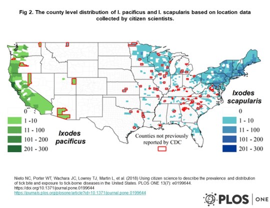
tick-bite-map

Tick bite map of US

Limits of the Study

While the study was hugely successful, it did have some limits. For one, the sample of ticks was limited to only those areas where citizens were participating, therefore the maps may not show all areas with ticks.

In addition, the researchers only tested for four of the many pathogens known to cause illness in humans.

Pathogens not tested for include: Multiple species of Borrelia including Mayonii, and Bisettii, which also cause Lyme-like illness; Ehrlichia chaffeensis, the cause of human monocytic erhlichiosis; Francisella tularensis, the cause of tularemia; Rickettsia rickettsii, the bacterial agent of Rocky Mountain spotted fever; multiple species of the protozoan pathogen Babesia, including duncani and divergens; and several viruses known to be transmitted by ticks including Bourbon virus, Colorado tick fever virus, Heartland virus, and Powassan virus.

Lack of funding for more studies

Lack of funding poses the biggest challenge to fully understanding the risks that ticks pose to the US population.

When asked, the CDC’s Ben Beard stated

“We’ve got national maps, but we don’t have detailed local information about where the worst areas for ticks are located.…The reason for that is there has never been public funding to support systematic tick surveillance efforts.”

It’s no secret that within weeks of the first Zika infection in 2016, Congress authorized $1.7 billion in funding, of which $397 million was made immediately available to rapidly develop an accurate test and begin work on a vaccine.

That same year, the federal budget allotted only $28 million for Lyme disease—the most common vector-borne disease in the US. (Note: while the CDC can study and report on diseases, it is not allowed to lobby Congress for funding.)

What did we learn?

The big take-away from the NAU study is that ticks are everywhere, and they are full of dangerous pathogens—not just Lyme disease. This study demonstrates that ticks are spreading in range, and they are carrying more pathogens than ever before.

Until we find better ways for the CDC to report illnesses, these type of risk maps, that are generated from the pathogens the ticks are carrying, will be the best predictor of disease.

Finally, we all need to get out there and tell our representatives the dangers that lurk in our backyards. Call them. Ask to meet with them. Bring them a copy of this report. Let them know we need an dramatic increase in funding for Lyme and tick-borne diseases.

Click here for more information about ticks:  https://www.lymedisease.org/lyme-basics/ticks/about-ticks/

LymeSci is written by Lonnie Marcum, a Licensed Physical Therapist and mother of a daughter with Lyme. Follow her on Twitter: @LonnieRhea Email her at: lmarcum@lymedisease.org .

References:

  1. Using citizen science to describe the prevalence and distribution of tick bite and exposure to tick-borne diseases in the United States, PLOSone, July 2018,  https://journals.plos.org/plosone/article?id=10.1371/journal.pone.0199644
  2. County-Scale Distribution of Ixodes scapularis and Ixodes pacificus (Acari: Ixodidae) in the Continental United States , Journal of Medical Entomology, Volume 53, Issue 2, 1 March 2016, Pages 349–386,  https://academic.oup.com/jme/article/53/2/349/2459744
  3. Entomological Society of America Position Statement on Tick-Borne Diseases Approved on July 29, 2015, Valid through July 29, 2019  http://www.entsoc.org/PDF/2015/ESA-PolicyStatement-TickBorneDiseases.pdf
  4. Report of the Disease Vectors, Surveillance, and Prevention Subcommittee to the Tick-Borne Disease Working Group  https://www.hhs.gov/ash/advisory-committees/tickbornedisease/reports/disease-vectors-2018-5-9/index.html

_________________

**Comment**

Canadian independent tick researcher, John Scott, stands in opposition to the climate change model affecting tick movement:  https://madisonarealymesupportgroup.com/2018/08/13/study-shows-lyme-not-propelled-by-climate-change/  A recent study shows that warm winters are lethal to I. scapularis (black-legged) ticks. In fact, overwinter survival dropped to 33% when the snow melted. This has been substantiated by other researchers as well. Scott & Scott, 2018, ticks and climate change, JVSM

Also, illogically, people on one hand admit that ticks are now “everywhere” but keep pushing the importance of geographical maps.  Why?  These maps are worthless & constantly changing.  Migratory birds are transiting ticks worldwide along with the fox, coyote, lizard, mouse, and 1,000 other carriers including humans traveling everywhere.

Why do we continue to push and believe in maps that have kept people from diagnosis and treatment for decades?

I’ll tell you why – it’s lining pockets with money.

Authorities look at the inaccurate and constantly changing maps and announce,
“You can’t have ______________, it doesn’t exist here.”

More data that actually hurts patients.

This tick border thing is a man-made constructed paradigm that has never been accurate, but it fits the CDC/NIH/IDSA narrative. http://steveclarknd.com/wp-content/uploads/2013/11/The-Confounding-Debate-Over-Lyme-Disease-in-the-South-DiscoverMagazine.com_.pdf (go to page 6 and read about Speilman’s maps which are faulty but have ruled like the Iron Curtain, and have been used to keep folks from being diagnosed and treated)

Dr. Masters fought this tooth and nail for his suffering patients in the South:  https://madisonarealymesupportgroup.com/2017/10/06/remembering-dr-masters-the-rebel-for-lyme-patients-who-took-on-the-cdc-single-handedly/  Even though Fier found borrelia in 2% of sampled lone star ticks and subsequently supported Masters’ Missouri Lyme, the CDC insisted that the EM rash was NOT diagnostic for LD for Missouri patients due to the fact that neither Ixodes dammini nor Ixodes pacificus were found there.  

Maps are continually used against patients and have ALWAYS been faulty:  https://madisonarealymesupportgroup.com/2017/08/24/canine-maps-better-than-the-cdcs-in-predicting-lyme-disease/ (nothing’s changed)

TIME TO PULL THE BLINDERS OFF AND LOOK AT THIS THING AS THE PANDEMIC IT TRULY IS, AND TIME TO FOCUS ON ISSUES THAT ACTUALLY RELIEVE PATIENT SUFFERING.


Case of Recurrent Fever & Multiple Splenic Infarcts (& Why Short Treatment Duration Often Doesn’t Work for Babesia)

https://journals.lww.com/infectdis/fulltext/2018/09000/An_85_Year_Old_Man_With_Recurrent_Fever_and.13.aspx  (See comment at end of article)

An 85-Year-Old Man With Recurrent Fever and Multiple Splenic Infarcts

Shuker, Orel, MSc; Subran, Mala, MBBS; Hardie, Rochelle, MBBS; Ghitan, Monica, MD; Chapnick, Edward K., MD; Lin, Yu Shia, MD

Infectious Diseases in Clinical Practice: September 2018 – Volume 26 – Issue 5 – p 300–302
doi: 10.1097/IPC.0000000000000643

An 85-year-old man with a history of benign prostatic hyperplasia, hyperlipidemia, and Guillain-Barré syndrome in 1989 presented in September 2017 for the third time in 2 months with intermittent fever, chills, and night sweats. In July, he presented with a 5-day history of intermittent fever, with a temperature of 103°F and chills. The urinalysis and chest x-ray were normal, and 2 sets of blood cultures showed no growth. A computed tomography (CT) scan of the abdomen and pelvis was performed (Fig. 1A). The leukocyte count was 4200/μL (69% neutrophils), hemoglobin (Hb) 11.0 g/dL, and platelet count 63,000/μL. Patient received 3 days of azithromycin and was discharged with 2 days of oral cefpodoxime and azithromycin to complete a 5-day course of treatment.

The patient was readmitted to the hospital 4 days later with fever of 104°F associated with chills and sweating. Intravenous vancomycin, cefepime, and metronidazole were given. The leukocyte count was 7800/μL, Hb 11.0 g/dL, and platelet count 386,000/μL. Blood cultures and CT of the abdomen and pelvis were negative. The patient was discharged from the hospital on day 4 without antibiotics when fever subsided.

In mid-September, 2 months after the initial presentation, the patient was readmitted to the hospital for recurrent fever for 10 days, with worsening fatigue, malaise, and anorexia. The leukocyte count was 6100/μL, Hb 7.6 g/dL, and platelet count 130,000/μL, with lactate dehydrogenase 849 U/L, alanine aminotransferase 53 U/L, aspartate aminotransferase 70 U/L, and alkaline phosphatase 41 U/L. The erythrocyte sedimentation rate was 115 mm/h, and C- reactive protein was 10 mg/dL. Blood cultures and echocardiogram were normal. Repeat CT scan of the abdomen and pelvis is shown in Figure 1B. The patient denied travel outside New York. He is originally from Greece and has lived in Brooklyn, NY, for 50 years. He did not recall any insect or tick bites, and no other family members had a similar illness. The patient denied any dental procedures in the past year. Patient’s home medications were finasteride and pravastatin. What is your diagnosis?

Part 2

Diagnosis: Babesia Infection Caused by Babesia microti

Our patient presented with intermittent fever of unknown origin, and tick-borne disease was not included in the differential diagnosis initially. Thus, peripheral smears for parasites were not done in his previous 2 admissions. The initial CT scan showed a normal liver and spleen size (Fig. 2A). A subsequent CT scan 2 months later revealed numerous new splenic infarcts with hepatosplenomegaly (Fig. 2B). As mentioned, blood cultures and echocardiogram were nondiagnostic. On further questioning, the patient recalled that he had visited family in Greenport, Long Island, for the fourth of July weekend, 5 days prior to the onset of his febrile illness. Peripheral blood smear showed intraerythrocytic and extraerythrocytic ring inclusions consistent with babesiosis (Fig. 3) with 0.3% parasitemia. Polymerase chain reaction for Babesia microti was positive. Our patient was immediately started on azithromycin plus atovaquone, and the temperature normalized within 48 hours. He was discharged home on hospital day 7.

Splenic infarction is a rare complication of Babesia infection. To date, only 2 cases of splenic infarction in association with Babesia infection in humans have been published in the literature.1 Interestingly, azithromycin as monotherapy at a higher dose resulted in a significant reduction in Babesia parasitemia and prolongation of survival when compared with controls in hamster models.2 Our patient’s initial improvement in fever and thrombocytopenia after his first hospitalization was likely due to azithromycin therapy. There has been evidence of drug resistance to azithromycin-atovaquone with relapse of Babesia infection in immunocompromised patients after initial exposure to azithromycin as monotherapy.3 However, our patient remained afebrile and has been doing well 3 months after completion of a 10-day course of azithromycin plus atovaquone therapy.

Babesiosis is a tick-borne infection caused by intraerythrocytic protozoa of the genus Babesia, transmitted by the Ixodes tick. Babesia microti is the most predominant strain in the Northeastern and upper Midwestern region of the United States.4 The most common route of transmission is via direct inoculation from Ixodes scapularis ticks. Blood transfusion and rarely transplacental transmission have also been documented.4 The peak acquisition of disease occurs between May and September. Although tick activity and tick-borne diseases are common in warmer months, I. scapularis is active throughout the year, when ambient-air temperature is greater than 4°C (40°F).5 Thus, babesiosis should be considered and investigated appropriately even during the months not typical for increased tick activity.

Clinical manifestations of Babesia infections are variable, depending on the Babesia species and the immune status of the host. Clinical presentation of babesiosis includes febrile hemolytic anemia due to parasite-mediated lysis of red blood cells in the circulation.6 Babesia infections may induce cycles of disease by varying antigen expression and by displaying new outer-surface proteins during the disease course. The antigenic variants are referred to as serotypes and prevent the elimination of the protozoa by the immune system. This may contribute to the recurring nature of relapsing fever.7

Immunocompetent individuals typically have subclinical illness, associated with low-level parasitemia (<4%) and may present with a gradual onset of nonspecific flulike symptoms. Babesiosis in immunocompromised patients is often severe, with a complication rate of 40% to 60%, including acute respiratory failure, congestive heart failure, renal failure, and disseminated intravascular coagulation.8,9 Patients may present with splenic infarction as in our patient. Proposed mechanisms of splenic infarction in human babesiosis include microthrombus formation and local release of vasoactive factors caused by red blood cell lysis leading to infarcted necrosis of splenic tissue.10 Moreover, during infection, proinflammatory cytokines are released, specifically tumor necrosis factor, interleukin 1, interleukin 6, and interferon, leading to increased expression of adhesion molecules on the surface of the vascular endothelium. This in turn results in cytoadherence of the infected erythrocytes to the vascular endothelium.10 The parasitized erythrocytes also lack the deformability needed to transit the splenic sinusoids, causing their sequestration by resident macrophages and obstruction of the vascular flow within the spleen.1

Our patient presented with fever of unknown origin and new splenic infarcts secondary to Babesia infection. Our case reinforces the importance of taking a detailed history including travel history when evaluating a patient with fever of unclear etiology. Our patient did not think that it was relevant to mention during his earlier admissions to the hospital that he traveled to Greenport, Long Island (Suffolk County), for a weekend. Furthermore, babesiosis should be included in the differential diagnosis in a patient who presents with nonspecific flulike symptoms, hematological manifestations, and new splenic infarcts on radiographic imaging. Physicians should ensure timely diagnostic testing and appropriate initiation of treatment in a patient with babesiosisand splenomegaly to prevent further progression to splenic infarct or rupture, which may lead to a fatal outcome.

ACKNOWLEDGMENTS

The authors thank Myrna Dyer from the Department of Microbiology for providing the blood smear figure and Keith Arbeeny from the Department of Radiology of Maimonides Medical Center in Brooklyn, NY, for providing the radiological imaging figures.

REFERENCES

1. Florescu D, Sordillo PP, Glyptis A, et al. Splenic infarction in human babesiosis: two cases and discussion. Clin Infect Dis. 2008;46(1):e8–e11.

2. Weiss LM, Wittner M, Wasserman S, et al. Efficacy of azithromycin for treating Babesia microti infection in the hamster model. J Infect Dis. 1993;168(5):1289–1292.

3. Krause PJ, Lepore T, Sikand VK, et al. Atovaquone and azithromycin for the treatment of babesiosis. N Engl J Med. 2000;343(20):1454–1458.

4. Vannier E, Krause PJ. Human babesiosis. N Engl J Med. 2012;366:2397–2407.

5. Duffy DC, Campbell SR. Ambient air temperature as a predictor of activity of adult Ixodes scapularis (Acari: Ixodidae). J Med Entomol. 1994;31(1):178–180.

6. Kavanaugh MJ, Decker CF. Babesiosis. Dis Mon. 2012;58:355–360.

7. Deitsch KW, Lukehart SA, Stringer JR. Common strategies for antigenic variation by bacterial, fungal and protozoan pathogens. Nat Rev Microbiol. 2009;7(7):493–503.

8. Hatcher JC, Greenberg PD, Antique J, et al. Severe babesiosis in long island: review of 34 cases and their complications. Clin Infect Dis. 2001;32(8):1117–1125.

9. Krause PJ, Gewurz BE, Hill D, et al. Persistent and relapsing babesiosis in immunocompromised patients. Clin Infect Dis. 2008;46(3):370–376.

10. Wozniak EJ, Lowenstine LJ, Hemmer R, et al. Comparative pathogenesis of human WA1 and Babesia microti isolates in a Syrian hamster model. Lab Anim Sci. 1996;46(5):507–515.

____________________

More on Babesia:  https://madisonarealymesupportgroup.com/2016/01/16/babesia-treatment/

https://madisonarealymesupportgroup.com/2018/05/31/widespread-babesiosis-in-canada/

https://madisonarealymesupportgroup.com/2018/04/08/zoonotic-babesia-microti-in-the-nw-u-s-evidence-for-the-expansion-of-a-specific-parasite-lineage/

https://madisonarealymesupportgroup.com/2018/03/22/what-is-air-hunger-anyway/

https://madisonarealymesupportgroup.com/2018/02/20/babesia-and-heart-issues/

https://madisonarealymesupportgroup.com/2018/10/05/variable-clinical-presentations-of-babesiosis/  This article put out by The Nurse Practitioner demonstrates clearly how their short treatment duration doesn’t work for immunocompromised patients, which is pretty much everyone with Lyme/MSIDS as we are typically infected with more than one thing, and even if it’s just Lyme with Babesia http://www.wildcondor.com/dr-horowitz-on-babesiosis.html Dr. Krause published in the New England Journal of Medicine that when a patient has Lyme and Babesia, Lyme is found three-times more frequently in the blood, proving Babesia suppresses the immune system. https://madisonarealymesupportgroup.com/2017/06/28/concurrent-babesiosis-and-lyme-in-patient/  Despite the lack of acknowledgment, there’s a lot of us with Lyme and Babesia and even more pathogens.  

The one drug, one disease paradigm does not work with Lyme/MSIDS.
**Complex Babesiosis is associated with severe anemia and high parasitaemia levels**
**Chronic Babesiosis can trigger cardiovascular, kidney, and liver problems**

Personally, I can attest to the importance of the anti-malarial medications my husband and I were both put on. We both had chest pressure and dizziness but the headaches I had were out of this world. My heart would also flop like a fish out of water and wake me up from a dead sleep – racing. Neither of us had night sweats and we presented differently. My husband developed hyper coagulation and anemia while I did not. Heparin helped him tremendously.

If you are having heart involvement and antibiotics typically used for Lyme are not touching it, please discuss Babesia with your health care provider (and Bartonella as well!). 

Also, go to the Babesia Treatment link above and print and fill out the Babesia checklist by Dr. Schaller.  These checklists are more helpful than testing.  Testing often misses cases.  

For us, treating for Babesia made a huge difference. I’m happy to report ALL of those symptoms are completely gone after a full year hitting it hard with anti-malarials. The best treatments overlap. Again, see Babesia Treatment link for ideas on what this looks like.


Variable Clinical Presentations of Babesiosis

https://journals.lww.com/tnpj/Pages/articleviewer.aspx?year=2018&issue=10000&article=00011&type=Fulltext

Variable clinical presentations of babesiosis

Paparone, Pamela, DNP, APN; Paparone, Philip W., DO

doi: 10.1097/01.NPR.0000545000.07640.11
Abstract: 
Human babesiosis continues to spread in multiple regions of the US. It is transmitted by Ixodes species ticks, as are Lyme disease and anaplasmosis. Its variable clinical presentations, together with serologic detection limitations, require that a high index of clinical suspicion be present for prompt diagnosis. This article discusses case examples showing the wide range of symptoms and presentations that are possible with babesiosis.
Human babesiosis is an infectious, malaria-like disease caused by intraerythrocytic protozoa of the genus Babesia, specifically Babesia microti and Babesia divergens.1-4 It is transmitted by Ixodes species ticks, as are Lyme disease and anaplasmosis (formerly known as ehrlichiosis). Babesia species are well-known pathogens in animals. During the past half century in the US, they have been increasingly recognized as pathogens in humans.1,5 Babesiosis may be acquired through the bite of an infected tick, a blood transfusion, or by transplacental transmission.2,6-8 (See Ixodes scapularis [blacklegged or deer ticks].)
Most infection passes undetected (because the patient may be unaware of the tick bite), especially in healthy adults.6,7 However, in immunocompromised patients—particularly those with hematologic disease and a history of splenectomy—Babesia infection may be severe and life-threatening.1

Epidemiology

The first reported case of babesiosis in the US was in 1968.9 It became a nationally notifiable disease in 2011, and among the 27 states where it was notifiable in 2013, there were 1,792 reported cases nationwide.5,10 Tick-borne and transfusion-associated cases of babesiosis occur in multiple parts of the country, including outside of areas of known endemicity.5 The number of reported cases is rising steadily in the US and worldwide, owing in part to increased medical awareness and improved diagnostic methods.1-3 (See Reported cases of babesiosis in the US.)

Health departments notify the CDC of babesiosis cases via the National Notifiable Diseases Surveillance System (NNDSS) using a standard case definition. In addition to basic demographic information (age, gender, and county of residence) provided via NNDSS, supplemental data (symptoms and history of transfusion) can be submitted to the CDC using a disease-specific case report form (CRF). Because babesiosis has been a reportable condition in some states for years, state-developed CRFs had already been in use to capture supplemental data.5

To promote standard data collection, the CDC developed a babesiosis CRF, which was approved by the Office of Management and Budget in August 2011 (www.cdc.gov/parasites/babesiosis/resources/50.153.pdf). Supplemental data, derived from the CDC’s or a state’s CRF, were merged manually with NNDSS records by matching a case ID number or demographic data. If case records had conflicting data, the more detailed record was considered correct.

As cases of babesiosis transmitted via tick bite or blood transfusion occur in multiple parts of the US, including outside of areas of known endemicity, ongoing national surveillance using the standard case definition will provide a foundation for developing evidence-based prevention and control measures to reduce the burden of the disease. In addition, mapping based on this surveillance allows for the identification of endemic areas, which aids the clinician in diagnosis.

Transmission and pathogenesis

The heightened recognition of tick-borne infection is derived largely from the increasing incidences of human babesiosis, anaplasmosis, and Lyme disease, both individually and together.11,12 Because these infections share the same rodent reservoir and tick vector hosts, they can be cotransmitted to human hosts.1,2,10,13-16 Coinfections involving various combinations of these pathogens are common and can be severe.12,14 The babesia parasite is suspected of causing proinflammatory cytokines that stimulate the production of nitric oxide, which may cause erythrocytic cellular damage when produced in excess.2

Diagnostic procedures and clinical management of the resulting disease syndrome are complicated by the diversity of pathogens involved and by the unusual diversity and duration of symptoms.

Clinical presentation

Common clinical features of babesiosis are similar to those of malaria and range in severity from asymptomatic to rapidly fatal. Most patients experience a viral infection-like illness with fever, chills, sweats, myalgia, arthralgia, anorexia, nausea, vomiting, or fatigue, and in some cases, patients may develop hemolytic anemia.1-4,10 Most symptomatic patients become ill 1 to 4 weeks after the bite of a B. microti-infected tick and 1 to 9 weeks (but up to 6 months in one reported case) after transfusion of contaminated blood products.6-8

A high index of clinical suspicion for babesiosis and the possible presence of other tick-borne infections are required for prompt diagnosis and proper treatment. Because the clinical findings are nonspecific, lab studies are necessary to confirm the diagnosis.

Diagnosis

Microscopic examination of blood smears is the current gold standard for detecting Babesia infection, while polymerase chain reaction testing has promising diagnostic value.1,2,16,17 Differentiating Babesia from malaria on peripheral smears can be difficult but rapidly resolved by the presence or absence of a history of travel.1 Peripheral smears for Babesia allow for same-day or, at the most, next-day confirmation of the diagnosis. The case examples described below demonstrate the range of symptoms and clinical presentations associated with babesiosis (with and without coinfection) that can challenge the NP.

Babesiosis is caused by parasites that infect red blood cells. Most US cases are caused by B. microti, which is transmitted by Ixodes scapularis ticks, primarily in the Northeast and Upper Midwest. Babesia parasites also can be transmitted via transfusion, anywhere, at any time of the year. In March 2018, the FDA approved the first B. microti blood donor screening tests. B. microti Arrayed Fluorescent Immunoassay detects antibodies to B. microti in human plasma, and B. microti Nucleic Acid Test detects B. microti DNA in human whole blood.18

Treatment

**Please see my comment at end of article**

Generally, treatment with atovaquone plus azithromycin is used for patients with mild-to-moderate babesiosis, whereas clindamycin plus quinine is recommended for patients with severe disease; both treatment regimens are given for 7 to 10 days.1-4 All four drugs are used FDA off-label for babesiosis; however, the dosage recommendations are supported by the clinical guidelines.1-4,19 The dosage regimen for atovaquone plus azithromycin for adult patients is atovaquone 750 mg orally every 12 hours, and azithromycin 500 to 1,000 mg orally on day 1 and 250 mg orally once daily for the subsequent days.1-4 Immunocompromised patients may require higher doses of azithromycin.2-4

The dosage regimen for clindamycin plus quinine for adult patients with severe disease is clindamycin 600 mg orally every 8 hours or clindamycin 300 to 600 mg I.V. infusion every 6 hours, and quinine 650 mg orally every 6 to 8 hours.1-4 Dose adjustments of quinine are needed for patients with severe chronic kidney disease.19,20 Of note, the only FDA-approved preparation of oral quinine currently available in the US is the 324 mg capsule.19,20 Previously, the dosage available in the US was a 325 mg capsule. The change in the quinine preparation from 325 mg to 324 mg may result in minor dose disparities between some guideline dosage recommendations that were published before the commercial preparation was changed.20,21

Although rare cases of resistance to atovaquone plus azithromycin have been reported, this combination is effective in most patients.2 Atovaquone is contraindicated in patients who develop or have a history of serious allergic or hypersensitivity reactions to the drug or any of the drug’s components. Azithromycin is contraindicated in patients with known hypersensitivity to azithromycin or any macrolide or ketolide antibiotic and also in patients with a history of cholestatic jaundice or hepatic dysfunction.19 Clindamycin is contraindicated in patients with a history of hypersensitivity to clindamycin or lincomycin. Quinine is contraindicated in patients with known hypersensitivity to quinine, mefloquine, or quinidine; prolonged QT interval; a glucose-6-phosphate dehydrogenase deficiency; or a history of myasthenia gravis or optic neuritis.19 Consult the manufacturer’s prescribing label for complete prescribing information for each drug.

Some patients, including those with severe illness, might require or benefit from supportive care, such as antipyretics, vasopressors (if the patient’s BP is low and unstable), blood transfusions, exchange transfusions (in which portions of a patient’s blood or blood cells are replaced with transfused blood components), mechanical ventilation, and dialysis. The NP should consider referral to an infectious-disease specialist for patients who are pregnant, have an underlying hematologic or oncologic problem, have had a splenectomy, are allergic to first-line antibiotic agents, or have had an unsatisfactory response to antibiotic therapy.

Red blood cell exchange transfusions are recommended for cases of severe babesiosis in patients with parasitemia of 10% or greater, severe anemia (hemoglobin less than 10 g/dL), or pulmonary, kidney, or liver impairment.2-4 Exchange transfusions are used to rapidly decrease parasitemia, correct anemia, and help remove toxic byproducts produced by the infection.2

Case examples

The case examples of patients with babesiosis show a wide range of symptoms and clinical presentations. The case examples below are cases that occurred in southeastern New Jersey, where the disease is endemic. All patients were hospitalized and treated in Atlantic County, New Jersey (see Summary of data from patients with babesiosis).

Case 1

Ms. A is a 78-year-old White female who was admitted with fever, chills, lethargy, fatigue, and marked changes in sensorium. She had a maximum temperature of 100.6° F (38.1° C); sepsis was considered for this patient. Multiple tick bites were found. Pertinent lab findings included lactate dehydrogenase (LDH), 528 units/L; aspartate aminotransferase (AST), 90 units/L; and alanine aminotransferase (ALT), 34 units/L. Her vitamin B12 and folate levels were normal.

Ms. A’s initial white blood cell (WBC) count was 5.0 × 10⁹/L, but over the first 3 days of hospitalization, it gradually dropped to 2.6 × 10⁹/L. Her hemoglobin dropped from 10.5 g/dL to a low of 8 g/dL, and her platelets were initially 39 × 109/L but gradually increased as she continued her course of treatment. Ms. A had 33% polymorphonuclear leukocytes, 2% bands, 49% lymphocytes, and 13% monocytes. Peripheral smear was positive for Babesia, and she had a Babesia immunoglobulin M (IgM) of 1:160 and Anaplasma (previously referred to as Ehrlichia) IgM of 1:320.

In view of Ms. A’s leukopenia and thrombocytopenia, anaplasmosis was suspected, and she was treated with doxycycline 100 mg I.V. infusion every 12 hours, atovaquone suspension 750 mg orally twice daily, and azithromycin 500 mg I.V. infusion every 24 hours. Doxycycline is the recommended treatment for anaplasmosis and was administered to cover the possibility of anaplasmosis in this patient. She was treated with that regimen for 5 days. She was then started on doxycycline twice daily, and azithromycin 500 mg daily (both oral) along with the atovaquone suspension of 750 mg twice daily for a 14-day course of therapy. Ms. A made a dramatic improvement in her mentation and resolution of her lethargy.

Case 2

Ms. C is a 90-year-old White female with a chief complaint of rectal bleeding. On admission, her lab studies revealed severe anemia with a hemoglobin of 7.6 g/dL and hematocrit of 22.6%. Her platelet count was 103 × 109/L and peripheral smear was positive for Babesia. Ms. C had spiking temperatures 100° F to 101° F (37.8° C to 38.3° C). Her rectal bleeding was controlled with an octreotide infusion to which she responded well (the bleeding ceased). Her peripheral smear was positive for Babesia, and she was placed on an oral dose of azithromycin 500 mg on day 1 and then 250 mg daily and atovaquone suspension 750 mg twice daily to complete a 10-day course.

Case 3

Mr. E is a 57-year-old White male admitted with fever, malaise, and chills. His temperature had risen to 101° F (38.3° C). His AST and ALT were 64 and 54 units/L, respectively, and gradually rose to a peak of 90 and 87 units/L, respectively, during his 5-day hospital stay. Mr. E’s WBC count decreased from his initial hospital results to 2.9 x 10⁹/L with a hemoglobin of 9.2 g/dL. His platelets were initially 60 × 109/L but dropped to 34 × 109/L at their lowest level. In view of his elevated liver enzymes, leukopenia, and thrombocytopenia, anaplasmosis was highly suspected, and he was started on doxycycline 100 mg I.V. infusion every 12 hours.

Mr. E’s peripheral smear was positive for Babesia. He was started on oral clindamycin 600 mg every 8 hours and oral quinine 650 mg three times daily. Acute hearing deterioration occurred, and the quinine was discontinued. Mr. E’s regimen was then switched to oral azithromycin 500 mg on day 1 and then 250 mg daily and oral atovaquone 750 mg twice daily. He went on to complete only 7 days of therapy, and his elevated liver enzymes and thrombocytopenia resolved. The suspected anaplasmosis was not confirmed, as the Anaplasma IgM was negative. However, Mr. E’s leukopenia and thrombocytopenia resolved on the above regimens.

Case 4

Mr. J is an 81-year-old White male who was admitted with increasing lethargy, weakness, chills, and blurred vision. He had a history of coronary artery disease and hypertension. His hemoglobin on admission was 12.1 g/dL, and his hematocrit was 35.4%. His WBC count was 5.3 × 10⁹/L. By day 2, his hemoglobin had dropped to 9.9 g/dL with a hematocrit of 29%. His platelets were initially 54 × 109/L and dropped to 46 × 109/L, but on therapy, rose to 191 × 109/L.

Mr. J had 82% polymorphonuclear leukocytes, 10% lymphocytes, and 6% monocytes. On the day of admission, a peripheral smear was positive for Babesia. Subsequently, serologic studies demonstrated an Anaplasma IgG of 1:256; the IgM was negative. Babesia serologies were greater than 1:320, both IgG and IgM. Anaplasmosis was suspected with Mr. J’s confirmed babesiosis, and he was started on azithromycin 500 mg I.V. infusion every 24 hours and doxycycline 100 mg twice daily.

At discharge on day 10, Mr. J was switched to clindamycin orally three times a day and quinine orally three times a day because of intolerance to azithromycin, and he completed a 14-day course of therapy. He convalesced satisfactorily. His hemoglobin at discharge was 12.5 g/dL and WBCs 7.4 × 10⁹/L; platelets improved to 137 × 109/L.

Case 5

Mr. K is an 85-year-old White male who was admitted with fever and chills intermittently, recurring for several days prior to admission. He had a history of hairy cell leukemia, splenectomy, permanent pacemaker insertion for atrioventricular block, gouty arthritis, prostatic hypertrophy, and polymyalgia rheumatica. In the ED, Mr. K had an immediate peripheral smear for Babesia, and the intraerythrocytic parasite was demonstrated. He had been working on a golf course for the week prior to admission.

A second peripheral smear was positive for intraerythrocytic parasites with 10.4% of his red blood cells infected. Findings were also positive for Howell-Jolly bodies, which are erthrocytic nuclear remnants associated with asplenia or decreased splenic function. Mr. K was started on oral azithromycin 500 mg on day 1 and then 250 mg daily and atovaquone 750 mg suspension twice daily. Due to the possibility of concurrent tick-borne infection, he was also started on oral doxycycline 100 twice daily.

Over the course of day 1, Mr. K’s platelet count dropped from 25 to 23 × 109/L, with blood urea nitrogen of 29 mg/dL and creatinine of 1.2 mg/dL. His WBC count dropped from 4.1 to 2.5 × 10⁹/L, and his hemoglobin dropped from 16 to 13 g/dL. He had 20% bands, 5% atypical lymphocytes, 47% polymorphonuclear leukocytes, and 23% lymphocytes. Mr. K remained on doxycycline, azithromycin, and atovaquone suspension for 8 days when he was discharged home.

Mr. K was readmitted the following day when he complained of the inability to ambulate and generalized weakness. He had peripheral smear positivity with babesiosis and was serologically positive for anaplasmosis with both IgM and IgG. Mr. K had continued on the prescribed antibiotic regimen up until his readmission that day. Due to the persistence of parasitemia despite adequate therapy, he was changed to clindamycin 600 mg I.V. infusion every 8 hours, and quinine was also being administered.

Unfortunately, Mr. K developed gastric distress and a generalized erythematous coalescing rash, which prompted the discontinuation of the clindamycin and quinine. His WBC count was 2.2 × 10⁹/L, and his hemoglobin was 9.5 g/dL. Platelets had risen to 43 × 109/L, and he had 43% polymorphonuclear leukocytes, 10% bands, 42% lymphocytes, and 5% monocytes.

Because of the persistence of parasitemia, Mr. K underwent exchange transfusion. At that point, he had been restarted on azithromycin 500 mg I.V. infusion every 24 hours and atovaquone suspension 750 mg orally twice daily. Azithromycin and atovaquone were continued for 5.5 weeks, at which time he was parasite smear negative for Babesia. Subsequently, a Babesia peripheral smear remained negative.

Discussion of case examples

Case 1 shows the unusual effect of babesiosis on the sensorium in the older adult, as any infectious process can. The patient’s cognitive function was dramatically improved following treatment, despite the marked changes in mentation on admission. A coinfection with Anaplasma was suspected. In general, all cases of babesiosis need to be tested for late Lyme disease, via Western blot, although not immediately addressed.1,2,4

Patients with concurrent babesiosis and anaplasmosis—suspected or serologically positive—are treated with doxycycline, which is equally effective for Lyme disease, early or late. Generally, the greater number of concurrent tick-borne infections and the higher the parasitimia load, the more toxic the presentation.1,12

Case 2 shows the need to check the peripheral smear for Babesia despite the rectal bleeding issue on admission. This diagnostic test could have easily been omitted, causing a delay in the diagnosis. Such a delay in older adult patients that results in delayed treatment can put these patients at greater risk for severity of babesiosis. Generally, the combination of clindamycin and quinine has a much higher probability of intolerance and adverse reactions. This combination is not the treatment of first choice for babesiosis. Pertaining to anaplasmosis, the triad of leukopenia, transaminase elevation (mild or moderate), and thrombocytopenia demands empiric treatment with doxycycline prior to serologic confirmation.1,2,4

A peripheral smear for Babesia is rapidly interpreted, is inexpensive, and should be requested in evaluating all patients with any degree of anemia—especially during the spring and summer months in endemic areas. Serologic studies are variable in developing positivity and are generally less readily available.

Case 3 illustrates the importance of suspecting and investigating the possibility of babesiosis and anaplasmosis coinfection in a patient presenting with a tick-borne illness.

Case 4 demonstrates that no additional lab studies—other than peripheral smear for Babesia—are needed to confirm the diagnosis of babesiosis.

Case 5 exemplifies the therapeutic challenge and refractory response to treatment of babesiosis in patients with the comorbidities of a hematologic disease and/or splenectomy.

Patient education

Heightened awareness of babesiosis as well as prompt diagnosis and treatment are essential to prevention. Both patients and the general public need to become more aware of the existence of the disease and other tick-borne infections, especially individuals who live in or travel to regions where babesiosis is found. The NP can play an active and important role in providing patient education about the disease. The basic points of information to communicate include:

  • What babesiosis is and its potential to be a life-threatening illness
  • How individuals acquire babesiosis (tick bite, transfusion, or, rarely, vertical transmission)
  • Where in the world babesiosis is found
  • Signs and symptoms of babesiosis
  • Note that many individuals do not have any symptoms and do not get sick
  • Importance of seeing a healthcare provider if babesiosis is suspected
  • Treatability of babesiosis and need for prompt diagnosis and treatment.22,23

Individuals who live in or travel to endemic areas should avoid tick-infested areas; apply repellents and wear long pants and long-sleeved shirts when outdoors; shower soon after being outdoors; and check their entire body for ticks.3 When outdoors, they should walk on cleared trails, stay in the center of the trail, and minimize contact with leaf litter, brush, and overgrown grasses (where ticks are most likely to be found). If a tick is found attached to a person’s body, it should be properly removed as soon as possible.

The CDC offers a printable, one-page fact sheet for patients and the general public that details the basic information for babesiosis awareness in addition to the link for the CDC guide to proper removal of a tick attached to a person (www.cdc.gov/parasites/babesiosis/resources/babesiosis_fact_sheet.pdf).

Conclusion

This article illustrates the need for the NP to appreciate the variable clinical presentations of babesiosis to facilitate prompt diagnosis, provide proper therapeutic management, and avoid the poor outcomes associated with this disease. Staying knowledgeable of babesiosis is essential. It is important for the NP to understand that infected patients may not recall a tick bite and that clinical presentations may not only be variable but also nonspecific, ranging from subclinical to severe. The possibility of coinfection with other tick-borne illnesses (Lyme disease and anaplasmosis) must be considered. Furthermore, the NP needs to assume an active role in patient education to affect babesiosis awareness and prevention.

Ixodes scapularis (blacklegged or deer ticks)

The images below are of the Ixodes scapularis ticks, also known as blacklegged or deer ticks. From left to right, the male (M) with a dorsal scutum (also known as a shield on the hard-bodied tick) that covers the entire back on the male, the female (F) with only a portion of the back covered by the dorsal scutum, the nymph (N), and the larva (L).

Figure

Sourse: Procop GW, Church DL, Hall GS, et al. Koneman’s Color Atlas and Textbook of Diagnostic Microbiology. 7th edition. Philadelphia, PA: Wolters Kluwer Health, 2016.

Reported cases of babesiosis in the US1,2,22,23

Most cases of babesiosis in the US occur in seven states, five of which are located in the Northeast (MA, CT, RI, NY, and NJ) and two in the upper Midwest (MN and WI). The geographic range of babesiosis has expanded beyond these highly endemic areas and it is now reported all along the northeastern seaboard and inland, ranging from Maine to Maryland.

Sporadic cases of babesiosis have been reported in other areas of the US including the West Coast. Additionally, transfusion-associated cases of babesiosis can occur anywhere in the country. Congenital transmission of babesiosis has also been reported.

REFERENCES

1. Sanchez E, Vannier E, Wormser GP, Hu LT. Diagnosis, treatment, and prevention of Lyme disease, human granulocytic anaplasmosis, and babesiosis: a review. JAMA. 2016;315(16):1767–1777.

2. Vannier EG, Diuk-Wasser MA, Ben Mamoun C, Krause PJ. Babesiosis. Infect Dis Clin North Am. 2015;29(2):357–370.

3. Vannier E, Krause PJ. Human babesiosis. N Engl J Med. 2012;366(25):2397–2407.

4. Wormser GP, Dattwyler RJ, Shapiro ED, et al The clinical assessment, treatment, and prevention of Lyme disease, human granulocytic anaplasmosis, and babesiosis: clinical practice guidelines by the Infectious Diseases Society of America. Clin Infect Dis. 2006;43(9):1089–1134.

5. Centers for Disease Control and Prevention. Babesiosis surveillance—18 States, 2011. MMWR Morb Mortal Wkly Rep. 2012;61(27):505–509.

6. Levin AE, Krause PJ. Transfusion-transmitted babesiosis: is it time to screen the blood supply. Curr Opin Hematol. 2016;23(6):573–580.

7. Leiby DA. Babesiosis and blood transfusion: flying under the radar. Vox Sang. 2006;90(3):157–165.

8. Herwaldt BL, Linden JV, Bosserman E, Young C, Olkowska D, Wilson M. Transfusion-associated babesiosis in the United States: a description of cases. Ann Intern Med. 2011;155(8):509–519.

9. Scholtens RG, Braff EH, Healey GA, Gleason N. A case of babesiosis in man in the United States. Am J Trop Med Hyg. 1968;17(6):810–813.

10. Centers for Disease Control and Prevention. Notice to readers: final 2013 reports of nationally notifiable infectious diseases. MMWR Morb Mortal Wkly Rep. 2014;63(32):702–715.

11. Western KA, Benson GD, Gleason NN, Healy GR, Schultz MG. Babesiosis in a Massachusetts resident. N Engl J Med. 1970;283(16):854–856.

12. Diuk-Wasser MA, Vannier E, Krause PJ. Coinfection by Ixodes tick-borne pathogens: ecological, epidemiological, and clinical consequences. Trends Parasitol. 2016;32(1):30–42.

13. Herwaldt BL, McGovern PC, Gerwel MP, Easton RM, MacGregor RR. Endemic babesiosis in another eastern state: New Jersey. Emerg Infect Dis. 2003;9(2):184–188.

14. Thompson C, Spielman A, Krause PJ. Coinfecting deer-associated zoonoses: Lyme disease, babesiosis, and ehrlichiosis. Clin Infect Dis. 2001;33(5):676–685.

15. Paparone PW, Glenn WB. Lyme disease with concurrent ehrlichiosis. J Am Osteopath Assoc. 1994;94(7):568–570, 573, 577.

16. Hildebrandt A, Gray JS, Hunfeld KP. Human babesiosis in Europe: what clinicians need to know. Infection. 2013;41(6):1057–1072.

17. Wang G, Wormser GP, Zhuge J, et al Utilization of a real-time PCR assay for diagnosis of Babesia microti infection in clinical practice. Ticks Tick Borne Dis. 2015;6(3):376–382.

18. U.S. Food & Drug Administration. FDA approves first tests to screen for tickborne parasite in whole blood and plasma to protect the U.S. blood supply. 2018. http://www.fda.gov/newsevents/newsroom/pressannouncements/ucm599782.htm.

19. Facts and Comparisons. Drug. Facts and Comparisons 2013. St. Louis, MO: Wolters Kluwer Health; 2013.

21. Gelfand JA, Vannier EG. Clinical manifestations, diagnosis, treatment, and prevention of babesiosis. UptoDate. 2017. http://www.uptodate.com.

22. Joseph JT, Purtill K, Wong SJ, et al Vertical transmission of Babesia microti, United States. Emerg Infect Dis. 2012;18(8):1318–1321.

23. Centers for Disease Control and Prevention. Tickborne diseases of the United States. A reference manual for health care providers. 2017. http://www.cdc.gov/lyme/resources/TickborneDiseases.pdf.

____________________

**Comment**

According to Dr. Horowitz, an experienced LLMD (Lyme literate doctor), Babesia is a tenacious tick-borne infection that persists.  Experience has shown him patients often need 9-12 months of treatment, a far cry longer than what is suggested here.

For treatment options, please see:  https://madisonarealymesupportgroup.com/2016/01/16/babesia-treatment/

All patients of tick-borne infections need follow-up – years later from treatment.

While the research shows again and again the persistent symptoms of patients, the “powers that be” continue to treat this short-term and seemingly ignore the vast population out here struggling (and it’s far greater than the purported 5-10% of the patient population, I assure you).

The one drug, one disease paradigm also doesn’t work with most patients as we are often coinfected:  https://madisonarealymesupportgroup.com/2017/05/01/co-infection-of-ticks-the-rule-rather-than-the-exception/  This link shows that 45% of tested ticks were coinfected and carried up to 5 different pathogens. This directly translates to human infection and a survey substantiates this: https://madisonarealymesupportgroup.com/2014/11/14/studies-show-why-its-tough-to-treat-lyme-and-co/ The most common co-infections in the LDo study were Babesia (32%), Bartonella (28%), and Ehrlichia (15%) while a study by Dr. Janet Sperling in Canada found that the most common were Bartonella (36%), Babesia (19%), and Anaplasma (13%).

There is also the issue of tick bites igniting latent infections already within the human body such as Epstein Barr, numerous herpes viruses, and even Bartonella. Yet, patients are struggling with these – sometimes all at once.  Is it any wonder we are sicker than dogs?http://www.wildcondor.com/dr-horowitz-on-babesiosis.html Dr. Krause published in the New England Journal of Medicine that when a patient has Lyme and Babesia,Lyme is found three-times more frequently in the blood, proving Babesia suppresses the immune system. https://madisonarealymesupportgroup.com/2017/06/28/concurrent-babesiosis-and-lyme-in-patient/  Besides the fact it is a misnomer to think it novel that a patient has concurrent Lyme and Babesiosis, it is also a huge mistake to base treatment on geographical area as time and time again, entomologists are finding ticks in places they just shouldn’t be and ticks that shouldn’t be carrying pathogens, carrying them. Also, using logic, until every bird, fox, squirrel, lizard, deer, and every other rodent on the earth read the memo that they are not supposed to cross state and country boundaries, ticks are going to continue to defy the box “experts” put them into. And, there are other ways for pathogens to travel across state lines:https://doi.org/10.1111/tid.12741

Abstract
The potential for transmission of Babesia microti by blood transfusion is well recognized. Physicians may be unaware that products used for transfusion may be collected from geographically diverse regions. We describe a liver transplant recipient in South Carolina who likely acquired B. microti infection from a unit of blood collected in Minnesota.  Also, one must be careful of the “history of tick bite,” as well, as many never see the tick or subsequent bite, and fail to get a rash. A nymphal tick is nearly impossible to see. Lyme/MSIDS is a CLINICAL diagnosis.

So much research begging to be done.


Tickology Video Series – Everything You Want to Know About Ticks & Prevention

Entomologist Larry Dapsis, Deer Tick Project Coordinator, of Cape Cod Cooperative Extension presents information about numerous types of ticks and the diseases they carry in the following Tickology video series.

Tickology

 Approx. 9 Min

Tick Identification & Ecology

Take aways:

  1. Female American Dog Tick is easy to spot as she has a creamy white wide spot up by the head.
  2. Female Lone Star tick has a bright white spot in the center of her back.
  3. Female Deer Tick has a bright red abdomen.
  4. A lot of this info is shared again in part 3 below where I have more notes.

 Approx. 12:30 Min.

Tick Borne Diseases

Take aways:

  1. He considers the American Dog Tick more of a nuisance than a threat.  I disagree.  Just ask anyone who’s ever had RMSF or Tularemia, both of which can kill you.
  2. The Deer Tick (Black legged tick) is endemic in 80 countries and has been here for thousands of years.
  3. Lyme is found in 49 out of 50 states in the U.S. (absent only in Hawaii)
  4. In 2016 the CDC adjusted Lyme prevalence to 300,000 new cases of Lyme a year.
  5. Martha’s Vineyard has more cases than anywhere in the universe.
  6. Risk of infection is year round.
  7. Largest risk is from the nymph as they are smaller and the bite is difficult to detect.   He is finding about 25% to be infected with Lyme.  50% of adults are infected.
  8. In Massachusetts, children ages 5-9 have the highest rates of infection.  Adults aged 50-70 has a surge of infection as well.
  9. Babesiosis, similar to Malaria, can be passed via blood transfusion with 50% of Massachusetts cases found in the south eastern part of the state and virtually found in some degree in every county in the state.
  10. Anaplasmosis (HGA) can look similar to Lyme and is more broadly distributed in Mass.
  11. All these diseases are steadily increasing.  95% of cases are aged 65 and older.
  12. Borrelia miyamotoi, related to Lyme, is a relapsing fever.  3% of Cape Cod ticks have it but is expected to increase.
  13. Powassan can put you in the hospital with brain swelling.  They did surveillance and found Powassan in 4 out of 6 site sites with infection rates as high as 10% in the tick population.  In reading the literature, he feels it has been on Cape Cod for thousands of years but it hasn’t been on medical radar.

  Approx. 8 Min.

Lone Star Tick – The New Tick in Town

Part 3 of the Tickology video project.

Take aways:

  1. The Lone Star Tick, normally considered a Southern tick, is in Cape Cod, and has moved North, and yes, is in Wisconsin.
  2. The adult female has a white dot on her back
  3. These ticks can run and are aggressive, fast & will actually chase you.  
  4. While he mentions a warming climate, independent Canadian tick researcher, John Scott, states emphatically temperature has nothing to do with tick expansion:  https://madisonarealymesupportgroup.com/2018/08/13/study-shows-lyme-not-propelled-by-climate-change/
  5. He claims Lone Star ticks have been established in Sandy Neck Beach Park and Shining Sea Bike Trail for a long time – it’s just nobody was looking for them.  I suspect this to be true for many other areas as well.
  6. He claims these areas are “perfect flyways” for migratory birds for transporting ticks.
  7. Lone Star ticks prefer intermediate size hosts.  He put out video surveillance and picked up wild turkeys in areas where these ticks were established.  Rabbits & coyotes are good hosts as well
  8. The adult female lays a cluster of 4,000-5,000 eggs,  which leaves a high concentration of larvae in late summer.  He claims when you find one, it could be a matter of minutes and you could have 200-300 bites.
  9. He claims Lone Star tick larvae do not transmit pathogens.
  10. The adults; however, can transmit Erlichiosis, STARI, Tularemia and Alpha Gal or meat allergy (all animal products).
  11. He claims you will not find deer ticks in an open lawn.  I was told otherwise by Susan Paskewitz, chair of the Department of Entomology at UW–Madison, whose crew is finding them in fields where kids are playing sports, and it’s here as well: https://newyork.cbslocal.com/2018/05/07/ticks-lyme-disease-cdc-putnam-county/
  12. He is finding Lone Star ticks in open spaces.  They don’t mind the heat.  Deer ticks will seek out leaf litter and/or snow when conditions are harsh.

 Approx. 13:22 Min

Permethrin Treated Clothing & Footwear

Take aways:

  1. Natural Pyrethrum is from the Aster Family, & is an extract from a type of chrysanthemum.  It has quick knockdown against insects but no residual control.  Breaks down in sunlight quickly.
  2. They manipulated it so now it has 4 weeks of residual control.
  3. You only use it on clothing and footwear.  He feels treating footwear to be crucial.  If a tick is on a treated surface with permethrin for 60 seconds it will die.  He feels strongly that using this product will reduce your exposure tick bites by upwards of 90%.  It is active thru 6 washings or 45 days which ever comes first.
  4. Pre-treated tick repellent clothing is also available.  EPA testing has shown it is active through 70 washings.  You can also send your clothing to “Insect Shield,” and they will treat your clothing and send it back with the 70 washing claim.  He says it’s about $10 per clothing item.
  5. It’s not the molecule that makes the poison, it’s the dosage.  As far as permethrin goes, there is low mammal toxicity except for cats.  It is 2,250 times more toxic to ticks than to humans.  According to the EPA, permethrin-treated clothing poses no harm to infants, children, pregnant women, or nursing mothers.
  6. Permethrin has low skin absorption and is metabolized quickly.
  7. National Research Council looked at long term exposure on the military wearing permethrin saturated clothing from head to foot for 18 hours a day for 10 years and found no reason for an adverse effect.
  8. The active ingredient is the same ingredient used for treating scabies and head lice and parents smear it on their kids from head to toe.
  9. He demonstrates how to apply it onto clothing and footwear.  Scroll to 10:00.  Make sure to wash these treated cloths away from other clothes.  Remember sunlight breaks it down so it lasts through 6 washings for 45 days, which ever comes first.
  10. He sprays the inside of the legs in case a tick gets underneath.  I tuck my pants into my white sprayed socks so ticks can not get inside.

 Approx. 6 Min

Skin Repellents

Take aways:

  1. The big distinction between repellents is the EPA registration.  Deet, Picaridan, IR 3535, and Oil of Lemon Eucalyptus have EPA registration with data on file for any claim being made.
  2. Go here for the EPA selection guide:  https://www.epa.gov/insect-repellents/find-repellent-right-you  (Fill in the questionnaire)
  3. Go to www.npic.orst.edu for pesticide information.
  4. Go to capecodextension.org for short factual answers on products.
  5. Naturals are not EPA registered so there is no data proving effectiveness.  Not all repel ticks.  Buyer beware.

__________________

For more on tick prevention:  https://madisonarealymesupportgroup.com/2017/05/11/tick-prevention-and-removal-2017/

https://madisonarealymesupportgroup.com/2018/06/06/mc-bugg-z/

https://madisonarealymesupportgroup.com/2018/05/27/study-conforms-permethrin-causes-ticks-to-drop-off-clothing/  “All tested tick species and life stages experienced the ‘hot-foot’ effect after coming into contact with permethrin-treated clothing,” Eisen said. 

https://madisonarealymesupportgroup.com/2018/04/03/fire-good-news-for-tick-reduction/  Study found a 78-98% reduction in ticks.
http://journals.plos.org/plosone/article?id=10.1371/journal.pone.0112174 These data indicate that regular prescribed burning is an effective tool for reducing tick populations and ultimately may reduce risk of tick-borne disease.