Archive for the ‘Autism’ Category

Aluminum in the Brain in Multiple Sclerosis: Regulatory and Funding Agencies Silent, Complicit

https://jameslyonsweiler.com/2018/08/18/aluminum-in-the-brain-in-multiple-sclerosis-regulatory-and-funding-agencies-silent-complicit/

By James Lyons Weiler in Cures August 18, 2018

https://jameslyonsweiler.com

MEDICAL SCIENCE proceeds along a hierarchy of evidence; often, patients are studied individually (case studies), or a small collection of patients are examined and characterized together (case series studies). Case series studies typically have smallish sample sizes and it is generally understood that larger studies will be necessary to determine more accurately the characteristics being studied.

In a new case series study, brain tissue from 14 donors with a diagnosis of MS was studied in a case series by Mold et al (2018) using transversely heated graphite furnace atomic absorption spectrometry. The study found high aluminum content (>10 ug/g dry weight) in all areas of the brain studied, with some areas exceeding 50 ug/g. They found aluminum both with cells and in the interstitium between cells. They found aluminum co-localised with structures known to be present in Secondary Progressive Multiple Sclerosis (SPMS) in the frontal cortex of one donor with SPMS.

There are a number of critical lines of evidence that make this fundamental finding critically important. Patients with MS have lower amounts of aluminum in their hair, suggesting depressed detoxification, and higher amounts are found in urine on chelation challenge testing (Fulgenzi et al., 2014). Chelation with EDTA is known to significantly reduce aluminum intoxication (Fulgenzi et al. 2014), and consumption of silica-rich mineral waters also increase urinary excretion of aluminum from patients with SPMS (Jones et al., 2017).

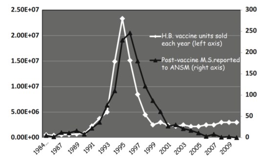
Extremely plausible direct mechanisms of the cause of MS from aluminum are known and animal studies routinely induced MS using aluminum hydroxide injections. So much evidence exists that points to aluminum as a source of strange new conditions of unknown causes, such as MMF and Gulf War Syndrome, one would think that calls to reconsider the use of aluminum in vaccines would be answered. The same team had previously found high amounts of aluminum in the brains of people with autism, and and in patients with Alzheimer’s disease. The latter result, while also important, is not surprising, as it has long been known that amyloid is part protein and part aluminum. Finally, when France brought on HepB vaccination, cases of MS following vaccination increased; when they stopped recommending the HepB vaccine, which contains aluminum hydroxide, the rate of HepB vaccine-associated MS cases dropped to near zero.

francems

Complicity

What is surprising is the lack of action on the part of the US FDA to put an end to the use of this dangerous metal in vaccines, and that the NIH is not funding more studies like this. NIH should fund studies to determine how to most safely remove aluminum from anyone exposed via vaccines; brain stem amyloidosis is a non-trivial concern. Approaches like ketogenic diet, silica-rich mineral waters, hyperbaric oxygen, EDTA, intranasal insulin and intranasal deferoxamine (to prevent brain stem amyloidosis) should all be tested in randomized clinical trails in clinical populations known to be afflicted with aluminum intoxication (autism, Alzheimer’s, MS).

What is also surprising is that the CDC and ACIP remain blithe to the morbidity and mortality their continued approval of vaccines that contain metals like aluminum and mercury. They make decisions on behalf of us all, and yet every member of ACIP with the exception of one military member has conflicts of interest with vaccine manufacturers. ACIP should review all of the literature on aluminum and make recommendations on how to phase it, and thimerosal, out of vaccines completely.

Further inaction on the part of these regulatory and funding agencies, and active denialism at this point will surely be seen by future generations as both callous disregard, and where conflicts of interest reside, complicity.

The study, conducted at the Keele University, was funded in part by the Children’s Medical Safety Research Institute.

References

Jones K et al. EBioMedicine. 2017 Urinary Excretion of Aluminium and Silicon in Secondary Progressive Multiple Sclerosis. 26:60-67. doi: 10.1016/j.ebiom.2017.10.028.

Fulgenzi A, Vietti D1, Ferrero ME. Aluminium involvement in neurotoxicity. Biomed Res Int. 2014;2014:758323. doi: 10.1155/2014/758323.

_______________

**Comment**

https://www.nationalmssociety.org/Symptoms-Diagnosis/Other-Conditions-to-Rule-Out/Lyme-Disease

Lyme disease can cause delayed neurologic symptoms similar to those seen in multiple sclerosis (MS) such as weakness, blurred vision caused by optic neuritis, dysesthesias (sensations of itching, burning, stabbing pain, or “pins and needles”), confusion and cognitive dysfunction, and fatigue. Lyme disease symptoms may also have a relapsing-remitting course. In addition, Lyme disease occasionally produces other abnormalities that are similar to those seen in MS, including positive findings on magnetic resonance imaging (MRI) scans of the brain and analysis of cerebrospinal fluid (CSF).

These similarities in symptoms and test results have led some people with MS to seek testing for the presence of antibodies to Borrelia, to determine if their neurologic symptoms are the result of Lyme disease or truly MS. The distinction is important because Lyme disease, especially when treated early, often responds to antibiotic therapy, whereas MS does not.

Studies examining Lyme disease & MS
Two studies have examined the overlap in diagnosis of MS and Lyme disease. The studies were conducted in parts of Long Island, New York, an area where Lyme disease is endemic, or regularly found.

In the first study, people who had Borrelia antibodies in their blood as well as a variety of neurologic symptoms considered to be “MS-like,” were evaluated with MRI, evoked potentials (EP) and CSF analysis, including a test for the presence of Borrelia antibodies in the spinal fluid.

While those with the MS-like illness had the highest incidence of abnormal MRIs and were the only ones among those studied to have abnormal EP and oligoclonal bands in their spinal fluid (indicating an abnormal immune response), they did not prove to have any Borrelia antibody in their spinal fluid.

The researchers concluded that the few patients with the MS-like symptoms probably had these symptoms due to MS and had also been exposed to the Borrelia bacterium.
A companion study looked for the presence of Borrelia antibodies in the blood of 100 people with the diagnosis of possible MS. Of 89 people who in fact turned out to have definite MS, only one had Borrelia antibodies. The researcher concluded that “…infection with Borrelia is infrequent in MS patients who live in an endemic area. Lyme disease is unlikely to be a significant factor in the differential diagnosis of MS.” Furthermore, the presence or antibodies to Borrelia does not prove that Borrelia is causing the neurological symptoms, only that there has been previous infection with the organism.

Just remember, “rare” is only “rare” if it isn’t you.


MCAS, Vaccination, & Lyme/MSIDS

I repost the following article because Lyme/MSIDS patients have chronically high inflammation within the body and many struggle with MCAS as well.  Please read the following article and consider your own symptoms.  Discuss these with your doctor.

For more on MCAS: https://madisonarealymesupportgroup.com/2017/04/17/mast-cell-activation-syndrome-lymemsids/

https://madisonarealymesupportgroup.com/2018/03/13/mcas-lyme-msids/

https://madisonarealymesupportgroup.com/2018/03/26/the-agony-of-mcas/

https://madisonarealymesupportgroup.com/2018/04/04/more-about-healing-from-mcas/

https://madisonarealymesupportgroup.com/2018/04/19/what-to-eat-when-youre-allergic-to-everything/

Mast Cell Disease and Vaccination: Is There Increased Risk?

https://thevaccinereaction.org/2018/07/mast-cell-disease-and-vaccination-is-there-increased-risk/

Mast Cell Disease and Vaccination: Is There Increased Risk?


Do you or does someone you know have severe symptoms of itching, rashes, flushing, stomach or other body pain, frequent diarrhea, nausea, fatigue, brain fog, headache and severe allergies to certain foods, medications or insect stings that may include fainting episodes or anaphylaxis? Although it has been classified as a rare immune system disorder, there are indications that Mast Cell Activation Syndrome (MCAS) may be more prevalent than previously thought and people can suffer for years without being correctly diagnosed.1 With severe allergies and chronic inflammatory diseases increasing in populations around the world, scientists are investigating the association between mast cell dysregulation and various brain and immune system disorders ranging from asthma, inflammatory bowel disease and chronic fatigue syndrome to ADHD, depression, autism and cancer.2 3 4 5

What are Mast Cells?

Mast cells are often described as the body’s sentinels because they modulate and orchestrate the immune response and play a critical role in innate and adaptive immunity, as well as maintaining homeostasis in the body.6 7 Mast cells and basophils are types of white blood cells (granulocytes) that are made in the bone marrow. While basophils circulate in the blood, mast cells reside in tissues, primarily connective and mucosal tissues near blood vessels and nerves of the skin, gastrointestinal, respiratory and genitourinary tracts, and the brain.8  9

Mast cells and basophils are part of the body’s first line of defense when responding to injury or foreign antigens, such as pathogenic bacteria, viruses, parasites, protozoa, fungi and toxins.10 During the immune system’s normal protective response to a perceived threat, mast cells can release over 200 potent pro-inflammatory mediators within seconds of activation, including histamine (amino acid product), heparin (anti-coagulant), tryptase (enzyme) and cytokines (cell signaling protein molecules).11 12

The activation of this normal protective inflammatory response to internal or external stress increases blood flow to the site of the infection or wound to aid in healing. Acute inflammatory responses are often accompanied by the four classic signs of redness, heat, swelling and pain, which eventually disappear as cells regenerate and inflammation resolves during the healing process.13 However, allergic inflammatory responses are also provoked by mast cell activation.14

What is Mast Cell Disease and MCAS?

Mast cell activation is common and helps combat threats to our health. However, when a dysregulated immune system overproduces mast cells or when mast cell activation is out of proportion to the perceived internal or external threat, it can compromise our health.

The most serious of mast cell activation diseases (MCAD) is systemic mastocytosis, which can develop when genetically altered mast cells infiltrate and accumulate in large numbers in major organ systems, such as the skin, liver and intestines.15 16 A rare form of MCAD is aggressive systemic mastocytosis, usually caused by somatic mutations in the KIT gene, which progresses rapidly and causes organ damage and failure.17 An even rarer form of mast cell disease is mast cell leukemia and mast cell sarcoma.18

Mast Cell Activation Syndrome (MCAS) is a common variation of mast cell disease and can be more or less severe depending upon epigenetic, environmental, lifestyle and other host factors. MCAS occurs when a normal amount of mast cells inappropriately and chronically release histamine and other pro-inflammatory mediators that can lead to persistent inflammation in one or more parts of the body. Over time, chronic inflammation can damage cells if inflammation cannot be resolved and every organ in the body can be affected.19

Systemic mastocytosis is estimated to affect 1 in 10,000 people. However, the prevalence of MCAS is not known and some researchers estimate the less serious forms of mast cell dysregulation could affect between 10 and 30 percent of populations.20 21

Symptoms of MCAS

Symptoms of MCAS can wax and wane and often various symptoms start in early childhood, although people generally do not get diagnosed for decades after symptoms appear. The effects of mast cell dysregulation can cause a plethora of diverse symptoms, depending on where the mast cells are activated in the body.

Common symptoms of MCAS can include fatigue; pruritus (severe itching); migraine; frequent nausea and diarrhea; allergic reactions to certain foods, medications, chemicals, insect bites and environmental antigens; chronic sinus congestion or dry cough; gastro-esophageal reflux disease (GERD); urinary tract infections; muscle cramping; feeling cold; sweating, especially at night; hair loss; dry eyes; conjunctivitis (pink eye); mouth ulcers (canker sores); dental decay; heart palpitations; inability to concentrate and brain fog; anxiety, depression and insomnia.22 23

When mast cells overeact to a benign substance as if it were a foreign antigen posing a serious threat, symptoms can be life threatening like when a person has an anaphylactic reaction to peanuts.24 Mast cell over-activation and release of large amounts of histamine in the body can be unpredictable, so people with MCAS are at risk of reacting to different foods, alcohol or medications at any time, leaving them uncertain as to when they might have another reaction to something they previously were not aware was a trigger for mast cell activation.25 Many people with MCAS carry an epi-pen with them in case of anaphylaxis.

Diagnosing mast cell disease is difficult and involves blood and urine testing and, less frequently, skin or bone marrow biopsy.26 Most medical doctors in general practice are not well informed about MCAS, while doctors specializing in functional medicine tend to be more familiar with symptoms. Functional medicine focuses on a personalized, integrative approach to investigating root causes of health problems by analyzing the unique genetic, epigenetic, biochemical, environmental and lifestyle factors that affect an individual’s immune function and influence the development of complex chronic diseases.

Because MCAS can present differently in different people, the goal is to identify individual triggers for mast cell activation, including food (such as gluten, dairy, baker’s yeast, shellfish, nuts, wheat, corn); or chemicals (alcohol, certain prescription medications, MSG, aspartame, artificial dyes, cleaning products); mold and spores; extreme heat or cold; vigorous exercise; stress or other potential triggers and avoid them.27 There are many unanswered questions about mast cell disease and few prescription drug treatments.28 There is evidence that certain flavonoids (such as Quercetin and Rutin) inhibit histamine release and expression of pro-inflammatory cytokines in mast cells. 29

MCAS and Chronic Disease

Because overactive mast cells release pro-inflammatory mediators causing widespread inflammation in the body, MCAS has been implicated in a number of diseases that involve chronic inflammation and immune dysfunction. There is evidence that MCAS is related to allergic/inflammatory diseases, autoimmune disorders, and autism spectrum disorder.30 31

Health problems that have been associated with MCAS include eczema, psoriasis, and other skin disorders; irritable bowel syndrome; small intestine bowel overgrowth (SIBO); interstitial cystitis (bladder pain syndrome); asthma; migraines; depression; anxiety; ADHD; Obsessive Compulsive Disorder (OCD); autoimmune diseases like rheumatoid arthritis, lupus and Hashimoto’s; cancer; peripheral neuropathy, multiple sclerosis; diabetes; obesity; endometriosis; infertility; fibromyalgia and postural orthostatic tachycardia syndrome (POTS), among others.32 33 34

Symptoms of Histamine Intolerance

Histamine is a neurotransmitter that facilitates communications between neurons throughout the nervous system. Histamine levels in the body help control the sleep and wake cycle and influence metabolism, thyroid function, reproduction and management of stress, as well as regulate body temperature, maintain fluid balance in the body and other important functions. Histamine can also increase permeability of the blood brain barrier.35

People with histamine intolerance lack sufficient levels of Diame oxidase (DAO), a gut enxyme, and histamine N-methyltransferase (HNMT), a liver enzyme, which break down and detoxify histamine in foods, medications or alcohol. When these enzymes fail to do their job, high levels of histamine circulate in the blood and cause histamine intoxication.36 Eating histamine-rich foods, drinking alcohol or taking prescription drugs that release histamine and or inhibit DAO or HNMT enzyme activity can cause high histamine levels and symptoms like diarrhea, headache, sinus congestion, heart palpitations, itching and flushing, low blood pressure and many other symptoms.

The symptoms of histamine intolerance and MCAS are similar and a person can have either histamine intolerance of MCAS or both. The main difference between the two is that histamine intolerance involves the triggering of high levels of histamine in the blood that cannot be efficiently detoxified, while MCAS involves dysregulated mast cells releasing not only histamine but multiple inflammatory and other types of mediators in tissues of the body.37

There is some evidence for genetic predisposition to histamine intolerance.  Like MCAS, histamine intolerance can be hard to diagnose even with blood and urine tests. Treatment for histamine intolerance focuses on avoiding histamine rich foods and alcohol or medications that block DAO or HNMT enzyme activity. Some people with histamine intolerance take DAO supplements to help the body break down histamine or take anti-histamines to control levels of histamine in the blood.38

MCAS and Autism Spectrum Disorder

Over the past decade, a number of reports and studies have linked Autism Spectrum Disorder (ASD) with immune dysregulation and chronic inflammation in the body, including in the brain.39 40 41  There is evidence that mast cell dysregulation is associated with Autism Spectrum Disorder (ASD).42 43

Some researchers have suggested that the relationship between immune response and brain function may be negatively affected when toxins cross the blood brain barrier during a critical point in neural development, causing neurotoxicity and immune dysregulation that disrupts the natural neuron pruning process and contributes to the development of autism spectrum disorders. 44 If the immune system is dysregulated, it can affect the formation and necessary removal of physical connections between neurons that is critical to maintaining healthy brain cell function.45

ASD children have a higher rate of allergies (30%) compared to neurotypical children (2.5%). Tufts University Professor Theodore Theoharides, PhD, MD, who has conducted extensive research into mast cell disorders, has published a series of studies on the association between MCAS and autism. The evidence he has provided suggests that overactive mast cells in the brain and gut triggered by non-allergic stimulus can lead to brain inflammation and chronic brain dysfunction with symptoms diagnosed as autism. Evidence that mast cells play a role in ASD is also supported by the fact that the hypothalamus, which regulates behavior and language, houses the majority of mast cells in the brain and people with ASD often have problems associated with language and behavior.46

What Are Co-Factors for Developing MCAS?

Currently, MCAS is not considered to be a genetically inherited disease but there is evidence for epigenetic predisposition to development of MCAS as it tends to run in families, albeit with varying degrees of severity and presentations in individuals within the same family.47 Perinatal stress, environmental exposures, DNA methylation, somatic genetic mutations and interactions between microbiota and mast cells have been proposed as contributing co-factors.48 49 50

According to University of Minnesota Professor Lawrence Afrin, MD, an oncologist and leading mast cell authority, mast cell disease can present with different manifestations and outcomes for each person because every person is unique:

“Conveying a new understanding that all mast cell disease features inappropriate mast cell activation, the new top level mast cell activation disease (MCAD) encompasses various types of rare mastocytosis and likely prevalent mast cell activation syndrome (MCAS). The apparent uniqueness in each patient with MCAD of constitutively activating mutational patterns in KIT and other mast cell regulatory elements likely is the principal driver of not only the specific clinical presentation, and therapeutic response profile, in each patient but also the great heterogeneity across this population.”51  

Inflammation and Vaccination

When the immune system repeatedly mounts an inappropriate acute inflammatory response to antigens or non-allergic substances, it can lead to unwanted chronic inflammation in the body that is common to a number of immune and neuroimmune system disorders.52 53 Vaccination stimulates an inflammatory immune response that promotes production of antibodies and the acquisition of artificial active immunity.54 However, unlike naturally acquired immunity that involves a normal inflammatory response producing both innate (cellular) and humoral (adaptive) immunity, most vaccines manipulate the immune system in way that only stimulates production of vaccine strain antibodies and humoral immunity.55

Because vaccine acquired artificial immunity is temporary, many vaccines contain adjuvants, such as aluminum or squalene, to stimulate a strong inflammatory response in the body, which involves mast cell activation.56 There is mounting scientific evidence that when individuals cannot tolerate hyper-stimulation of the immune system, the atypical inflammatory response to vaccination can remain unresolved, become chronic, and lead to allergy and autoimmunity.57

Vaccines contain many ingredients, including chemicals, virus like protein particles and heavy metals, such as aluminum adjuvants and mercury (Thimerosal) preservatives, as well as other substances that can cause inflammation.58 59 Mercury can activate and destabilize mast cells,60 61 which disrupts the blood brain barrier and makes it easier for mercury to enter the brain where it can remain for long periods of time.62 Even a low concentration of mercury has been shown to activate mast cell mediators in the brain. Scientists have demonstrated that Thimerosal-derived ethylmercury is a mitochondrial toxin and may damage mitochondrial DNA.63

Polysorbate 80 is a chemical emulsifier added to some vaccines. Polysorbate 80 has the ability to help deliver substances across the protective blood brain barrier and into the brain.64 When toxins enter the brain, mast cells are activated and cause inflammation.65 Polysorbate 80 has also been shown to increase histamine levels in animal studies.66

The first vaccine found to cause acute and chronic brain inflammation (acute and chronic encephalopathy) and permanent brain dysfunction was smallpox vaccine created by Edward Jenner in 1796.67 The first vaccine found to cause acute and chronic encephalopathy with permanent brain dysfunction that ranged from learning disabilities and behavior disorders to profound mental retardation was whole cell pertussis vaccine licensed in 1915 and combined in 1949 with diphtheria and tetanus vaccine to create DPT vaccine.68 69 Whole cell pertussis vaccine ingredients include pertussis toxin, endotoxin, aluminum and mercury.70

Cases of pertussis vaccine-related brain inflammation followed by development of autism were first described in the book DPT: A Shot in the Dark published in 1985.71 72

Is Mast Cell Disease a Risk Factor for Vaccine Reactions?

Mast cells play an important part in keeping the body healthy, but when they malfunction, can cause system wide chronic inflammation in the body that interferes with quality of life or can even cause death.

Dr. Afrin recently related the story of a patient,

“who in the first year of his life had been perfectly normal and then, within hours of his first DTP vaccine at age one, developed into just a terrible multi-system inflammatory mess, including essentially acute onset autism.” When he was 20 years old, biopsies tested positive for mast cells. He was subsequently treated for MCAS with remarkable improvement.73

Most babies in the U.S. are being given 25 doses of nine different vaccines (or more) by their first birthday and can receive eight or more vaccines simultaneously.74 As mentioned previously, there are ingredients in vaccines that provoke inflammatory responses in the body that involve mast cell activation.75

Although for the past several decades, most pediatricians and public health officials have rejected the possibility of a relationship between vaccination and the development of allergic and autoimmune disorders,76 the apparent increase in mast cell dysregulation in highly vaccinated populations deserves more in-depth investigation.

The two outstanding questions are:

  • Does repeated atypical manipulation of the immune system with multiple vaccines in early life trigger MCAS or development of histamine intolerance in genetically or epigenetically predisposed individuals?
  • Are individuals with undiagnosed MCAS or histamine intolerance at greater risk for suffering vaccine reactions, particularly if they have a personal or family history of allergy or autoimmunity?
There is urgent need for more basic science research into how and why MCAS and histamine intolerance occurs and whether vaccination is a co-factor in increasing individual risks for mast cell dysregulation.

References:

1 Cáceres M. Should We Be Concerned About Vaccines Made in China? The Vaccine Reaction Apr. 3, 2016.
2 Buckley C. China’s Vaccine Scandal Threatens Public Faith in Immunizations? The  New York Times Apr.  18, 2016.
3 Qiu J, Hu H, Zhou S, Liu Q.  Vaccine scandal and crisis in public confidence in China. The Lancet 387(10036); 2382 June 11, 2016.
4 Xinhua China sentences man for illegal vaccine trade. Xinhuanet Feb. 20, 2018.
5 TVR Staff. DPT Vaccines Recalled in China. The Vaccine Reaction Nov. 30, 2017.
6 Yiwen C. China’s FDA Says 650,000 Vaccines Failed National Standards. Sixth Tone Nov. 6, 2017.
7 Hernández JC. In China, Vaccine Scandal Infuriates Parents and Tests Government. The New York Times July 23, 2018.
8 Bloomberg New and Associated Press. Bio-tech execs taken away by Chinese police after 250,000 doses of defective vaccine sold. National Post July 24, 2018.
9 Lo K. Changsheng Bio-tech, the vaccine maker behind China’s latest public health scare. South China Morning Post July 24, 2018.
10 Liu A. Chinese rabies vaccine maker ordered to stop production over forged data. Fierce Pharma July 17, 2018.
11 Meixler E. Outrage in China Over Latest Vaccine Safety Scandal. TIME July 23, 2018.
12 Jourdan A, Ruwitch J. China police probe vaccine maker after scandal sparks fury. Reuters July 22, 2018.
13 Strong treatment needed to remedy vaccine system: China Daily editorial. China Daily July 23, 2018.
14 Palmer E. China drug exports to U.S. rise but companies struggle with quality. Fierce Pharma Aug. 30, 2016.
15 Marsteller D. As drug making goes global, oversight found lacking. USA Today Oct. 21, 2012.

Netflix Currently Showing “Brain on Fire”

https://www.netflix.com/title/80128245

Stricken with seizures, psychosis and memory loss, a young New York Post reporter visits doctor after doctor in search of an elusive diagnosis.

  Approx. 9:30 Min

UK Interview with Susannah Cahalan who was diagnosed with a brain disorder called Autoimmune Encephalitis (AE)…7th Feb 2013

After reading the book a while back, I decided to watch the movie.  It’s a heart-wrenching story of another patient that almost got lost in the cracks and was misdiagnosed from everything from bipolar disorder to alcohol withdrawal.  It’s also another example of how a true physical problem can present like mental illness.

For more on this topic:  https://madisonarealymesupportgroup.com/2017/10/03/treat-the-infection-psychiatric-symptoms-get-better/

https://madisonarealymesupportgroup.com/2018/02/20/mysterious-disease-where-the-body-attacks-the-brain-more-common-than-initially-thought/  The Mayo Clinic’s new study, published in February in the journal Annals of Neurology, suggests that cases of autoimmune encephalitis aren’t nearly as rare as researchers once believed. By drawing on data from the Rochester Epidemiology Project, a medical records database in Olmsted County, Minnesota, the researchers were able to estimate that roughly 1 million people across the globe had autoimmune encephalitis at some point in their life. Each year, roughly 90,000 people may develop AE, they estimated.  “No prior studies evaluated this,” Eoin Flanagan, the lead author on the paper and an autoimmune neurology specialist at the Mayo Clinic, said in a statement.  Kelley, who is working on his own forthcoming study of the frequency of AE in young people, said his work echoes Flanagan’s findings.  “You can’t diagnose something you don’t know about, or that you don’t recognize,” Kelley told Business Insider.

In children, infections like strep throat appear to be a trigger of AE.  Susan Schulman, a pediatrician in New York, told Business Insider last year that she had seen hundreds of cases of a related condition, called PANS (pediatric acute-onset neuropsychiatric syndrome), in her patients. Her first case, in 1998, was a five-year old girl from Brooklyn who flew into a panic about keeping special holiday clothes separate from her regular clothes.  “She was driving her mother crazy,” Schulman said last year. At first, she believed the girl had childhood obsessive-compulsive disorder, but medication made the child’s symptoms worse. She later returned to Schulman’s office with a nasty case of strep throat and strangely, after Schulman treated the strep with antibiotics, the OCD symptoms vanished.

The reason we need to be aware of this issue is Lyme/MSIDS can also be a trigger:  https://madisonarealymesupportgroup.com/2017/10/01/panspandas-steroids-autoimmune-disease-lymemsids-the-need-for-medical-collaboration/

https://madisonarealymesupportgroup.com/2017/04/11/hidden-invaders-infections-can-trigger-immune-attacks-on-kids-brains-provoking-devastating-psychiatric-disorders/

https://madisonarealymesupportgroup.com/2017/06/30/child-with-lymemsidspans-told-by-doctors-she-made-it-all-up/

https://madisonarealymesupportgroup.com/2018/06/14/depression-the-radical-theory-linking-it-to-inflammation/

Study Links Food Allergy to Autism Spectrum Disorder in Children

https://www.public-health.uiowa.edu/news-items/study-links-food-allergy-to-autism-spectrum-disorder-in-children/

Study links food allergy to autism spectrum disorder in children

Published on June 15, 2018

Wei-Bao-130x182Assistant Professor Wei Bao of the Department of Epidemiology in the University of Iowa College of Public Health.

A new study from the University of Iowa finds that children with autism spectrum disorder (ASD) are more than twice as likely to suffer from a food allergy than children who do not have ASD.

Wei Bao, assistant professor of epidemiology at the UI College of Public Health and the study’s corresponding author, says the finding adds to a growing body of research that suggests immunological dysfunction as a possible risk factor for the development of ASD.

“It is possible that the immunologic disruptions may have processes beginning early in life, which then influence brain development and social functioning, leading to the development of ASD,” says Bao.

The study analyzed the health information of nearly 200,000 children gathered by the U.S. National Health Interview Survey (NHIS), an annual survey of American households conducted by the U.S. Centers for Disease Control and Prevention. The children were between the ages of 3 and 17 and the data were gathered between 1997 and 2016.

The study found that 11.25 percent of children reportedly diagnosed with ASD have a food allergy, significantly higher than the 4.25 percent of children who are not diagnosed with ASD and have a food allergy.

Bao says his study could not determine the causality of this relationship given its observational nature. But previous studies have suggested possible links—increased production of antibodies, immune system overreactions causing impaired brain function, neurodevelopmental abnormalities, and alterations in the gut biome. He says those connections warrant further investigation.

“We don’t know which comes first, food allergy or ASD,” says Bao, adding that another longitudinal follow-up study of children since birth would be needed to establish temporality.

He says previous studies on the association of allergic conditions with ASD have focused mainly on respiratory allergy and skin allergy, and those studies have yielded inconsistent and inconclusive results. The new study found 18.73 percent of children with ASD suffered from respiratory allergies, whereas only 12.08 percent of children without ASD had such allergies, and 16.81 percent of children with ASD had skin allergies, well above the 9.84 percent of children without ASD.

“This indicates there could be a shared mechanism linking different types of allergic conditions to ASD,” says Bao.

Bao says the study is limited in that the NHIS depends on respondents to voluntarily self-report health conditions, so the number of children with ASD or allergies may be misreported by those taking the survey. But he says the large number of respondents and ethnic and gender cross-representation of the survey are major strengths.

The study, “Association of Food Allergy and Other Allergic Conditions with Autism Spectrum Disorder in Children,” was published online in the June 8 issue of JAMA Network Open. The first author is Guifeng Xu, PhD candidate in the UI College of Public Health and graduate research assistant in the UI Roy J. and Lucille A. Carver College of Medicine. Additional co-authors include Linda G. Snetselaar, professor of epidemiology in the UI College of Public Health; Jin Jing, professor of maternal and child health at the Sun Yat-Sen University in China; Buyun Liu, postdoctoral researcher in the UI College of Public Health; and Lane Strathearn, professor of pediatrics in the Carver College of Medicine.

This story originally appeared in Iowa Now:  https://now.uiowa.edu/2018/06/ui-study-links-food-allergy-autism-spectrum-disorder-children

______________

For More:  https://madisonarealymesupportgroup.com/2018/06/15/canadian-data-more-autism-where-vaccine-coverage-is-highest/

https://madisonarealymesupportgroup.com/2017/09/19/autism-aluminum-adjuvant-link-corroborated/

https://madisonarealymesupportgroup.com/2018/06/01/immunoexcitotoxicity-as-the-central-mechanism-of-etiopathology-treatment-of-autism-spectrum-disorders-a-possible-role-of-fluoride-aluminum/

https://madisonarealymesupportgroup.com/2017/10/26/clinical-trial-shows-most-kids-with-autism-are-not-born-with-it/

https://madisonarealymesupportgroup.com/2018/06/25/success-of-blood-test-for-autism/


Success of Blood Test For Autism

https://www.sciencedaily.com/releases/2018/06/180619122434.htm

Success of blood test for autism affirmed

First physiological test for autism proves high accuracy in second trial

June 19, 2018
Rensselaer Polytechnic Institute

One year after researchers published their work on a physiological test for autism, a follow-up study confirms its exceptional success in assessing whether a child is on the autism spectrum. A physiological test that supports a clinician’s diagnostic process has the potential to lower the age at which children are diagnosed, leading to earlier treatment. Results of the study, which uses an algorithm to predict if a child has autism spectrum disorder (ASD) based on metabolites in a blood sample, published online today, appear in the June edition of Bioengineering & Translational Medicine.

“We looked at groups of children with ASD independent from our previous study and had similar success. We are able to predict with 88 percent accuracy whether children have autism,” said Juergen Hahn, lead author, systems biologist, professor, head of the Rensselaer Polytechnic Institute Department of Biomedical Engineering, and member of the Rensselaer Center for Biotechnology and Interdisciplinary Studies (CBIS). “This is extremely promising.”

It is estimated that approximately 1.7 percent of all children are diagnosed with ASD, characterized as “a developmental disability caused by differences in the brain,” according to the Centers for Disease Control and Prevention. Earlier diagnosis is generally acknowledged to lead to better outcomes as children engage in early intervention services, and an ASD diagnosis is possible at 18-24 months of age. However, because diagnosis depends solely on clinical observations, most children are not diagnosed with ASD until after 4 years of age.

Rather than search for a sole indicator of ASD, the approach Hahn developed uses big data techniques to search for patterns in metabolites relevant to two connected cellular pathways (a series of interactions between molecules that control cell function) with suspected links to ASD.

“Juergen’s work in developing a physiological test for autism is an example of how the interdisciplinary life science-engineering interface at Rensselaer brings new perspectives and solutions to improve human health,” said Deepak Vashishth, CBIS director. “This is a great result from the larger emphasis on Alzheimer’s and neurodegenerative diseases at CBIS, where our work joins multiple approaches to develop better diagnostic tools and biomanufacture new therapeutics.”

The initial success in 2017 analyzed data from a group of 149 people, about half of whom had been previously diagnosed with ASD. For each member of the group, Hahn obtained data on 24 metabolites related to the two cellular pathways — the methionine cycle and the transsulfuration pathway. Deliberately omitting data from one individual in the group, Hahn subjected the remaining dataset to advanced analysis techniques and used results to generate a predictive algorithm. The algorithm then made a prediction about the data from the omitted individual. Hahn cross-validated the results, swapping a different individual out of the group and repeating the process for all 149 participants. His method correctly identified 96.1 percent of all typically developing participants and 97.6 percent of the ASD cohort.

The results were impressive and created, said Hahn, a new goal: “Can we replicate this?”

The new study applies Hahn’s approach to an independent dataset. To avoid the lengthy process of gathering new data through clinical trials, Hahn and his team searched for existing datasets that included the metabolites he had analyzed in the original study. The researchers identified appropriate data from three different studies that included a total of 154 children with autism conducted by researchers at the Arkansas Children’s Research Institute. The data included only 22 of the 24 metabolites he used to create the original predictive algorithm, however Hahn determined the available information would be sufficient for the test.

The team used their approach to recreate the predictive algorithm, this time using data of the 22 metabolites from the original group of 149 children. The algorithm was then applied to the new group of 154 children for testing purposes. When the predictive algorithm was applied to each individual, it correctly predicted autism with 88 percent accuracy.

Hahn said the difference between the original accuracy rate and that of the new study can likely be attributed to several factors, the most important being that two of the metabolites were unavailable in the second dataset. Each of the two metabolites had been strong indicators in the previous study.

Overall, the second study validates the original results, and provides insights into several variants on the approach.

“The most meaningful result is the high degree of accuracy we are able to obtain using this approach on data collected years apart from the original dataset,” said Hahn. “This is an approach that we would like to see move forward into clinical trials and ultimately into a commercially available test.”

Hahn was joined on the research by Rensselaer doctoral students Troy Vargason and Daniel P. Howsmon; Robert A. Rubin of Whittier College; Leanna Delhey, Marie Tippett, Shannon Rose, and Sirish C. Bennuri of the Arkansas Children’s Research Institute and the University of Arkansas for Medical Sciences; John C. Slattery, Stepan Melnyk, and S. Jill James of the University of Arkansas for Medical Sciences; and Richard E. Frye of Phoenix Children’s Hospital. The research was partially funded by the National Institutes of Health.

Materials provided by Rensselaer Polytechnic Institute. Note: Content may be edited for style and length.

Journal Reference:

Daniel P. Howsmon, Troy Vargason, Robert A. Rubin, Leanna Delhey, Marie Tippett, Shannon Rose, Sirish C. Bennuri, John C. Slattery, Stepan Melnyk, S. Jill James, Richard E. Frye, Juergen Hahn. Multivariate techniques enable a biochemical classification of children with autism spectrum disorder versus typically-developing peers: A comparison and validation study. Bioengineering & Translational Medicine, 2018; DOI: 10.1002/btm2.10095

________________

**Comment**

Autism like Lyme/MSIDS is an epidemic and according to one Wisconsin LLMD, 80% of his Autistic patients are also infected with Lyme/MSIDS.

For more:

https://madisonarealymesupportgroup.com/2018/06/15/canadian-data-more-autism-where-vaccine-coverage-is-highest/

https://madisonarealymesupportgroup.com/2018/06/01/immunoexcitotoxicity-as-the-central-mechanism-of-etiopathology-treatment-of-autism-spectrum-disorders-a-possible-role-of-fluoride-aluminum/

https://madisonarealymesupportgroup.com/2017/10/26/clinical-trial-shows-most-kids-with-autism-are-not-born-with-it/