FIVE POSTDOCTORAL SCIENTISTS WILL FOCUS ON CHRONIC LYME
GREENWICH, Conn (Dec. 5, 2017)—Global Lyme Alliance (GLA), the leading 501(c)(3) dedicated to conquering Lyme and other tick-borne diseases through research, education and awareness, today announced the awarding of its first-ever fellowships to five young postdoctoral scientists whose work focuses on Post-Treatment Lyme Disease Syndrome (PTLDS) or chronic Lyme.
The three-year fellowships, made possible with the support of Deborah and Mark Blackman, will support five recent Ph.D. graduates with specific interest in understanding whether persistence of the bacteria Borrelia burgdorferi, the Lyme disease pathogen, or host evasion mechanisms are responsible for the continued symptoms experienced by patients treated for Lyme disease.
“The new fellows are hard-working and brilliant young scientists with fresh ideas who will tackle the mechanistic underpinnings of PTLDS,” said Mayla Hsu, Ph.D., GLA’s director of research and grants. “We’re delighted to be able to support these researchers at the beginning of their careers.”
The five are:
George Aranjuez, Ph.D., University of Central Florida, is studying the molecular mechanisms that Borrelia uses to survive during mammalian infection and how it evades the immune system.
Ashley Groshong, Ph.D., University of Connecticut, is examining the link between Borrelia protein metabolism and its ability to form persister cells thus evading antibiotic assault.
Matthew Muramatsu, Ph.D., University of Texas-Southwestern, is exploring how the genetics of persister Borrelia differ from that of replicating bacteria. His work will focus on how the transcription signals that start the persister pathway are regulated.
Bijaya Sharma, Ph.D., Tufts University, is studying whether immune deficiency is related to continued symptoms in Borrelia-infected mice. Her work explores the genetic factors in Borrelia that underlie bacterial persistence.
Xuran Zhuang, Ph.D., University of Maryland, will use tick microinjection to study the growth of persister bacteria and its genetic pathways in samples she recovers from patients.
The “Blackman-GLA Postdoctoral Fellowships” will total $1,125,000. Each fellow will receive $75,000 per year, for each of the three years, including travel expenses to Lyme disease conferences and an invitation to GLA’s annual closed-door scientific symposium, where they will participate in scientific discussions, present their findings and meet with program donors Deborah and Mark Blackman.
All applicants were required to submit a detailed scientific proposal for expert review. Annual reports on progress of project milestones will be required before second and final installments will be awarded.
“Supporting young researchers at the beginning of their careers shows the commitment of GLA and the Blackmans to nurture the development of a cadre of experts in the Lyme disease field,” said Scott Santarella, GLA’s CEO. “We also hope that the findings of these scientists will be potentially broadly applicable to other infectious diseases.”
In addition to the fellowships, GLA awards grants each year to researchers working on promising projects that best fulfill the organization’s goal to improve diagnostic testing and uncover more effective treatment protocols. Proposals received for the 2017-18 research grant cycle represented a broad range of interests ranging from tick ecology to co-infections, from new treatment approaches to basic biology of Borrelia infection in the mouse model, in both its acute and chronic stages. The 2017-18 grantees will be announced before the end of the year.
Global Lyme Alliance (GLA) is the leading 501(c)(3) dedicated to conquering Lyme disease through research, education and awareness. GLA has gained national prominence for funding the most urgent and promising research in the field, while expanding education and awareness programs for the general public and physicians. Learn more at GLA.org.
Interview With Professor Laane About the Suppression of Microscopy for Lyme Diagnostics
Written by Huib Kraaijeveld
Around 2003 the WHO encouraged research into microscopy as a direct test for the Borrelia spirochete, the pathogen causing Lyme disease. When a promising new and simple technique was discovered in 2013, it was however violently attacked. Not on the science itself, which is the normal procedure in science, but personally. Now retired professor microbiology Morten Laane was fired after he gave a lecture at a scientific conference in 2014. Moreover, his laboratory was closed down, the website of the scientific journal was hacked and the article disappeared. An exclusive interview.
“If experts treated airplane accidents in the same manner as an average medical scientist studies disease, I would book my next travel to the USA by ship.”
Professor Laane
Courtesy of Under Our Skin
Who is professor Laane?
Born in July 1940, in the small city of Toensberg, Morton Laane grew up during the second World War. “My mother visited her sister in the city of Bergen the ninth of april 1940 the day war broke out in Norway. They survived the severe attacks by the German warships. My mother travelled along the coast by a Norwegian local ship to the small city of Toensberg where the family lived close to the Oslo fjord. This was not a very smart idea, as several ships were attacked and destroyed by German submarines.
My father was in Finmark, the arctic part of Norway, together with a substantial number of Norwegian soldiers as a medical officer. When I was born, my mother, who had professional training in handling weapons, sat alone with a hidden gun in a small flat close to the German headquarter across the street. When the free artic part capitulated a couple of months after the start of the German invasion, he was first arrested by the Germans, later released and went back to Toensberg.
In 1947 my father showed me how to detect the Syphilis spirochete by very simple microscopy. Numerous sailors lived in this city known for its substantial commercial fleet. Back then, Syphilis infection was not uncommon in sailors.
My father had a small, old Leitz brass microscope of high quality. A tiny sample from the patient was mixed with an equal part of drawing ink on the glass slide made for microscopy. Drawing ink consists of extremely tiny black particles (coal). They do not penetrate into the bacteria. Normally they are almost invisible. Light pass through the unstained bacteria against a total black background.
This started my interest for microscopy. Later my father specialised in psychiatry. He had no use of his microscope and I got it as a gift. Looking back, almost everything in my life started as a hobby.
I became scientific assistent in general experimental genetics. My boss, Dr. Øistein Stromnaes worked with the fungus Penicillium. Nobody had seen its chromosomes before, but I discovered a simple method to count them in the light microscope.
So I was offered a research fellowship a the University of Bergen, I continued my research there and my PhD dissertation took place in 1971. The committee evaluated my theses as very good and suggested that I might have the qualifications of a personal professor position for life time and further develop new methods in genetics.
One of my discoveries regarding Penicillium brought me in contact with Lynn Margulis, then Lynn Sagan, wife of the famous Carl Sagan. Her research on cellular evolution and endosymbiosis led to the understanding that the energy-converting organelles, the mitochondria and chloroplasts, are actually bacterial symbionts living inside eukaryotic cells. This discovery earned her the reputation of being the most famous scientist in evolutionary biology after Charles Darwin.
My discovery was connected with her spirochete research, which focused on the symbiotic nature of the relationship between spirochetes and their hosts, in particular the noted ability of these bacteria to form dormant “round bodies” that are capable of reactivation. Nobody in the world knew more about spirochetes than Lynn.*
Later I discovered a somewhat similar structure in the slime mould (Physarum) which was much used for experimental research. I got in touch with Professor Ivar Giaever, a Nobel laureate in Physics and we cooperated for many years supervising Master and Ph.D. Students.”
Microscopic Lyme diagnostics
Microscopy is considered the Golden Standard in diagnostics for Syphilis. Given his background and expertise in microscopy, it made sense to Laane to try and find a method to detect the Borrelia in infected blood.
Laane: “My coworkers and I had published a series of papers in the Norwegian journal ‘Biolog’ regarding a problematic sheep disease called “alveld”. The papers contained unusual and spectacular images of possible toxic blue green algae (bacteria) that were suspected to be connected with this disease.
The Biological Institute then asked for cooperation regarding Lyme infections in order to compare microscopy and molecular tests. The Institute was responsible for administration regarding formal permissions to do medical research. It turned out that no reliable data were obtained from the molecular studies. The institute was responsible for this part. Microscopy showed, however, characteristic spirochetes in a number of patients together with other organisms such as Babesia.
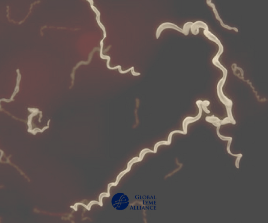
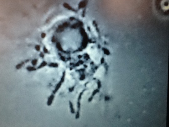
The syphilis and Borrelia spirochetes share many properties. They cause permanent infections in humans which cause long-lasting, multi-stage diseases. They exist in more than one form – sometimes in the long spirochete form, but also in a round “cyst-like” form. The spirochete form can be made visible by simple methods and observed with a microscope.
Borrelia spirochetes in dry smears and in isotonic solutions appear as very thin cells, only 0.2 micrometers wide, and as very long – in the range of 50 to 100 micrometers. These are visible above the resolution limit for light microscopy – due to what we call light interference – by slight defocusing. In dry preparations, a focus error of more than 1/1000 mm renders them invisible under the microscope. In wet preparations, the cells swell substantially.
My colleague and I published the results of our part of the project due to our firm belief that microscopy is useful for detecting these infections. Beforehand we were assured by the Institute that formal permissions were granted by the health authorities. It turned out that the Institute, somehow had forgot to point out in a formal enough manner that a substantial part of the project included microscopy by us!
Our project leader was educated as a medical doctor, and had later became a biologist. He was also Head of the Institute to which the project was connected. We, my colleague and I, got a copy of the first page of the application accepted and signed by the Health authorities. We did not, unfortunately, see the entire application before it was too late, as the project description was incomplete in that it did not express without any doubts that about half of the project involved extensive use of microscopy to analyze samples.
The project leader knew of my cooperation with Oeystein Brorson, the extremely clever technician who was able to grow Borrelia from infected blood samples. We had agreed that the application should be written to state that extensive microscopy was needed for the project as a way to confirm the results of molecular tests.
The Institute received around 2 million euro (NOK 20 mln) and bought two DNA machines. The test project was thus an official Institute project, not an application by a group of three scientists. In hindsight, we should have asked to see the complete application, but we did not and went on the trust for our project leader. This turned out to be a mistake, as we were later blamed for not reading the regulations for medical research and additional legal papers.
The trust that caused us to not read the full application was labelled as “intention,” to mean we intended to subvert the regulations when we embarked on this project. This is simply not true.
Silenced for speaking
After publishing the 2013 article ‘A simple method for the detection of live Borrelia spirochetes in human blood using classical microscopy techniques’, professor Laane was invited to give a lecture at the 2014 Norvect conference in Oslo. An English patient saved the pdf, so you can still read it, via the link provided.
I was present at that conference and still remember how nervous he was. The reason was that several medical professors complained to his university. He was threatened with losing his job, if he would speak at the conference.
In fact, he did not literally speak – as you can see in the movie below – but used performing arts to show the slides of the spirochetes. Professor Laane was fired anyway and his laboratory was closed down.
Laane: “As for being forbidden to speak, Waldemar Broegger, a Geology professor in the late half of the nineteenth century was forbidden to speak about Charles Darwin’s evolution theory. Lynn Margulis was originally ridiculed for her theories on the evolutionary origins of mitochondria and chloroplasts and later received numerous awards for this discovery. As both of these scientists were found to be right and later became famous, I feel I am in good company.
The 2013 publication of ‘A simple method for the detection of live Borrelia spirochaetes in human blood using classical microscopy techniques’ in the journal Biological and Biomedical Reports resulted in much opposition by conservative medical doctors and some scientists, most of whom had little or no microbiology experience working with spirochetes in a laboratory environment.
The article and research was criticized because the cellular objects we were observing were presumed to be “artifacts,” meaning objects of some other origin that just magically appeared in our samples. It’s extremely important to point out that all of our principal research was performed under highly controlled conditions, and our results were confirmed using more than one microscopy method. The critics of our work have yet to explain how these supposed “artifacts” we observed were able to reproduce and even move like spirochetes, which we observed them doing, and even more importantly, the critics have failed to explain how an “artifact” could possibly contain nucleic acids from Borrelia.
Yet, in that same year Dr. Alan MacDonald found out independently the same as me regarding how to detect chronic Lyme infections in human blood. Before this the assumed borrelia bacteria had been found by a Norwegian microscopist, who had an extremely ill son from Lyme.
MacDonalds arguments in these two videos (part one and part two) are brief, but very much to the point. He has later developed methods to detect single Borrelia bacteria direct in a microscope slide by exact molecular methods.
Oeystein Brorson is also mentioned by MacDonald. He was the researcher who grew all known strains of Borrelia in Norway, sent me samples and cooperated with Lynn Margulis and me, until he got disabled due to disease and had to leave his job.
After my lab was closed down, also the website of the scientific journal that published our article was so severely hacked that it stayed offline for three years. Once it came back up, our article had dissapeared.
The hacking of the Journal ‘Biological and Biomedical Reports’ seems to be done with a person knowing more than my closest enemies. And was someone with access to a quite advanced compilator system. Why this was done might well have been political or personal prestige.
I have never seen this happening on any topic in science before.”
Proof or propaganda?
In 2016 the Norwegian Health Department (FHI) published the study ‘Validate or falsify: Lessons learned from a microscopy method claimed to be useful for detecting Borrelia and Babesia organisms in human blood’. It stated,
“microscopy by the LM-method identified structures claimed to be Borrelia- and/or Babesia in 66% of the blood samples of the patient group and in 85% in the healthy control group”.
The TV2 reporter in the infamous documentary ‘Deceit or Borrelia’ misquoted this sentence by claiming that “it was proven that Dark Field microscopy produces 85% false positives”.
There are however suspicions that the Norwegian study was set up to discredit professor Laane’s work, using contaminated blood.
Laane:
“Officially, on their website The National Institute of Public Health claimed that the only known case of Babesia in Norway was a veterinarian that have had his spleen removed, and else that Babesia was unknown in the Norwegian population.
But I found a faked sample of Babesia. It was blood from a cow (or ox). It was mixed with human blood from a socalled control person and sent to me by mail.
Of course, this sample clotted impossible to see anything in the sample by microscopy except lumps of erytrocytes and fibrine fibers. If I had got the original sample unmixed, I would have found them at once. The FHI showed me a few images from a preparation they had made themselves, BEFORE it was mixed with human blood. The Babesia images there were the same as I found in several so-called control samples.
When I showed them, they looked a little worried – like school children not telling the truth to their teacher – but they would not admit that control samples contained Babesia merozoites.”
Borrelia stained with the Mysterud-Laane method, blue color.
Live biofilm of Borrelias developing in a mixture of blood and sodium citrate (this patient was tested positive also by molecular methods by American laboratory).
Serology
In the introduction to their 2016 article, the FHI authors also state that Lyme serology has a 70-90% sensitivity in the earliest stages and a sensitivity of almost 100% in later stages, which suddenly seems to become the new mantra in several countries. This seems to have been taken from a 2016 article written by IDSA and CDC authors, which was criticised for using circular logic.
Laane:
“A recent report presented in Norwegian newspapers claim that inaccurate science, especially in medical topics, is common. Norwegian Professor Oeyvind Oesterud has written several short articles in Aftenposten, Norway’s largest newspaper about science and popular science papers.”
He points out that false conclusions are favoured by both journals and media on the criteria that discoveries should be spectacular. Unless they are not, they are not accepted. Also the official evaluation system for research grants favours this. Many of these discoveries can not be repeated. But it generates much money for the universities.
Such scientists believe in accepted science. Any deviation shown by experiment may be interpreted as incorrect or false. One may wonder why they publish at all. Regarding deviating disease data they are often neglected – according to what the medical scientist was taught as a student by his professor. The deviating results are never studier further and ignored as “errors”. Up to about half of recent medical papers may contain this.
The result is delayed progress, sometimes with big consequences for severely ill patients. Patients that could be saved. Disease problems are very complicated and there is need for research listening to what both the patients tell and meticulous analysis of unexpected data.
As a final thought:
“Travel by modern airplanes is now very safe. If experts treated airplane accidents in the same manner as an average medical scientist studies disease, I would book my next travel to the USA by ship.” Professor Laane
Exception or a pattern?
The European Union has recently provided a 2 million euro grant to a cooperation of three parties to develop a better Lyme test, as the current one is considered imperfect. At best.
So why are there several indication of the active repression and sabotage of new, better and direct ways to diagnose Lyme? Is professor Laane’s story unique or showing a doisturbing pattern?
“This never happened before on any topic in the history of science in Norway”, Laane said in his interview with the makers of ‘Under Our Skin Emergence’. Yet also in other countries scientists have been attacked for working on promising new and direct Lyme tests.
In 2014, French lab director Schaller was fined was fined with paying 280,000 euro to the government and was sentenced to nine months in jail. Also in 2014 the American Centers for Disease Control and Prevention (CDC) publicly attacked the credibility of Advanced Laboratory Services, which had developed a culture-based test for Lyme disease diagnosis. The basis for the CDC’s attack has since been proven wrong, yet the CDC has never retracted the article in which the errant criticism was made.
A direct and ‘no false-positive’ DNA test for Lyme was no longer made available to the general public, after its inventor was fired from a Connecticut hospital in 2010. And recently media attacked the tests of specialised labs in Germany, using undercover reporters and twisted patients’ stories, claiming they were either not ‘FDA approved’. As professor Ahern explained in her recent interview, none of the tests promoted by the CDC or your national Health agencies are ‘FDA approved’.
The next interview with Dr. Lee will look into what happened around the development and public accessibility of the DNA diagnostics mentioned above. It will be published very soon.
* Professor Laane and I will work together on a future article about the work of Lynn Margulis, as too few people seem to have heard of her important work.
Conquering Lyme Disease
Science Bridges the Great Divide
Brian A. Fallon, MD, and Jennifer Sotsky, MD
Columbia University Press
Lyme disease is the most common tick-borne illness in the United States, with more than 300,000 cases diagnosed each year. However, doctors are deeply divided on how to diagnose and treat it, giving rise to the controversy known as the “Lyme Wars.” Firmly entrenched camps have emerged, causing physicians, patient communities, and insurance providers to be pitted against one another in a struggle to define Lyme disease and its clinical challenges. Health care providers may not be aware of its diverse manifestations or the limitations of diagnostic tests. Meanwhile, patients have felt dismissed by their doctors and confused by the conflicting opinions and dubious self-help information found online.
In this authoritative book, the Columbia University Medical Center physicians Brian A. Fallon and Jennifer Sotsky explain that, despite the vexing “Lyme Wars,” there is cause for both doctors and patients to be optimistic. The past decade’s advances in precision medicine and biotechnology are reshaping our understanding of Lyme disease and accelerating the discovery of new tools to diagnose and treat it, such that the great divide previously separating medical communities is now being bridged. Drawing on both extensive clinical experience and cutting-edge research, Fallon, Sotsky, and their colleagues present these paradigm-shifting breakthroughs in language accessible to both sides. They clearly explain the immunologic, infectious, and neurologic basis of chronic symptoms, the cognitive and psychological impact of the disease, as well as current and emerging diagnostic tests, treatments, and prevention strategies. Written for the educated patient and health care provider seeking to learn more, Conquering Lyme Disease gives an up-to-the-minute overview of the science that is transforming the way we address this complex illness. It argues forcefully that the expanding plague of Lyme and other tick-borne diseases can be confronted successfully and may soon even be reversed.
ABOUT THE AUTHORS
Brian A. Fallon, MD, is director of the Lyme and Tick-Borne Diseases Research Center at the Columbia University Medical Center.
Jennifer Sotsky, MD, is a physician with a specialty in narrative medicine. She is a resident at Columbia University Medical Center.
Zika vs. Aluminum: Double Standards on Levels of Evidence and Media Liability
Millions of Dollars spent, Massive Media Coverage for Zika and Microcephaly – Based on One Autospy Report. Aluminum Found in Five Autistic Brains (N=5)… Media Crickets.
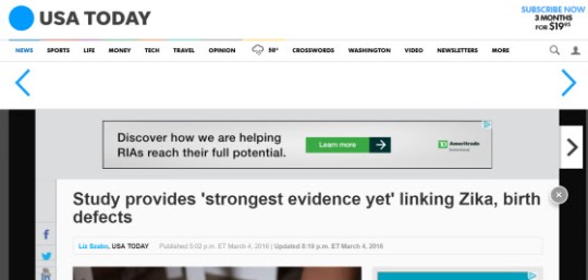
WHEN THE CDC announced that Zika virus had been found in 1 (ONE) brain of an aborted fetus from Brazil back in 2016, they heralded that 1 (ONE) data point as “The Srongest Evidence Yet”. Here’s the BBC News’s webpage from 10 Feb 2016:
Some have criticized the Exley study for not having “controls”. I’m sorry? There is not supposed to be ANY aluminum in children’s brains. Tested against the null hypothesis, yes, there is significantly more than zero.
CDC’s fetus with Zika virus came from a population in which Zika virus infection was high. Most cases of Zika virus infection during pregnancy did not lead to microcephaly. And microcephaly was highest in the northeastern part of Brazil, in poor women from the slums… who were being experimented on with whole-cell pertussis.
Yes, you read that right. WHOLE cell pertussis.
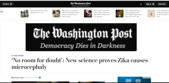
Well, it’s one thing to say the media is biased. After all, Exley’s study provides the strongest evidence to date that aluminum from vaccines is involved as a cause of autism. In fact, the study leaves no room for doubt, if we apply the same standard of the level of evidence used to support the idea that Congress had to pony up $1.1 Billion dollars for a vaccine against Zika.
But there is AMPLE room for doubt that Zika drove the microcephaly increase in Brazil.
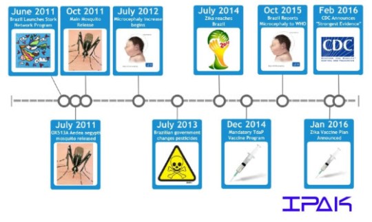
Here is a timeline of microcephaly in Brazil:
Note that the microcephaly increase actually started in July 2012,a year after the advent of the national Stork program, a prenatal care program that includes vaccination during pregnancy. And note that July 2012 is BEFORE Zika landed on the continent of S. America in July 2014. In December 2014, Brazil’s mandatory Tdap vaccination program started. But vaccination during pregnancy had already begun. The CDC’s autopsied fetus study was published in early 2016 – that’s the CDC’s best evidence to date.
So it turns out that Zika infection rates are seasonal in Brazil. So we’d expect another surge in microcephaly with the annual increase in Zika infections, right?
No, not at all. Why?
Zika does not cause microcephaly. The American taxpayer has been duped. And sprayed with pesticides to “protect” against Zika – in NYC even before any reported cases of Zika – with pesticides that are known to cause autism.
So when can we start asking: Is the media bias in the US now a causal factor in the autism epidemic? How can the media be held accountable?
What Can You Do?
You can write to the reporters on each of these stories and ask them to report on Dr. Exley’s study as providing “proof” that aluminum causes autism – be sure to send them the link to this blog.
If bona fide, objective reporters write to me, I’ll share a manuscript that was not published by PLOS One because they found it too “confusing”. The manuscript simply listed all of the reported possible causes of microcephaly and examined the available evidence at that time.
Lyme/MSIDS isn’t the only topic of media bias. The only way things are going to change is if we all play an active role and hold scientists and journalists accountable. I’ve often pointed out the obvious bias in the handling of Zika vs the handling of Lyme/MSIDS. While research for Lyme boasts 230 peer-reviewed studies showing borrelia persistence,Zika has 1 autopsy.
TheCaseforthePersistenceofLymeDiseaseAfterAntibioticTherapy The truth is that over 50% of the IDSA’s guidelines are based on “expert opinion” rather than “evidence-based medicine” as their publication suggests. A further 31% of the IDSA guidelines are based on observational studies. Only a meagre 29% of the IDSA Guidelines fit into “evidence-based medicine”.(22,23,24,25,27) Importantly, the IDSA’s own research supports these very findings.(22)
Please understand for those of you just tuning in – THIS IS A WAR of epic proportions.
The reason we find ourselves here even 40 years after Lyme was “officially” discovered is due to money, power, and collusion at the highest level of government. The government holds the patents on Borrelia burgdorferi (Bb), Lyme testing, the Lyme vaccine, and has been controlling the narrative for decades, even taking specific bands out of the test that shows infection due to their patent on the Lyme vaccine. https://www.google.com/patents/US8680236Gov. patent on OspA (outer surface protein) of Borrelia Burgdorferi (Bb). https://www.google.com/patents/US6087097Gov. patent on Lyme test based on OspA (outer surface protein) which causes the same disease it was meant to prevent which is the real reason it was yanked from the market. https://www.google.com/patents/US4721617 Gov. patent on Lyme Vaccine https://crymedisease.wordpress.com/2016/02/28/the-conspirators-they-own-the-patents-and-changed-the-testing/ A lengthy expose on all shenanigans for the stout of heart.