How DMSO Cures Eye, Ear, Nose, Throat and Dental Disease
https://www.midwesterndoctor.com/p/how-dmso-cures-eye-ear-nose-throat?
How DMSO Cures Eye, Ear, Nose, Throat and Dental Disease
Many of those “incurable” conditions respond remarkably to DMSO
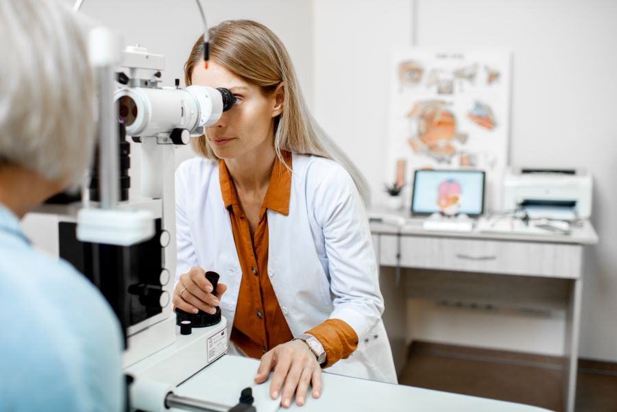
- DMSO can often significantly improve one’s vision, treat conditions such as macular degeneration, retinitis pigmentosa, and at times allow blind individuals to regain their sight. It is also often very helpful for sore and strained eyes and relieves excessive irritation and inflammation, along with many other eye conditions (e.g., cataracts).
- DMSO frequently treats a variety of ear conditions such as tinnitus, hearing loss, airplane ear, and a variety of infections inside the ear (e.g., otitis media).
- DMSO often is very helpful for sinusitis and a variety of infections of the nose and throat. Likewise, it is extremely helpful in dentistry, both for cleaning the mouth (e.g., by preventing bleeding gums), and by allowing the mouth to rapidly heal after dental surgeries.
- In this article, I will review the evidence supporting each of those uses, along with the data demonstrating the safety of these methods of DMSO administration and instructions on how to do them.
DMSO is a phenomenally effective medicine that can treat a wide variety of common, debilitating, or incurable conditions, which allowed it to rapidly take the country by storm (as both the public and the medical community saw its results and rapidly embraced it). Unfortunately, the widespread enthusiasm behind something that completely changed medicine and allowed people to care for themselves independently was unacceptable to the FDA. For the next two decades, the agency went to incredible lengths to suppress it (e.g., it actively defied Congress for over 16 years) and eventually made DMSO become a Forgotten Side of Medicine.
Note: extensive data shows that DMSO is a very safe substance with negligible toxicity.
In turn, one of the truly ironic things about this was that many of those who attacked DMSO later needed it. For example, the pioneer of DMSO discusses how Former President Lyndon Johnson sought his help in 1971 —after his FDA commissioner had just spent almost three years weaponizing the FDA against anyone wishing to use DMSO (which in turn set the stage for many of the police-state tactics the FDA would illegally use against natural medicine in the decades to come).
Note: in the previous article I erroneously stated this conversation took place in 1981 not 1971 (at which point LBJ was deceased).
I have now received hundreds of unbelievable reports from readers (which can be read here) of what DMSO did for them—many of which are almost identical to what people reported fifty years ago before the FDA wiped DMSO off the map.
For context, the majority of those reports were for the most common uses of DMSO, such as chronic pain, acute injuries, and arthritis (discussed further here). However, as discussed here, DMSO is also immensely valuable for a variety of circulatory and neurological disorders (e.g., varicose veins, hemorrhoids, Down Syndrome, and Parkinson’s)—all of which readers here reported significant improvement from. Likewise, (as discussed here) DMSO also helps various autoimmune conditions.
In this article, I will focus on another group of conditions DMSO was found to be extraordinarily effective—those within the head.
Note: headaches were covered in a previous article and will not be discussed here. (See link for article)
_______________
**Comment**
Another very in depth article about the power of DMSO to help eye, ear, nose, throat, and dental conditions.
For more:
- https://madisonarealymesupportgroup.com/2018/03/02/dmso-msm-for-lyme-msids/
- https://madisonarealymesupportgroup.com/2024/09/16/dmso-its-remarkable-properties/
- https://madisonarealymesupportgroup.com/2024/10/25/how-dmso-treats-incurable-autoimmune-and-contractile-disorders/
- dmso-eyes-ears-nose-mouth-throat-health-pdf