Archive for the ‘Anaplasmosis’ Category

Mild Winter Days? Watch Out for Ticks

Winter thaws bring out the ticks. Take precautions when heading outdoors on warm winter days to avoid tick bites. Wearing light-colored clothing and tucking pantlegs into socks can help. Credit: Pavla Zakova | Dreamstime.com

We asked disease ecologist Rick Ostfeld if we need to worry about ticks during the winter. His response is below. (TL; DR = yes)

Blacklegged ticks, which transmit the agents of Lyme disease, babesiosis, and anaplasmosis, disappear in winter, right? Well, not entirely. Although human encounters with these ticks are less likely in winter, there are plenty of ticks out in the environment lurking in a state of (almost) suspended animation.

Larval ticks that hatched the prior summer but failed to find an animal host can become dormant and remain on the forest floor in a quiescent state. The same is true of nymphal ticks, which are normally most active in spring and early summer. These two stages are unlikely to activate until day-length and temperature both increase dramatically. Adult stage ticks, in contrast, most actively seek hosts in the late fall.

The females that were able to engorge on host blood last fall are overwintering in soil pores or under leaf litter, while slowly converting host blood into eggs that they will lay next spring or summer. Some of the adults that did not find a host have died of starvation or other causes, but the unfed survivors will activate any winter or spring day with above-freezing temperatures. In the southern United States, these ticks can be active all year, but even in the northeastern and upper Midwestern regions, ticks can bite during any warmish spell in January, February, and beyond.

As part of The Tick Project, we invite participating households to mail us ticks found embedded in or crawling on people or pets, and we typically receive quite a few even in the coldest months. The riskiest seasons for diseases transmitted by blacklegged ticks are spring and summer, but risk never goes away entirely. And, our 25-years of data from Dutchess County, NY show that, as the climate warms, the ticks come out earlier in the year, advancing the dates of greatest risk.

_____________________

For More:  https://madisonarealymesupportgroup.com/2016/01/20/polar-vorticks/   TICK GUY, TOM MATHER, SHOWS HOW TICKS SURVIVE IN 3 DEGREES OVERNIGHT UNDER SNOW COVER.

And regarding all the clamor of “climate change,” independent Canadian tick researcher John Scott has proven it has absolutely nothing to do with tick proliferation and therefore the spread of Lyme/MSIDS:  https://madisonarealymesupportgroup.com/2018/11/07/ticks-on-the-move-due-to-migrating-birds-and-photoperiod-not-climate-change/

https://madisonarealymesupportgroup.com/2018/08/13/study-shows-lyme-not-propelled-by-climate-change/  Warm winters are lethal to I. scapularis (black-legged) ticks.  In fact, overwinter survival dropped to 33% when the snow melted.

So…..when people push the idea that warmer winters somehow make ticks more abundant you can explain with science on your side that –

Warmer winters actually kill ticks


North Dakota Tick Survey Sees Large Uptick of Samples. Here’s What the Health Department Found

https://www.thedickinsonpress.com/lifestyle/health/4556529-north-dakota-tick-survey-sees-large-uptick-samples-heres-what-health

North Dakota tick survey sees large uptick of samples. Here’s what the health department found.

ticks1_0

The American Dog tick was the most common tick surveyed in 2018 by the North Dakota Department of Health. This adult female is a known carrier of Rocky Mountain spotted fever.   submitted photo
BISMARCK – Those creepy, crawly creatures are living up to their nasty reputation. In brief, the summary of a second season of a statewide survey of ticks confirms what most people fear – a high percentage of the bloodsucking, pincer pests are carriers of disgusting diseases that are easily transmitted to hosts.


The North Dakota Department of Health first ventured into tick surveillance in 2017 by collecting and testing a relatively small sample of ticks. The ticks in the sample were provided with the help of veterinarians and zoos in the state. The program was significantly expanded in 2018 with active participation from 37 veterinarians, four zoos, North Dakota Game and Fish, Department of Agriculture Wildlife Services and at least one individual.

“They all submitted ticks, which was a huge help,”said Laura Cronquist, NDDOH division of disease control. “We had more coverage throughout the state in 2018, which was real nice.”

Of the 13,640 ticks collected and sent to the state laboratory for disease testing, nearly half were contributed by a single interested individual – John Heiser of Grassy Butte. In all, ticks were submitted from 25 counties in the state.

“We had more participation last year, which was awesome,” said Cronquist. “More ticks were submitted. Our project is really unique for our area.”

The state’s most common tick is the American Dog tick. The dog tick and Rocky Mountain wood tick, which is also found within the state, are closely related. Both are known for their speciality, carrying the dreaded Rocky Mountain spotted fever virus.

Rocky Mountain spotted fever symptoms include stomach pain, nausea, vomiting, loss of appetite and muscle pain. Delayed treatment may lead to severe illness or death. Some Rocky Mountain spotted fever victims may suffer irreversible hearing loss, paralysis, mental disability and damage to blood vessels which could lead to amputation of extremities. Fourteen cases of Rocky Mountain spotted fever were reported in the state in 2017.

There’s another tick present in North Dakota with a well-deserved and ugly reputation too. A surprising number of them were collected in 2018.

“We ended up with more deer ticks from across the state,” noted Cronquist.

While the number of deer ticks submitted to the NDDOH was small in comparison to the number of other ticks, just 51, they were found in 22 of the 25 counties surveyed, including Ward County. Deer ticks are are known carriers of Lyme disease, 56 cases of which were reported in the state in 2017. In addition, deer ticks are believed to have transmitted 17 cases of anaplasmosis and one case of Powassan in 2017.

The symptoms of anaplasmosis are similar to Rocky Mountain spotted fever but can include chills, cough and confusion. Severe cases can lead to difficulty breathing, hemorrhage, kidney failure and disrupt various functions of the nervous system.

Fortunately, Powassan transmission from ticks remains quite rare in North Dakota. It is closely related to West Nile disease. A tick can transmit Powassan in as little as 15 minutes after biting a human. About 10 percent of Powassan cases result in death. According to the NDDOH report on the 2018 study, statistics show that approximately half of Powassan survivors have permanent neurologic conditions including headaches, muscle wasting and memory problems.

The Lone Star tick, whose range is primarily the southern and eastern United States, and has been linked to red meat allergies, is believed to be expanding its range. However, Lone Star ticks remain extremely rare in North Dakota.

“That’s correct,” said Cronquist. “Two years ago two were found. Last year just one and it’s unknown how they got here. They are not all that concerning yet.”

The state was divided into eight regions for the tick surveillance survey. Tick pools from each region, consisting of up to 20 ticks each, were tested for the presence of several diseases. Of the 176 pools made up of American Dog ticks and Rocky Mountain wood ticks, 106 tested positive for disease carrying pathogens. Half of the deer tick pools tested positive.

According to the NDDOH report, ticks can transfer some pathogens to their hosts in as little as 15 minutes. Some pathogens require that the tick to be attached from 24 to 48 hours. When a tick bites into flesh it inserts a feeding tube into the incision which enables the transmission of disease.

Complete results and information regarding the 2018 tick surveillance project can be found on the NDDOH website.

_____________________

For more:  https://madisonarealymesupportgroup.com/2018/07/22/citizen-scientists-help-track-tick-borne-illness-exposure/

https://madisonarealymesupportgroup.com/2018/04/10/canadian-citizen-scientists-helping-with-tick-surveillance/

https://madisonarealymesupportgroup.com/2018/10/26/scientists-high-school-students-find-new-pathogens-hiding-in-indiana-ticks/

https://madisonarealymesupportgroup.com/2018/10/22/tick-project-takes-a-deeper-look-at-disease/

CAPC Study: Lyme Disease Spreading to Regions Once Thought Low-risk

http://veterinarynews.dvm360.com/capc-study-lyme-disease-spreading-regions-once-thought-low-risk

CAPC study: Lyme disease spreading to regions once thought low-risk

Condition in dogs could signal increasing threat to people, researchers say.

Jan 19, 2019

By dvm360.com staff

DVM360 MAGAZINE

(andriano cz/stock.adobe.com)

The Companion Animal Parasite Council (CAPC) recently released a study that shows that Lyme disease is spreading to regions not previously thought to be at risk for tick-borne disease. States such as Illinois, Iowa, North Dakota, Ohio, Michigan, West Virginia and Tennessee have all seen an increase in the prevalence of Lyme disease, according to a media release discussing the study, which CAPC conducted from January 2012 to December 2016. Results from the study were recently published in Environmetrics.

“The results of this milestone study show increasing risk for Lyme disease in endemic areas and pinpoint regions in the U.S. where Lyme is spreading—areas not historically considered endemic,” says Michael Yabsley, PhD, a professor in the Department of Population Health, College of Veterinary Medicine and Warnell School of Forestry and Natural Resources at the University of Georgia. “This expanding risk of Lyme disease demands heightened vigilance in protecting both our pets and our families.”

New research from CAPC found that the prevalence of Lyme disease is trending up in areas previously thought to be at a lower risk for tick-borne diseases. (Image courtesy of CAPC)

The study was motivated by the increase in Lyme disease cases in the U.S. and, in particular, in states not traditionally considered Lyme-endemic, the release states. Results suggest that:

  • Canine prevalence rates for Lyme disease are rising.
  • Lyme prevalence rates are increasing most in areas where the pathogen has encroached recently.
  • Lyme prevalence in dogs is rising in states traditionally not considered to be of high Lyme risk, suggesting that human risk may also be increasing in these areas, including regions in Illinois, Iowa, North Dakota, Ohio, Michigan and Tennessee.
  • Significant increases in canine Lyme prevalence have been seen in some areas that are not yet reporting significant human incidence. Researchers speculate that canine prevalence is more sensitive to changes in Lyme risk and could serve as an early warning system for changes in human risk.

The study was created to investigate regional trends in the prevalence of antibodies to Borrelia burgdorferi, the disease-causing bacterium of Lyme disease, according to the release. To conduct the research, the CAPC team analyzed more than 16 million Lyme tests from domestic dogs in the U.S. over 60 months. The serologic data was provided by IDEXX Laboratories.

“CAPC research shows the risk for Lyme disease is not static. The way it’s changing varies spatially across the country,” says Christopher McMahan, associate professor in the department of mathematical sciences at Clemson University, in the release.

Crucial in the fight against Lyme, Yabsley says, is year-round tick protection. Different species of ticks are active all 12 months of the year, and ticks that transmit Lyme are active at different times in the year in different regions, the release states. For instance, as you move further south, adult ticks are more active in the winter.

“I’ve been practicing for over 34 years in Nashville where many people don’t think Lyme disease is a concern. But I’ve seen canine Lyme increasing in Tennessee for several years and regularly test and vaccinate for the disease,” says Craig Prior, BVSC, CVJ, a veterinarian and former owner of VCA Murphy Road Animal Hospital in Nashville, Tennessee. “Many people tend to believe that if they don’t go on hikes or spend time in wooded areas, they aren’t at risk for Lyme. Ticks are everywhere—including suburban and gated communities where deer, raccoons, opossum, birds and other hosts frequent back yards. That’s why CAPC recommends year-round tick prevention for dogs—and cats—and regular screening to protect dogs from this debilitating disease that can be extremely hard to treat.

On petdiseasealerts.org, CAPC now provides monthly forecasts for Lyme disease and other tick-borne diseases. It also provides access to monthly canine test results in prevalence maps, a CAPC resource available free online at petsandparasites.org. With more than 21 million canine B. burgdorferi antibody test results collected between 2012 and 2017 in dogs, these maps allow veterinarians, physicians, pet owners and travelers to assess the risk of exposure across the United States and Canada.

__________________

**Comment**

Wisconsin prevalence rate for Lyme, Anaplasmosis, and Ehrlichiosis in pets:  https://capcvet.org/maps/#2012/all/lyme-disease/dog/united-states/wisconsin/  As you can see there are many counties where NO DATA has been collected.  Please remember maps are a very loose guide to what’s happening out there and have been used against patients for decades – denying them accurate diagnosis and treatment.  FYI:  Dane County is at HIGH risk for all 3.

According to independent Canadian tick researcher, John Scott, the reason for this tick proliferation is due to migrating birds and photoperiod, NOT climate change:  https://madisonarealymesupportgroup.com/2018/08/13/study-shows-lyme-not-propelled-by-climate-change/

https://madisonarealymesupportgroup.com/2018/11/07/ticks-on-the-move-due-to-migrating-birds-and-photoperiod-not-climate-change/

Also, infected dogs spread infections as well as ticks when they cross borders:  https://madisonarealymesupportgroup.com/2018/03/09/infected-dogs-with-tbis-spreading-infection-across-borders/  Think of pets as luggage that can and do carry pathogens right into your home.  Please do not allow your dog on your bed or furniture and make sure you use tick prevention on all pets.


2018 Review of Previous Pathogen Transmission Time Studies in Deer Ticks

https://www.ncbi.nlm.nih.gov/pubmed/29398603

2018 Mar;9(3):535-542. doi: 10.1016/j.ttbdis.2018.01.002. Epub 2018 Jan 31.

Pathogen transmission in relation to duration of attachment by Ixodes scapularis ticks.

Abstract

The blacklegged tick, Ixodes scapularis, is the primary vector to humans in the eastern United States of the deer tick virus lineage of Powassan virus (Powassan virus disease); the protozoan parasite Babesia microti (babesiosis); and multiple bacterial disease agents including Anaplasma phagocytophilum (anaplasmosis), Borrelia burgdorferi and Borrelia mayonii (Lyme disease), Borrelia miyamotoi (relapsing fever-like illness, named Borrelia miyamotoi disease), and Ehrlichia muris eauclairensis (a minor causative agent of ehrlichiosis).

With the notable exception of Powassan virus, which can be transmitted within minutes after attachment by an infected tick, there is no doubt that the risk of transmission of other I. scapularis-borne pathogens, including Lyme disease spirochetes, increases with the length of time (number of days) infected ticks are allowed to remain attached. This review summarizes data from experimental transmission studies to reinforce the important disease-prevention message that regular (at least daily) tick checks and prompt tick removal has strong potential to reduce the risk of transmission of I. scapularis-borne bacterial and parasitic pathogens from infected attached ticks.

The most likely scenario for human exposure to an I. scapularis-borne pathogen is the bite by a single infected tick. However, recent reviews have failed to make a clear distinction between data based on transmission studies where experimental hosts were fed upon by a single versus multiple infected ticks. A summary of data from experimental studies on transmission of Lyme disease spirochetes (Bo. burgdorferi and Bo. mayonii) by I. scapularis nymphs indicates that the probability of transmission resulting in host infection, at time points from 24 to 72 h after nymphal attachment, is higher when multiple infected ticks feed together as compared to feeding by a single infected tick.

In the specific context of risk for human infection, the most relevant experimental studies therefore are those where the probability of pathogen transmission at a given point in time after attachment was determined using a single infected tick. The minimum duration of attachment by single infected I. scapularis nymphs required for transmission to result in host infection is poorly defined for most pathogens, but experimental studies have shown that Powassan virus can be transmitted within 15 min of tick attachment and both A. phagocytophilum and Bo. miyamotoi within the first 24 h of attachment. There is no experimental evidence for transmission of Lyme disease spirochetes by single infected I. scapularis nymphs to result in host infection when ticks are attached for only 24 h (despite exposure of nearly 90 experimental rodent hosts across multiple studies) but the probability of transmission resulting in host infection appears to increase to approximately 10% by 48 h and reach 70% by 72 h for Bo. burgdorferi. Caveats to the results from experimental transmission studies, including specific circumstances (such as re-attachment of previously partially fed infected ticks) that may lead to more rapid transmission are discussed.

________________

**Comment**

There are a number of problematic issues with this study:

  1. This is a review of previous studies.  There is nothing NEW here.  
  2. It’s important to note that ticks typically carry more than just borrelia and transmission times have not taken this fact into account: https://madisonarealymesupportgroup.com/2017/05/01/co-infection-of-ticks-the-rule-rather-than-the-exception/ and https://www.lymedisease.org/lyme-basics/co-infections/about-co-infections/  Infection with more than one pathogen is associated with more severe illness.https://madisonarealymesupportgroup.com/2018/10/30/study-shows-lyme-msids-patients-infected-with-many-pathogens-and-explains-why-we-are-so-sick/  For the first time, Garg et al. show a 85% probability for multiple infections including not only tick-borne pathogens but also opportunistic microbes such as EBV and other viruses.  This is a BIG DEAL.  Finally, a study showing what we face as patients in the real world.  They also never take into account nematodes (worms), mycoplasma, tularemia, and/or Bartonella.  These are infections many if not most patients have to contend with.  Some have been bioweaponized.
  3. They assume that the most likely scenario is for a person to be bitten by one tick.  Assuming makes an ass out of u and me.  When you take into account the latest information on the Asian tick, you quickly realize the probability of coming into contact with hundreds if not thousands of ticks at one time:  https://madisonarealymesupportgroup.com/2018/09/12/three-surprising-things-i-learned-about-asian-longhorned-ticks-the-tick-guy-tom-mather/  While human infection has yet to be found in the U.S., this tick is responsible for plenty of misery in Asia:  https://madisonarealymesupportgroup.com/2018/06/12/first-longhorned-tick-confirmed-in-arkansas/  It spreads SFTS (sever fever with thrombocytopenia syndrome), “an emerging hemorrhagic fever,” but the potential impact of this tick on tickborne illness is not yet known. In other parts of the world, it has been associated with several tickborne diseases, such as spotted fever rickettsioses, Anaplasma, Ehrlichia, and Borrelia, the causative agent of Lyme Disease.
  4. While they discuss the probability of multiple tick attachment, they never discuss the issue of partially fed ticks, where spirochetes would be in the salivary glands – leading to quicker transmission: http://iai.asm.org/content/61/6/2396.full.pdf  Ticks can spontaneously detach – and the authors of this study found that they did so 15% of the time in mice.  They also state that about a tenth of questing nymphs appear distended with partially fed sub-adult ticks being common.
  5. While the current review states, “There is no experimental evidence for transmission of Lyme disease spirochetes by single infected I. scapularis nymphs to result in host infection when ticks are attached for only 24 h (despite exposure of nearly 90 experimental rodent hosts across multiple studies), this study shows transmission can occur in under 16 hours:  https://www.ncbi.nlm.nih.gov/pmc/articles/PMC4278789/
  6. https://madisonarealymesupportgroup.com/2017/04/14/transmission-time-for-lymemsids-infection/  Within this video, microbiologist Holly Ahern discusses the numerous problems with animal Bb transmission studies.  Transmission Time:  Only one study done on Mice. At 24 hours every tick had transmitted borrelia to the mice; however, animal studies have proven that transmission can occur in under 16 hours and it occurs frequently in under 24 hours.  No human studies have been done and https://www.dovepress.com/lyme-borreliosis-a-review-of-data-on-transmission-time-after-tick-atta-peer-reviewed-article-IJGM  no studies have determined the minimum time it takes for transmission.  And, never forget the case of the little girl who couldn’t walk or talk after a tick bite attachment of 4-6 hours:  https://madisonarealymesupportgroup.com/2016/12/07/igenex-presentation/
  7. They continue to blame Lyme/MSIDS on the black legged tick as the sole perp when experience and studies show there’s more potential transmitters at play:  https://madisonarealymesupportgroup.com/2018/11/07/are-mosquitoes-transmitting-lyme-disease/https://madisonarealymesupportgroup.com/2016/07/23/german-study-finds-borrelia-in-mosquitos/https://madisonarealymesupportgroup.com/2019/01/17/remember-deer-keds-study-shows-bartonella-causing-deer-ked-dermatitis-in-humans/
Please, quit doing reviews of previous data and do something new using better laboratory techniques!  We don’t need MORE of the same thing.

Multistate Infestation with the Exotic Disease Vector Tick Haemaphysalis Longhornis – U.S., Aug. 2017- Sept. 2018

https://www.cdc.gov/mmwr/volumes/67/wr/mm6747a3.htm

Multistate Infestation with the Exotic Disease–Vector Tick Haemaphysalis longicornis — United States, August 2017–September 2018

C. Ben Beard, PhD1; James Occi, MA, MS2; Denise L. Bonilla, MS3; Andrea M. Egizi, PhD4; Dina M. Fonseca, PhD2; James W. Mertins, PhD3; Bryon P. Backenson, MS5; Waheed I. Bajwa, PhD6; Alexis M. Barbarin, PhD7; Matthew A. Bertone, PhD8; Justin Brown, DVM, PhD9; Neeta P. Connally, PhD10; Nancy D. Connell, PhD11; Rebecca J. Eisen, PhD1; Richard C. Falco, PhD5; Angela M. James, PhD3; Rayda K. Krell, PhD10; Kevin Lahmers, DVM, PhD12; Nicole Lewis, DVM13; Susan E. Little, DVM, PhD14; Michael Neault, DVM15; Adalberto A. Pérez de León, DVM, PhD16; Adam R. Randall, PhD17; Mark G. Ruder, DVM, PhD18; Meriam N. Saleh, PhD14; Brittany L. Schappach10; Betsy A. Schroeder, DVM19; Leslie L. Seraphin, DVM3; Morgan Wehtje, PhD3; Gary P. Wormser, MD20; Michael J. Yabsley, PhD21; William Halperin, MD, DrPH22 (View author affiliations)

Summary

What is already known about this topic?

Haemaphysalis longicornis is a tick indigenous to Asia, where it is an important vector of human and animal disease agents, which can result in human hemorrhagic fever and substantive reduction in dairy production.

What is added by this report?

During 2017–2018, H. longicornis has been detected in Arkansas, Connecticut, Maryland, New Jersey, New York, North Carolina, Pennsylvania, Virginia, and West Virginia on various species of domestic animals and wildlife, and from two humans.

What are the implications for public health practice?

The presence of H. longicornis in the United States represents a new and emerging disease threat. Characterization of the tick’s biology and ecology are needed, and surveillance efforts should include testing for potential indigenous and exotic pathogens.

Haemaphysalis longicornis is a tick indigenous to eastern Asia and an important vector of human and animal disease agents, resulting in such outcomes as human hemorrhagic fever and reduction of production in dairy cattle by 25%. H. longicornis was discovered on a sheep in New Jersey in August 2017 (1). This was the first detection in the United States outside of quarantine. In the spring of 2018, the tick was again detected at the index site, and later, in other counties in New Jersey, in seven other states in the eastern United States, and in Arkansas. The hosts included six species of domestic animals, six species of wildlife, and humans. To forestall adverse consequences in humans, pets, livestock, and wildlife, several critical actions are indicated, including expanded surveillance to determine the evolving distribution of H. longicornis, detection of pathogens that H. longicornis currently harbors, determination of the capacity of H. longicornis to serve as a vector for a range of potential pathogens, and evaluation of effective agents and methods for the control of H. longicornis.

H. longicornis is native to eastern China, Japan, the Russian Far East, and Korea. It is an introduced, and now established, exotic species in Australia, New Zealand, and several island nations in the western Pacific Region. Where this tick exists, it is an important vector of human and animal disease agents. In China and Japan, it transmits the severe fever with thrombocytopenia syndrome virus (SFTSV), which causes a human hemorrhagic fever (2), and Rickettsia japonica, which causes Japanese spotted fever (3). Studies in Asia identified ticks infected with various species of Anaplasma, Babesia, Borrelia, Ehrlichia, and Rickettsia, and all of these pathogen groups circulate zoonotically in the United States (4,5). In addition, parthenogenetic reproduction, a biologic characteristic of this species, allows a single introduced female tick to generate progeny without mating, thus resulting in massive host infestations. In some regions of New Zealand and Australia, this tick can reduce production in dairy cattle by 25% (6). Before 2017, H. longicornis ticks were intercepted at U.S. ports of entry at least 15 times on imported animals and materials (James W. Mertins, U.S. Department of Agriculture [USDA], personal communication).

The USDA Animal and Plant Inspection Service coordinated cooperative efforts through telephone conference calls with various local, state, and federal agricultural and public health agencies. Through these efforts, enhanced vector and animal surveillance were implemented to detect additional tick infestations. Suspect archival specimens that were available among previously collected ticks were also examined. Ticks were identified definitively by morphology at the USDA National Veterinary Services Laboratories or by DNA sequence analysis (molecular barcoding) at Rutgers University Center for Vector Biology, Monmouth County (New Jersey) Mosquito Control Division; College of Veterinary Medicine, University of Georgia; and Center for Veterinary Health Sciences, Oklahoma State University. By definition, a “report” is any new morphologic or molecular identification of H. longicornis ticks with a new county or host species from that county, identified from August 2017 through September 2018. Subsequent repeat collections are not reported here.

From August 2017 through September 2018, vector and animal surveillance efforts resulted in 53 reports of H. longicornis in the United States, including 38 (72%) from animal species (23 [61%] from domestic animals, 13 [34%] from wildlife, and two [5%] from humans), and 15 (28%) from environmental sampling of grass or other vegetation using cloth drags or flags* or carbon dioxide–baited tick traps. With the exception of one report from Arkansas, the remaining reports of positively identified ticks are from eight eastern states: New Jersey (16; 30%), Virginia (15; 28%), West Virginia (11; 21%), New York (three; 6%), North Carolina (three; 6%), Pennsylvania (two; 4%), Connecticut (one; 2%), and Maryland (one; 2%) (Figure). Among the 546 counties or county equivalents in the nine states, ticks were reported from 45 (8%) counties (1.4% of all 3,109 U.S. counties and county equivalents) (Table 1). Excluding 15 reports of positive environmental sampling using flagging, dragging, or carbon dioxide traps, the remaining 38 reports reflect collection of ticks from infested host species (Table 2). Surveillance efforts did not include testing the ticks or hosts for pathogens. No cases of illness in humans or other species were reported. Concurrent reexamination of archived historical samples showed that invasion occurred years earlier. Most importantly, ticks collected from a deer in West Virginia in 2010 and a dog in New Jersey in 2013 were retrospectively identified as H. longicornis.

Discussion

Cooperative efforts among federal, state, and local experts from agricultural, public health, and academic institutions during the last year have documented that a tick indigenous to Asia is currently resident in several U.S. states. The public health and agricultural impacts of the multistate introduction and subsequent domestic establishment of H. longicornis are not known. At present, there is no evidence that H. longicornis has transmitted pathogens to humans, domestic animals, or wildlife in the United States. This species, however, is a potential vector of a number of important agents of human and animal diseases in the United States, including Rickettsia, Borrelia, Ehrlichia, Anaplasma, Theileria, and several important viral agents such as Heartland and Powassan viruses. Consequently, increased tick surveillance is warranted, using standardized animal and environmental sampling methods.

The findings in this report are subject to at least two limitations. First, the findings are limited by the variable surveillance methods used to identify the geographic and host distribution of H. longicornis. These methods included both passive and active surveillance. Conclusions about the geographic and host distribution might reflect the biases in the collection and submission of samples to states and USDA and the paucity of available information. Second, the data in this report reflect the collection of specimens that were positively identified by morphology or molecular barcoding. These represent sentinels that H. longicornis is present in different U.S. states and regions, and not a comprehensive assessment of the distribution of H. longicornis in the United States. The absence of positive samples from many states and counties might reflect the absence of infestation, absence of sampling, or failure to recover the tick. Even in states where H. longicornis has been found, the available data do not describe the actual extent or intensity of infestation.

The biology and ecology of H. longicornis as an exotic species in the United States should be characterized in terms of its vector competence (ability to transmit a pathogen) and vectorial capacity (feeding habits, host preference, climatic sensitivity, population density, and other factors that can affect the risk for pathogen transmission to humans) for tickborne pathogens known to be present in the United States (5). Surveillance for H. longicornis should include adequate sampling of companion animals, commercial animals, wildlife, and the environment. Where H. longicornis is detected, there should be testing for a range of indigenous and exotic viral, bacterial, and protozoan tickborne pathogens potentially transmitted by H. longicornis. Given the similarity between SFTSV and Heartland virus, a tickborne phlebovirus (https://www.cdc.gov/heartland-virus/index.html), further evaluation of the potential role of H. longicornis in transmission of this disease agent among animal reservoirs and possibly to humans is warranted. A broad range of interventions should be evaluated, including insecticide and acaricide sensitivity testing. Many state and federal agencies are developing and disseminating information for stakeholders, including development of hotlines, and some states are identifying ticks submitted by the public. The recently documented occurrence of H. longicornis in the United States presents an opportunity for collaboration among governmental, agricultural, public health agencies and partners in academic public health, veterinary sciences, and agricultural sciences to prevent diseases of potential national importance before onset in humans and other animal species.

Top

Acknowledgments

Wes Watson, Andrew D. Haddow, Naomi Drexler, Gleeson Murphy, Harry Savage, Howard Ginsberg, Kim Cervantes, field and laboratory personnel.

Corresponding author: C. Ben Beard, cbeard@cdc.gov, 970-221-6418.


* Drags consist of white cloth (usually 1 m2) that have a wooden leading frame and are dragged by a cord through grass or a leafy forest floor. Flags are similar but are used to brush uneven surfaces such as small bushes in wooded areas. Drags and flags are used to sample the environment for ticks trying to locate a host.

Carbon dioxide traps consist of dry ice–filled small boxes with holes that allow the CO2 to escape which are placed on a white cloth or mat in a grassy area or forest floor. Ticks, attracted by the CO2, crawl on to the cloth or mat surface, which is inspected for ticks after a period of time.

Top

References

  1. Rainey T, Occi JL, Robbins RG, Egizi A. Discovery of Haemaphysalis longicornis (Ixodida: Ixodidae) parasitizing a sheep in New Jersey, United States. J Med Entomol 2018;55:757–9. CrossRef PubMed
  2. Luo L-M, Zhao L, Wen H-L, et al. Haemaphysalis longicornis ticks as reservoir and vector of severe fever with thrombocytopenia syndrome virus in China. Emerg Infect Dis 2015;21:1770–6. CrossRef PubMed
  3. Mahara F. Japanese spotted fever: report of 31 cases and review of the literature. Emerg Infect Dis 1997;3:105–11. CrossRef PubMed
  4. Kang J-G, Ko S, Smith WB, Kim H-C, Lee I-Y, Chae J-S. Prevalence of Anaplasma, Bartonella and Borrelia species in Haemaphysalis longicornis collected from goats in North Korea. J Vet Sci 2016;17:207–16. CrossRef PubMed
  5. Rosenberg R, Lindsey NP, Fischer M, et al. Vital signs: trends in reported vectorborne disease cases—United States and territories, 2004–2016. MMWR Morb Mortal Wkly Rep 2018;67:496–501. CrossRef PubMed
  6. Heath A. Biology, ecology and distribution of the tick, Haemaphysalis longicornis Neumann (Acari: Ixodidae) in New Zealand. N Z Vet J 2016;64:10–20. CrossRef PubMed
Return to your place in the textFIGURE. Counties and county equivalents* where Haemaphysalis longicornis has been reported (N = 45) — United States, August 2017–September 2018

The figure is a map showing the counties and county equivalents where Haemaphysalis longicornis has been reported (N = 45), in the United States, during August 2017–September 2018.* 
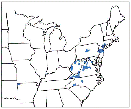
Benton County, Arkansas; Fairfield County, Connecticut; Washington County, Maryland; Bergen, Hunterdon, Mercer, Middlesex, Monmouth, Somerset, and Union Counties, New Jersey; Davidson, Polk, and Rutherford Counties, North Carolina; Richmond, Rockland, and Westchester Counties, New York; Bucks and Centre Counties, Pennsylvania; Albemarle, Augusta, Carroll, Fairfax, Giles, Grayson, Louisa, Page, Pulaski, Rockbridge, Russell, Scott, Smyth, Staunton City, Warren, and Wythe Counties, Virginia; Cabell, Hardy, Lincoln, Mason, Marion, Monroe, Putnam, Ritchie, Taylor, Tyler, Upshur Counties, West Virginia.

Top

TABLE 1. Percentage of Haemaphysalis longicornis–infested counties or county equivalents in infested states — nine states, August 2017–September 2018Return to your place in the text
State No. of counties* per state No. (%) of counties* with H. longicornis on host or in environment
Arkansas 75 1 (1)
Connecticut 8 1 (13)
Maryland 24 1 (4)
New Jersey 21 7 (33)
New York 62 3 (5)
North Carolina 100 3 (3)
Pennsylvania 67 2 (3)
Virginia 134 16 (12)
West Virginia 55 11 (20)
Total 546 45 (8)

* Counties or county equivalents

TABLE 2. Distribution of Haemaphysalis longicornis, by host and species — nine states, August 2017–September 2018Return to your place in the text
Host category, no. (% of total)/Species No. (% of host category)
Domestic animal, 23 (61)
Cat 1 (4)
Cow 4 (17)
Dog 12 (52)
Goat 2 (9)
Horse 2 (9)
Sheep 2 (9)
Total 23 (100)
Wildlife, 13 (34)
Coyote 1 (8)
White-tailed deer 7 (54)
Gray fox 1 (8)
Groundhog 1 (8)
Virginia opossum 2 (15)
Raccoon 1 (8)
Total 13 (100)
Human, 2 (5) 2 (100)
Total 38 (100)

Beard CB, Occi J, Bonilla DL, et al. Multistate Infestation with the Exotic Disease–Vector Tick Haemaphysalis longicornis — United States, August 2017–September 2018. MMWR Morb Mortal Wkly Rep 2018;67:1310–1313. DOI: http://dx.doi.org/10.15585/mmwr.mm6747a3.

____________________

**Comment**

In the section discussing the species and the other pathogens it’s been known to transmit, Theileria was mentioned. Theileria is a malarial-like pathogen similar to Babesia:

https://en.wikipedia.org/wiki/Theileria_microti

Babesia IS also spread by ticks and is a frequent coinfection with Lyme.

An important difference from malaria is that T. microti does not infect liver cells. Additionally, the piroplasm is spread by tick bites (Ixodes scapularis, the same tick that spreads Lyme disease), while the malaria protozoans are spread via mosquito. Finally, under the microscope, the merozoite form of the T. microti life cycle in red blood cells forms a cross-shaped structure, often referred to as a “Maltese cross“, whereas malaria forms more of a diamond ring structure in red blood cells.[3]

Much is yet to be discovered about the Asian tick that clones itself and can drain cattle of its blood.  For more:  https://madisonarealymesupportgroup.com/2018/09/12/three-surprising-things-i-learned-about-asian-longhorned-ticks-the-tick-guy-tom-mather/

One of the biggest discoveries by Mather was how the ticks line up on stalks of grass resembling grains of wheat.  When anything touches this, it’s like a tick cluster bomb and ticks go everywhere.  Not just one or two, mind you, but hundreds at one time.  See link for pictures.

https://madisonarealymesupportgroup.com/2018/03/01/asian-ticks-mysteriously-turn-up-in-new-jersey/

https://madisonarealymesupportgroup.com/2018/10/03/1st-person-bitten-by-east-asian-longhorned-tick/

https://madisonarealymesupportgroup.com/2018/11/05/hawk-found-carrying-asian-long-horned-tick-the-one-that-drains-cattle-of-all-their-blood/