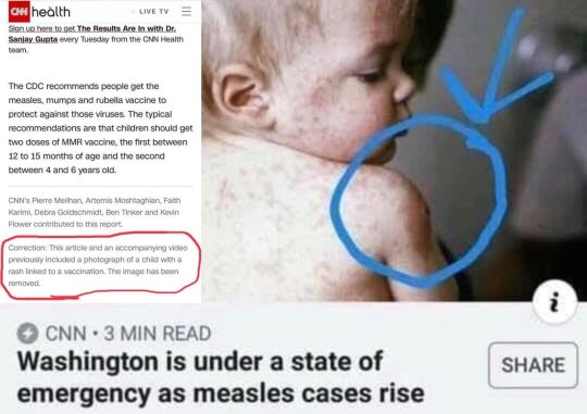
https://www.healthnutnews.com/cnn-forced-to-apologize-for-measles-scare-piece-using-pic-of-child-with-rash-from-bad-mmr-reaction/

Measles

What they failed to mention entirely is that horrible lump in the picture is actually from a MMR VACCINE.

Oops

Vaccine proponents also fail to mention that fully vaccinated people still contract the disease they’ve been vaccinated for:   https://www.usatoday.com/story/news/nation-now/2018/04/07/measles-cases-nevada-california/495735002/

For a great read on Measles:  https://www.healthnutnews.com/measles-a-rash-of-misinformation/

A few excerpts:

Natural infection with wild measles creates long-lasting viral-specific and viral-neutralizing antibodies that are not acquired following vaccine-introduced infection. There are numerous documented cases of measles occurring in highly vaccinated communities which can be attributed primarily to short-term efficacy (secondary vaccine failure).  This has important implications considering the fact that measles has an increased rate of complications in adults when compared to school age children….

Wild measles exposure occurs through contact with the human respiratory tract. The measles vaccine introduces a lab altered, live-virus through an unnatural route of exposure.  This weakened, man-made virus can bury deep into the tissues and create a slow infection in practically any area of the body including the gastro-intestinal (GI) tract and central nervous system (CNS). The consequences of these vaccine-induced infections may not show up for months, years or decades later.

A vaccine induced form of SSPE known as Measles Inclusion-Body Encephalitis (MIBE) has been documented in children months to years following measles vaccination.  Could the rapid rise in chronic inflammatory bowel and neurological disorders be caused by these slow infections? How many doctors would ever think to investigate the possibility that these illnesses may be with a distant vaccination?  To further complicate the issue, in a phenomenon known as recombination, the measles virus can combine with other live viruses in the vaccine to create a novel virus with unknown effects.

Lyme/MSIDS patients are in a battle for their health.  They suffer from dysfunctional immune systems that are in a war of epic proportions.  Please do your reading before considering any and all vaccines.  Every single patient I know who got vaccinated suffered a relapse and a worsened condition.


https://www.ncbi.nlm.nih.gov/pubmed/30655046

2019 Jan 14. pii: S0929-693X(18)30263-X. doi: 10.1016/j.arcped.2018.11.013. [Epub ahead of print]

Opsoclonus in a child with neuroborreliosis: Case report and review of the literature.

Abstract

Opsoclonus consists of massive erratic rapid eye jerks. They may occur in isolation or in association with myoclonus and ataxia, i.e., opsoclonus-myoclonus syndrome (OMS). We report the case of a 9-year-old girl who suffered from headaches for several days and was shown to have opsoclonus and left peripheral facial palsy. Work-up excluded the diagnosis of neuroblastoma, but CSF analysis showed aseptic meningitis, and serology for Borrelia burgdorferi (Lyme) was positive. The outcome was favorable with complete regression of symptoms after treatment with ceftriaxone 2g/day for 3 weeks. Although rare, the diagnosis of Lyme neuroborreliosis must be raised in the presence of isolated opsoclonus, particularly if the clinical picture is incomplete and if other features, such as peripheral facial palsy and pleocytosis in the CSF, are present.

___________________

**Comment**

To see what Opsoclonus looks like:

Again, researchers shouldn’t be writing that this manifestation is “rare,” because nobody’s truly keeping track of this.  For decades folks have been undiagnosed due to horrifically abysmal testing that misses half of all cases.  

Facial palsy and aseptic meningitis are huge clues for Lyme

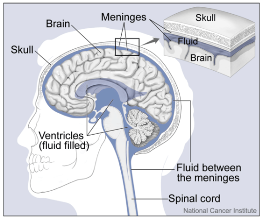
Aseptic meningitis is when the lining of the brain becomes inflamed.  In this case, due to a Borrelia burgdorferi (Bb) infection.  This explains the headaches.

577px-Brain_and_Nearby_StructuresBy Alan Hoofring  https://visualsonline.cancer.gov/details.cfm?imageid=4279, Public Domain, https://commons.wikimedia.org/w/index.php?curid=34938301

As the picture shows, the meninges cover the entire brain so the headache caused by this is going to cover the entire head.  This is what I personally had that took years of antibiotics to rid.  I wondered if there would ever be a day without this type of headache.

I’ve spoken with many Lyme/MSIDS patients who have had exactly the same thing.  It is not rare.  The pain is unbelievable, and ONLY treatment took it away.  Ibuprophen will not touch this Mother one little bit.  CBD, systemic enzymes, NOTHING will touch this if you have a Bb infection.  The infection must be dealt with first.

For those of you with this presentation, please also consider Chiari, especially if you’ve had this type of headache for some time:  https://madisonarealymesupportgroup.com/2016/04/02/chiari/  Within a week I met numerous Lyme/MSIDS patients with a Chiari diagnosis.

Lyme/MSIDS can cause numerous other eye issues:

https://madisonarealymesupportgroup.com/2017/07/21/growing-list-of-eye-problems-in-lyme-disease/  The authors described patients with tick-transmitted diseases presenting with the following ophthalmologic findings:

  • Follicular conjunctivitis
  • Periorbital edema and mild photophobia
  • Bell’s palsy, cranial nerve palsies and Horner syndrome
  • Argyll Robertson pupil
  • Keratitis
  • Optic neuritis, papilledema, papillitis and neuroretinitis
  • Myositis of extraocular muscles and dacryoadenitis
  • Episcleritis, anterior and posterior scleritis
  • Anterior, intermediate, posterior and panuveitis
  • Retinal vasculitis, cotton wool spots and choroiditis
  • Retinitis, macular edema and endophthalmitis
Now add Opsoclonus to this growing list

https://madisonarealymesupportgroup.com/2015/09/16/bizarre-symptoms-msids/

https://madisonarealymesupportgroup.com/2018/08/08/dr-jay-davidson-video-on-nematodes-in-the-eye/


https://wwwnc.cdc.gov/eid/article/25/4/18-1541_article

Volume 25, Number 4—April 2019

Distribution, Host-Seeking Phenology, and Host and Habitat Associations of Haemaphysalis longicornis Ticks, Staten Island, New York, USA

Danielle M. Tufts1Comments to Author , Meredith C. VanAcker1, Maria P. Fernandez, Anthony DeNicola, Andrea Egizi, and Maria A. Diuk-Wasser

Abstract

Haemaphysalis longicornis, an invasive Ixodid tick, was recently reported in the eastern United States. The emergence of these ticks represents a potential threat for livestock, wildlife, and human health. We describe the distribution, host-seeking phenology, and host and habitat associations of these ticks on Staten Island, New York, a borough of New York City.

https://zookeys.pensoft.net/article/30448/

A pictorial key to differentiate the recently detected exotic Haemaphysalislongicornis Neumann, 1901 (Acari, Ixodidae) from native congeners in North America

expand article infoAndrea M. Egizi, Richard G. Robbins, Lorenza Beati, Santiago Nava, Colleen R.Evans, James L. Occi, Dina M. Fonseca

Abstract

Until recently, only two haemaphysaline species, Haemaphysalis chordeilis (Packard, 1869) and Haemaphysalis leporispalustris (Packard, 1869), were known to occur in the United States, and neither was considered to be of significant medical or veterinary importance. In 2017–2018 established populations of the Asian longhorned tick, Haemaphysalis longicornis Neumann, 1901, were detected in the eastern US for the first time. Haemaphysalis longicornis has the potential to be a significant threat to human and animal health, and the urgent need to determine the full extent of its distribution and host range requires availability of a straightforward and practical guide to differentiate it from native species. We created a pictorial dichotomous key to all stages of Haemaphysalis spp. known to occur in North America with scanning electron photomicrographs of all H. longicornis life stages, including rarely seen males, to aid researchers in differentiating these species. The largely Neotropical species Haemaphysalis juxtakochi Cooley, 1946, with established populations in Mexico and sporadic detections in the US on migrating birds is also included.

_________________

**Comment**

Please see the second link above for pictures of the Asian Longhorned tick.

Remember, this tick is not new but has become a concern due to vast numbers found on domestic sheep & in field collections in the U.S.

The Asian Longhorned tick transmits severe fever with thrombocytopenia syndrome virus in Asia (and also transmits trasstadially & transovarially to other ticks and mice).  It is unknown if these ticks also transmit pathogens endemic to the U.S.  They have been found in the following 8 states:  New York (Bronx, Suffolk, Rockland, and Westchester), Pennsylvania, Connecticut, New Jersey, Maryland, West Virginia, Virginia, North Carolina, and Arkansas.  They have been found on many mammals including deer, cats, dogs, cows, sheep, red tailed hawk, and a few humans.

Major concerns of Asian Longhorned ticks:
  • feed on a wide range of hosts
  • produce asexually
  • survival – making most regions suitable to sustain them
  • all 3 stages were found on white-tailed deer
  • co-feeding pathogen transmission has been documented in studies, which would enable them to acquire other pathogens when feeding alongside other infected ticks.

For more:  https://madisonarealymesupportgroup.com/2018/09/12/three-surprising-things-i-learned-about-asian-longhorned-ticks-the-tick-guy-tom-mather/

https://madisonarealymesupportgroup.com/2018/11/05/hawk-found-carrying-asian-long-horned-tick-the-one-that-drains-cattle-of-all-their-blood/

https://madisonarealymesupportgroup.com/2018/10/03/1st-person-bitten-by-east-asian-longhorned-tick/

https://madisonarealymesupportgroup.com/2019/01/14/multistate-infestation-with-the-exotic-disease-vector-tick-haemaphysalis-longhornis-u-s-aug-2017-sept-2018/


WLN January 2019 Newsletter

WLN-Logo-copy-300x84

at_a_glance_wln_newsletter_1_29_19

Highlights:

http://www.jneurosci.org/content/38/50/10672.full

Characterization of Brain Dysfunction Induced by Bacterial Lipopeptides That Alter Neuronal Activity and Network in Rodent Brains

Kwang-Min Kim, Alsu I. Zamaleeva, Youn Woo Lee, M. Rafiuddin Ahmed, Eunkyung Kim, Hye-Ryeon Lee, Venkata Raveendra Pothineni, Juan Tao, Siyeon Rhee, Mithya Jayakumar, Mohammed Inayathullah, Senthilkumar Sivanesan, Kristy Red-Horse, Theo D. Palmer, Jon Park, Daniel V. Madison, Ho-Young Lee and Jayakumar Rajadas

Abstract

The immunopathological states of the brain induced by bacterial lipoproteins have been well characterized by using biochemical and histological assays. However, these studies have limitations in determining functional states of damaged brains involving aberrant synaptic activity and network, which makes it difficult to diagnose brain disorders during bacterial infection. To address this, we investigated the effect of Pam3CSK4 (PAM), a synthetic bacterial lipopeptide, on synaptic dysfunction of female mice brains and cultured neurons in parallel. Our functional brain imaging using PET with [18F]fluorodeoxyglucose and [18F] flumazenil revealed that the brain dysfunction induced by PAM is closely aligned to disruption of neurotransmitter-related neuronal activity and functional correlation in the region of the limbic system rather than to decrease of metabolic activity of neurons in the injection area. This finding was verified by in vivo tissue experiments that analyzed synaptic and dendritic alterations in the regions where PET imaging showed abnormal neuronal activity and network. Recording of synaptic activity also revealed that PAM reorganized synaptic distribution and decreased synaptic plasticity in hippocampus. Further study using in vitro neuron cultures demonstrated that PAM decreased the number of presynapses and the frequency of miniature EPSCs, which suggests PAM disrupts neuronal function by damaging presynapses exclusively. We also showed that PAM caused aggregation of synapses around dendrites, which may have caused no significant change in expression level of synaptic proteins, whereas synaptic number and function were impaired by PAM. Our findings could provide a useful guide for diagnosis and treatment of brain disorders specific to bacterial infection.

SIGNIFICANCE STATEMENT It is challenging to diagnose brain disorders caused by bacterial infection because neural damage induced by bacterial products involves nonspecific neurological symptoms, which is rarely detected by laboratory tests with low spatiotemporal resolution. To better understand brain pathology, it is essential to detect functional abnormalities of brain over time. To this end, we investigated characteristic patterns of altered neuronal integrity and functional correlation between various regions in mice brains injected with bacterial lipopeptides using PET with a goal to apply new findings to diagnosis of brain disorder specific to bacterial infection. In addition, we analyzed altered synaptic density and function using both in vivo and in vitro experimental models to understand how bacterial lipopeptides impair brain function and network.