Archive for the ‘research’ Category

Cochrane – No Longer a Collaboration

For those of you just tuning in, Cochrane is an international network of scientists promoting evidence-based medicine.  In early September, the board, by a narrow majority, ended the membership of Cochrane founder, Peter C Gotzsche.  Then, four board members promptly resigned in protest.  The trouble started brewing for Gotzsche when he spoke out against a favorable Cochrane analysis of the HPV vaccine because he felt it overlooked side effects.  https://madisonarealymesupportgroup.com/2018/09/18/evidence-based-medicine-group-in-turmoil-after-expulsion-of-co-founder/

https://blogs.bmj.com/bmj/2018/11/08/peter-c-gotzsche-cochrane-no-longer-a-collaboration/

Cochrane—no longer a Collaboration

by Peter C Gøtzsche

November 8, 2018

Peter C Gøtzsche discusses his expulsion from the Cochrane Collaboration

My expulsion from Cochrane’s Governing Board and the Cochrane Collaboration is well known.

As one of its founding fathers, I had great aspirations for the charity and have realised many of them. I have published many important Cochrane reviews, e.g. on mammography screening and health checks, contributed to developing the methods and reporting of research, and documented the harms of overuse of drugs. My greatest contribution to science was to get access to clinical trial data at the European Medicines Agency, a feat that many people said would be impossible. [1]

When CEO Mark Wilson dropped the word “collaboration” from the charity’s registered name, it was a sign of things to come. Wilson changed the organisation from a grass roots scientific organisation, where centres worked autonomously and embraced diversity, into a highly centralised business, focused on ”brand” and “logos,” with “one voice, one position.” I could not abide by this oppressive culture.

When Iain Chalmers started the Collaboration 25 years ago, he wrote in the invitational letter to 50 people, including me, that the collaboration is

”committed to opposing any tendency for it to become dominated by any nation, institution, or individual.” Unfortunately, Cochrane has gone in that direction.

Academic freedom has gone, scientific debates are unwelcome, and transparency is a thing of the past. Cochrane’s public statements deny this, but I am a witness on the inside and Cochrane’s show trial against me is illustrative. [2,3]

Eighteen months ago, I was elected to Cochrane’s Governing Board with the most votes of the 11 candidates. My aim was to stop the rot—what I saw as a moral slide—and I challenged the leadership on core issues and on the way it was managing the charity.

I wrote a policy a year ago that would prevent Cochrane authors from having a commercial interest in the interventions they were assessing. The Cochrane leadership stalled.

I tried to block the CEO from micromanaging centres, so researchers could be free to operate autonomously with their own funding, but failed.

I tried to uphold Cochrane’s values of publicly challenging science. But when my team challenged Cochrane’s HPV vaccine review, we came under heavy criticism.This was scientific censorship. [2, 4, 5]

The Board and the CEO believe that public debates undermine Cochrane’s reputation. I disagree. Scientific debates further science, to everyone’s benefit.

Cochrane no longer lives up to its core values of collaboration, openness, transparency, accountability, democracy and keeping the drug industry at arm’s length.

In my view, the Board and the CEO should resign. They should resign from their posts and call for independent elections to be held and respect the requests from 31 centre directors and many others for an independent investigation of the “process” against me. It’s the only way to restore this great organisation to what it once was.

The ultimate excuse for ousting me from Cochrane was my “seriously bad behaviour.” If questioning our leadership, holding the drug industry to account and criticising bad science qualifies as “seriously bad behaviour,” then I am proud to own it.

I have submitted a complaint to the Charity Commission and hope the Commission and the funder of the UK groups will help to accomplish highly needed changes in the leadership and set-up of Cochrane.

Peter C Gøtzsche is a Danish physician and medical researcher

Competing interests: None declared.

References:

1 Gøtzsche PC, Jørgensen AW. Opening up data at the European Medicines Agency. BMJ 2011;342:d2686.

2 Demasi M. Cochrane – A sinking ship? 2018; 16 Sept. https://blogs.bmj.com/bmjebmspotlight/2018/09/16/cochrane-a-sinking-ship/.

3 Hammerstein D. Regenerate Cochrane to strengthen the production of trusted evidence for the common good of public health. 2018; 8 Oct. http://www.nogracias.eu/2018/10/08/regenerate-cochrane-to-strengthen-the-production-of-trusted-evidence-for-the-common-good-of-public-health-by-david-hammerstein/.

4 Jørgensen L, Gøtzsche PC, Jefferson T. The Cochrane HPV vaccine review was incomplete and ignored important evidence of bias. BMJ Evidence-Based Medicine 2018; 27 July. http://dx.doi.org/10.1136/bmjebm-2018-111012.

5 Jørgensen L, Gøtzsche PC, Jefferson T. The Cochrane HPV vaccine review was incomplete and ignored important evidence of bias: Response to the Cochrane editors. https://ebm.bmj.com/content/early/2018/07/27/bmjebm-2018-111012.responses#the-cochrane-hpvvaccine-review-was-incomplete-and-ignored-important-evidence-of-bias-response-to-the-cochraneeditors 2018; 17 September.

_________________

**Comment**

“Science” has become little more than big pharma propaganda.  Here’s why:

https://madisonarealymesupportgroup.com/2017/01/28/sit-down-science/  Industry funded “science” has tainted our world and turned science-based evidence into science-biased propaganda. Universities are laundering money through foundations to intentionally hide relationships, while scientists secretly nurture their relationships with corporate executives.  Negative outcomes go unpublished, the peer review process is so weak only studies that challenge industry interests are heavily scrutinized (usually by scientists hired by corporate public relations firms). Media is paid handsomely to ensure the public that “the science is settled,” especially when corporate liability is a primary concern.  Raw data is held captive, conflicts of interest are not fully disclosed and studies are designed to specifically obtain a desired outcome.

This is also happening regarding Lyme/MSIDS research:  https://madisonarealymesupportgroup.com/2017/01/13/lyme-science-owned-by-good-ol-boys/

  • Sin Lee, a pathologist and scientist who directs Milford Molecular Diagnostics, has received numerous publication rejections when he attempted to rebut the oft repeated dogma that has ruled the medical world for decades regarding tick borne illness.  


  • Marcia Herman, Giddens, adjunct professor at Gillings School of Global Public Health at the University of North Carolina states, “There has never been a well-designed study to examine this issue,” in regard to 80% getting the EM rash.  Yet, the rash is still the gold standard in diagnosis.


  • Christian Perronne, physician on the infectious diseases faculty at the University of Versailles-St Quentin, France, states, “If you try to publish a little bit different from the guidelines, it’s anti-science.”


  • Lyme literate physician Raphael Stricker goes on record, “The primer propagates one of the biggest myths about Lyme disease diagnosis instead of acknowledging the dreadful state of 30-year-old Lyme serology and the need for better testing.”


  • Benjamin Luft, one of the physicians who wrote the original Lyme guidelines in 2000 admitted that he now agrees that Lyme disease can persist making it infinitely more difficult to treat as time progresses and that the cabal’s continual emphasis on “false positives” is a red herring – that early treatment has great benefit.


  • Raymond Dattwyler, a 1994 CDC panelist that helped write the LD guidelines, stated, “Twenty years ago I would’ve said they’re fine. Now I say,

‘oh shit, we were wrong.’ It doesn’t look as good as we thought it was.”

Yet, The Cabal continues to play cacophony but insists it’s music.

iur

We are sick, not stupid.


Study Finds Increased Lyme Test Sensitivity With Additional Antigens

https://www.ncbi.nlm.nih.gov/pubmed/30344068

Diagn Microbiol Infect Dis. 2018 Oct 3. pii: S0732-8893(18)30435-8. doi: 10.1016/j.diagmicrobio.2018.09.012. [Epub ahead of print]

Evaluation of in vivo expressed Borrelia burgdorferi antigens for improved IgM serodiagnosis of early Lyme disease.

Brandt KS1, Ullmann AJ1, Molins CR1, Horiuchi K1, Biggerstaff BJ1, Gilmore RD2.

Abstract
Improved serologic tests are needed for accurate diagnosis and proper treatment of early stage Lyme disease. We evaluated the 3 antigens currently used for 2-tiered IgM immunoblot testing (FlaB, OspC, and BmpA) in combination with 3 additional antigens (BBA65, BBA70, and BBA73) and measured the sensitivity and specificity against a serum repository of positive and negative controls. Using 3 statistical methods for positivity cutoff determinations and scoring criteria, we found increased sensitivities for early Lyme disease when 2 of 6 antigens were positive as compared with the 2 of 3 antigen IgM criteria currently used for second-tier immunoblot scoring. Specificities for negative controls were comparable or superior to using 2 of 3 antigens. These results indicate that IgM sensitivity and specificity of serological testing for Lyme disease in the early stages of illness can be improved by employing antigens that target the initial host antibody responses.


Four Essential Oils for Stopping Bartonella From Taking Over Your Brain

Four Essential Oils for Stopping Bartonella from Taking Over Your Brain

lavaPublished on October 30, 2018

Greg Lee (Founder of the Two Frogs Healing Center)

For people with neurological Bartonella symptoms of swelling and anxiety

My nephew invited us to his wedding in Hawaii. As we were booking our trip, the Kilauea volcano started spewing lava into residential neighborhoods. People had no other choice and had to evacuate as their homes and cars were burned by the spreading lava.

How is flowing lava similar to neurological Bartonella infections in people with Lyme disease?

Just like a hot lava eruption, a Bartonella infection can slowly burn through your body

Bartonella is a rod shaped, gram-negative bacteria that can be transmitted to humans via insect bites1, animal scratches and bites2, organ transplant3, needle sticks4, and blood transfusion5. At least thirteen different species of Bartonella are known to infect humans6. Bartonella has been shown to infect endothelial cells, macrophages, red blood cells7, and the lymphatic system8. Bartonella can spread through the bloodstream via the lymphatic system9. Bartonella manipulates the production of vascular endothelial growth factor10 (VEGF) and Interleukin-811 (IL-8) to make it easier for it to spread via new blood vessels through the skin and the body. Unfortunately, Bartonella can also infect the nervous system.

Bartonella has been detected in the cerebral spinal fluid of patients12

Patients with a Bartonella infection may present with multiple neurological symptoms including: confusion, encephalitis13, vision loss, neuroretinitis, optic neuropathy14, subarachnoid hemorrhage, cerebral embolism15, fever, vomiting, ataxia16, slurred speech, weakness17, convulsions18, chronic inflammatory demyelinating polyneuropathy19, depression, anxiety, mood swings, severe headaches, muscle spasms, decreased peripheral vision, diminished tactile sensation, and hallucinations20. Multiple patients have both Bartonella and Lyme disease in their nervous system21. Inflammation may play a role in Bartonella’s ability to spread into the brain.

Inflammatory compounds may help Bartonella spread into the nervous system

Patients diagnosed with Bartonella have elevated levels of IL-822, Interleukin-1023 (IL-10), and vascular endothelial growth factor24 (VEGF). Elevated levels of IL-825 and VEGF26 have been correlated with blood brain barrier increased permeability and dysfunction. Il-10 may help to protect the blood brain barrier27. Similarly, inflammatory compounds Interleukin-6 (IL-6), Interleukin- (IL-8), chemokine ligand 2 (CCL2), and CXCL13 are implicated in the spread of Lyme disease in the nervous system28. Another factor in persistent neurological infections may be due to drug resistant Bartonella strains that have been discovered.

Bartonella drug resistant strains have been discovered

Highly antibiotic resistant mutants of Bartonella bacilliformis have been found in a lab study29. Another study has found drug resistant forms of Bartonella henselae30.

Can essential oils help to reduce recurring neurological symptoms by preventing how Bartonella may spread into the nervous system?

Fortunately, there are four essential oils that lower the inflammatory compounds that Bartonella uses to spread through the body

In multiple studies, essential oils were effective at lowering inflammatory compounds and symptoms like anxiety that may be elevated in neurological Bartonella infections. Formulating these oils into microparticles called liposomes may help deliver these remedies deeper inside the brain. Many of these essential oils have been used safely for years in our food supply31. Formulating these essential oils into microparticles called liposomes may help them penetrate deeper inside of blood cells, endothelial cells and the nervous system where Bartonella likes to hide32.

Anti-Neurological Bartonella Essential Oil #1: Peppermint

In a mouse wound study, peppermint essential oil was effective at lowering VEGF and increasing IL-1033. Peppermint oil has had positive effects in reducing anxiety in human studies34. Do not apply peppermint oil undiluted to the feet of children under 12 years old, avoid large doses, it may cause heartburn, perianal burning, blurred vision, nausea and vomiting when taken internally. Peppermint essential oil use is contraindicated in children under 30 months old, and people should avoid the intake of peppermint oil with gallbladder disease, severe liver damage, gallstones, chronic heartburn35, and cases of cardiac fibrillation and in patients with a G6PD (Glucose-6-Phosphate Dehydrogenase) deficiency36. This oil is classified as Generally Recognized as Safe (GRAS) by the FDA37. Black cumin seed oil may also help to lower VEGF.

Anti-Neurological Bartonella Essential Oil #2: Black Cumin Seed

In lab studies, black cumin seed oil down regulated the expression of VEGF in endothelial cells38. In a rat study, this oil increased levels of tryptophan and reduced anxiety levels39. Black cumin seed oil is contraindicated in pregnancy and breastfeeding. It’s use is cautioned with diabetes medications, on hypersensitive, diseased or damaged skin, and in children under 2 years of age40. Mastic gum essential oil also lowers VEGF in experiments.

Anti-Neurological Bartonella Essential Oil #3: Mastic Gum

In a mouse lab study, mastic essential oil inhibited the release of VEGF41. In an outpatient study on Crohn’s disease, mastic gum decreased IL-6 and C-reactive protein (CRP)42. Citron essential oils lowered VEGF in a lab study.

Anti-Neurological Bartonella Essential Oil #4: Citron

In a lab study, citron essential oil lowered VEGF in endothelial cells43. These essential oils alone or in combination may help to reduce neurological symptoms caused by a spreading Bartonella infection in the nervous system.

Essential oils may help to reduce the spread of inflammation caused by neurological Bartonella infection

Similar to lava that is stopped by the cold waters of the ocean, essential oils that lower Bartonella inflammatory compounds may limit it’s spread in the brain and reduce neurological symptoms. Formulating these essential oils into microparticle liposomes may enhance their ability to penetrate into cells and stop Bartonella from invading the nervous system. Since these essential oils have cautions and contraindications on their use, work with a Lyme literate essential oil practitioner to develop a proper, safe, and effective strategy for your condition.


1 Billeter, S. A., M. G. Levy, B. B. Chomel, and E. B. Breitschwerdt. “Vector Transmission of Bartonella Species with Emphasis on the Potential for Tick Transmission.” Medical and Veterinary Entomology 22, no. 1 (March 2008): 1–15. https://doi.org/10.1111/j.1365-2915.2008.00713.x.

2 “Transmission | Bartonella | CDC.” Accessed July 22, 2016.http://www.cdc.gov/bartonella/transmission/.

3 Scolfaro, C., F. Mignone, F. Gennari, A. Alfarano, A. Veltri, R. Romagnoli, and M. Salizzoni. “Possible Donor-Recipient Bartonellosis Transmission in a Pediatric Liver Transplant.” Transplant Infectious Disease: An Official Journal of the Transplantation Society 10, no. 6 (December 2008): 431–33.https://doi.org/10.1111/j.1399-3062.2008.00326.x.

4 Breitschwerdt, Edward Bealmear. “Bartonellosis: One Health Perspectives for an Emerging Infectious Disease.” ILAR Journal 55, no. 1 (2014): 46–58. https://doi.org/10.1093/ilar/ilu015.

5 Núñez, M. Antonieta, Karla Contreras, M. Soledad Depix, Enrique Geoffroy, Nicolás Villagra, Sandra Mellado, and Ana M. Salinas. “[Prevalence of Bartonella henselae in blood donors and risk of blood transmission in Chile].” Revista Chilena De Infectologia: Organo Oficial De La Sociedad Chilena De Infectologia 34, no. 6 (December 2017): 539–43. https://doi.org/10.4067/S0716-10182017000600539.

6 Lamas, C., A. Curi, Mn Bóia, and Ers Lemos. “Human Bartonellosis: Seroepidemiological and Clinical Features with an Emphasis on Data from Brazil – a Review.” Memorias Do Instituto Oswaldo Cruz 103, no. 3 (May 2008): 221–35.

7 Breitschwerdt, Edward Bealmear. “Bartonellosis: One Health Perspectives for an Emerging Infectious Disease.” ILAR Journal 55, no. 1 (2014): 46–58. https://doi.org/10.1093/ilar/ilu015.

8 Choi, Alexander H., Michael Bolaris, Diana K. Nguyen, Eduard H. Panosyan, Joseph L. Lasky, and Gloria B. Duane. “Clinicocytopathologic Correlation in an Atypical Presentation of Lymphadenopathy with Review of Literature.” American Journal of Clinical Pathology 143, no. 5 (May 2015): 749–54.https://doi.org/10.1309/AJCPPSKWRX0GD8HJ.

9 Hong, Jiehua, Yan Li, Xiuguo Hua, Yajie Bai, Chunyan Wang, Caixia Zhu, Yuming Du, Zhibiao Yang, and Congli Yuan. “Lymphatic Circulation Disseminates Bartonella Infection Into Bloodstream.” The Journal of Infectious Diseases 215, no. 2 (January 15, 2017): 303–11. https://doi.org/10.1093/infdis/jiw526.

10 Kempf, V. A., B. Volkmann, M. Schaller, C. A. Sander, K. Alitalo, T. Riess, and I. B. Autenrieth. “Evidence of a Leading Role for VEGF in Bartonella Henselae-Induced Endothelial Cell Proliferations.”Cellular Microbiology 3, no. 9 (September 2001): 623–32.

11 McCord, Amy M., Sandra I. Resto-Ruiz, and Burt E. Anderson. “Autocrine Role for Interleukin-8 in Bartonella Henselae-Induced Angiogenesis.” Infection and Immunity 74, no. 9 (September 2006): 5185–90.https://doi.org/10.1128/IAI.00622-06.

12 Samarkos, Michael, Vasiliki Antoniadou, Aristeidis G. Vaiopoulos, and Mina Psichogiou. “Encephalopathy in an Adult with Cat-Scratch Disease.” BMJ Case Reports 2018 (March 5, 2018).https://doi.org/10.1136/bcr-2017-223647.

13 Samarkos, Michael, Vasiliki Antoniadou, Aristeidis G. Vaiopoulos, and Mina Psichogiou. “Encephalopathy in an Adult with Cat-Scratch Disease.” BMJ Case Reports 2018 (March 5, 2018).https://doi.org/10.1136/bcr-2017-223647.

14 Habot-Wilner, Zohar, Omer Trivizki, Michaella Goldstein, Anat Kesler, Shiri Shulman, Josepha Horowitz, Radgonde Amer, et al. “Cat-Scratch Disease: Ocular Manifestations and Treatment Outcome.” Acta Ophthalmologica, March 5, 2018. https://doi.org/10.1111/aos.13684.

15 Yuan, Y., M. Shen, and X. G. Gao. “[Presented with subarachnoid hemorrhage and then blood culture negative infective endocarditis: a case report and literature review].” Beijing Da Xue Xue Bao. Yi Xue Ban = Journal of Peking University. Health Sciences 49, no. 6 (December 18, 2017): 1081–86.

16 Barnafi, Natalia, Natalia Conca, Cecilia von Borries, Isabel Fuentes, Francisca Montoya, and Elisa Alcalde. “[Central nervous system infection by Bartonella henselae associated with a choroid plexus papilloma].” Revista Chilena De Infectologia: Organo Oficial De La Sociedad Chilena De Infectologia 34, no. 4 (August 2017): 383–88. https://doi.org/10.4067/s0716-10182017000400383.

17 Teoh, Laurence S G, Hamish H Hart, May Ching Soh, Jonathan P Christiansen, Hasan Bhally, Martin S Philips, and Dominic S Rai-Chaudhuri. “Bartonella Henselae Aortic Valve Endocarditis Mimicking Systemic Vasculitis.” BMJ Case Reports 2010 (October 21, 2010). https://doi.org/10.1136/bcr.04.2010.2945.

18 Balakrishnan, Nandhakumar, Marna Ericson, Ricardo Maggi, and Edward B. Breitschwerdt. “Vasculitis, Cerebral Infarction and Persistent Bartonella Henselae Infection in a Child.” Parasites & Vectors 9, no. 1 (2016): 254. https://doi.org/10.1186/s13071-016-1547-9.

19 Mascarelli, Patricia E, Ricardo G Maggi, Sarah Hopkins, B Robert Mozayeni, Chelsea L Trull, Julie M Bradley, Barbara C Hegarty, and Edward B Breitschwerdt. “Bartonella Henselae Infection in a Family Experiencing Neurological and Neurocognitive Abnormalities after Woodlouse Hunter Spider Bites.”Parasites & Vectors 6 (April 15, 2013): 98. https://doi.org/10.1186/1756-3305-6-98.

20 Breitschwerdt, Edward B., Patricia E. Mascarelli, Lori A. Schweickert, Ricardo G. Maggi, Barbara C. Hegarty, Julie M. Bradley, and Christopher W. Woods. “Hallucinations, Sensory Neuropathy, and Peripheral Visual Deficits in a Young Woman Infected with Bartonella Koehlerae ▿.” Journal of Clinical Microbiology49, no. 9 (September 2011): 3415–17. https://doi.org/10.1128/JCM.00833-11.

21 Podsiadły, Edyta, Tomasz Chmielewski, and Stanisława Tylewska-Wierzbanowska. “Bartonella Henselae and Borrelia Burgdorferi Infections of the Central Nervous System.” Annals of the New York Academy of Sciences 990 (June 2003): 404–6.

22 McCord, Amy M., Sandra I. Resto-Ruiz, and Burt E. Anderson. “Autocrine Role for Interleukin-8 in Bartonella Henselae-Induced Angiogenesis.” Infection and Immunity 74, no. 9 (September 2006): 5185–90.https://doi.org/10.1128/IAI.00622-06.

23 Huarcaya, Erick, Ciro Maguina, Ivan Best, Nelson Solorzano, and Lawrence Leeman. “Immunological Response in Cases of Complicated and Uncomplicated Bartonellosis during Pregnancy.” Revista Do Instituto De Medicina Tropical De Sao Paulo 49, no. 5 (October 2007): 335–37.

24 Kempf, V. A., B. Volkmann, M. Schaller, C. A. Sander, K. Alitalo, T. Riess, and I. B. Autenrieth. “Evidence of a Leading Role for VEGF in Bartonella Henselae-Induced Endothelial Cell Proliferations.”Cellular Microbiology 3, no. 9 (September 2001): 623–32.

25 Kossmann, T., P. F. Stahel, P. M. Lenzlinger, H. Redl, R. W. Dubs, O. Trentz, G. Schlag, and M. C. Morganti-Kossmann. “Interleukin-8 Released into the Cerebrospinal Fluid after Brain Injury Is Associated with Blood-Brain Barrier Dysfunction and Nerve Growth Factor Production.” Journal of Cerebral Blood Flow and Metabolism: Official Journal of the International Society of Cerebral Blood Flow and Metabolism 17, no. 3 (March 1997): 280–89. https://doi.org/10.1097/00004647-199703000-00005.

26 Zhang, Zheng Gang, Li Zhang, Quan Jiang, Ruilan Zhang, Kenneth Davies, Cecylia Powers, Nicholas van Bruggen, and Michael Chopp. “VEGF Enhances Angiogenesis and Promotes Blood-Brain Barrier Leakage in the Ischemic Brain.” Journal of Clinical Investigation 106, no. 7 (October 1, 2000): 829–38.

27 Lin, Ronggui, Fei Chen, Shi Wen, Tianhong Teng, Yu Pan, and Heguang Huang. “Interleukin-10 Attenuates Impairment of the Blood-Brain Barrier in a Severe Acute Pancreatitis Rat Model.” Journal of Inflammation (London, England) 15 (2018): 4. https://doi.org/10.1186/s12950-018-0180-0.

28 Ramesh, Geeta, Peter J. Didier, John D. England, Lenay Santana-Gould, Lara A. Doyle-Meyers, Dale S. Martin, Mary B. Jacobs, and Mario T. Philipp. “Inflammation in the Pathogenesis of Lyme Neuroborreliosis.”The American Journal of Pathology 185, no. 5 (May 2015): 1344–60.https://doi.org/10.1016/j.ajpath.2015.01.024.

29 Gomes, Cláudia, Sandra Martínez-Puchol, Lidia Ruiz-Roldán, Maria J. Pons, Juana del Valle Mendoza, and Joaquim Ruiz. “Development and Characterisation of Highly Antibiotic Resistant Bartonella BacilliformisMutants.” Scientific Reports 6 (September 26, 2016): 33584. https://doi.org/10.1038/srep33584.

30 Biswas, Silpak, Ricardo G. Maggi, Mark G. Papich, and Edward B. Breitschwerdt. “Molecular Mechanisms of Bartonella Henselae Resistance to Azithromycin, Pradofloxacin and Enrofloxacin.” Journal of Antimicrobial Chemotherapy 65, no. 3 (March 1, 2010): 581–82. https://doi.org/10.1093/jac/dkp459.

31 Hyldgaard, Morten, Tina Mygind, and Rikke Louise Meyer. “Essential Oils in Food Preservation: Mode of Action, Synergies, and Interactions with Food Matrix Components.” Frontiers in Microbiology 3 (January 25, 2012). https://doi.org/10.3389/fmicb.2012.00012.

32 Sherry, Mirna, Catherine Charcosset, Hatem Fessi, and Hélène Greige-Gerges. “Essential Oils Encapsulated in Liposomes: A Review.” Journal of Liposome Research 23, no. 4 (December 2013): 268–75.https://doi.org/10.3109/08982104.2013.819888.

33 Modarresi, Mohammad, Mohammad-Reza Farahpour, and Behzad Baradaran. “Topical Application of Mentha Piperita Essential Oil Accelerates Wound Healing in Infected Mice Model.” Inflammopharmacology, July 6, 2018. https://doi.org/10.1007/s10787-018-0510-0.

34 Stea, Susanna, Alina Beraudi, and Dalila De Pasquale. “Essential Oils for Complementary Treatment of Surgical Patients: State of the Art.” Evidence-Based Complementary and Alternative Medicine : ECAM 2014 (2014). https://doi.org/10.1155/2014/726341.

35 “Peppermint Safety Info | National Association for Holistic Aromatherapy.” Accessed April 1, 2017. http://naha.org/naha-blog/peppermint-safety-info/.

36 Tisserand, Robert, and Rodney Young. Essential Oil Safety: A Guide for Health Care Professionals. 2 edition. Edinburgh: Churchill Livingstone, 2013.

37 “CFR – Code of Federal Regulations Title 21.” Accessed October 28, 2018.https://www.accessdata.fda.gov/scripts/cdrh/cfdocs/cfcfr/CFRSearch.cfm?fr=182.20.

38 M. Baharetha, Hussein, Zeyad Nassar, Abdalrahim Aisha, Abd Kadir M.O, Zhari Ismail, and Amin Malik Shah Abdul Majid. “Essential Oil of Nigella Sativa Inhibits Angiogenesis via Down-Regulation of VEGF Expression,” 2015. https://doi.org/10.4172/2375-4273.C1.009.

39 Perveen, Tahira, Saida Haider, Sumera Kanwal, and Darakhshan Jabeen Haleem. “Repeated Administration of Nigella Sativa Decreases 5-HT Turnover and Produces Anxiolytic Effects in Rats.”Pakistan Journal of Pharmaceutical Sciences 22, no. 2 (April 2009): 139–44.

40 Tisserand, Robert, and Rodney Young. Essential Oil Safety: A Guide for Health Care Professionals. 2 edition. Edinburgh: Churchill Livingstone, 2013. p. 793.

41 Loutrari, Heleni, Sophia Magkouta, Anastasia Pyriochou, Vasiliki Koika, Fragiskos N. Kolisis, Andreas Papapetropoulos, and Charis Roussos. “Mastic Oil from Pistacia Lentiscus Var. Chia Inhibits Growth and Survival of Human K562 Leukemia Cells and Attenuates Angiogenesis.” Nutrition and Cancer 55, no. 1 (2006): 86–93. https://doi.org/10.1207/s15327914nc5501_11.

42 Kaliora, Andriana C, Maria G Stathopoulou, John K Triantafillidis, George VZ Dedoussis, and Nikolaos K Andrikopoulos. “Chios Mastic Treatment of Patients with Active Crohn’s Disease.” World Journal of Gastroenterology : WJG 13, no. 5 (February 7, 2007): 748–53. https://doi.org/10.3748/wjg.v13.i5.748.

43 “Effects of Citron Essential Oils on Normal Human Epidermal Keratinocytes Stimulated with Vitamin D3 and TNF-A.” Journal of the American Academy of Dermatology 76, no. 6 (June 1, 2017): AB110.https://doi.org/10.1016/j.jaad.2017.04.436.

___________________

For more on essential oils:  https://madisonarealymesupportgroup.com/2017/10/13/oregano-cinnamon-and-clove-found-to-have-high-anti-persister-activity-for-bb/

https://madisonarealymesupportgroup.com/2018/10/26/essential-oils-as-treatment-against-lyme-disease/

https://madisonarealymesupportgroup.com/2018/08/02/can-these-essential-oils-help-lyme-patients-overcome-chronic-candida/

I have personally used 1-2 drops of DMSO in a capsule with EO’s instead of the liposomal form with success.  I can smell/taste the DMSO so I know even at that low dose it’s gone systemic, driving the EO’s deep into the body, yet, it can’t be smelled by others at this dose!  Also, I use black seed oil as a carrier as well, which is listed as #2 in the article.  If you haven’t read about the usage of DMSO, please go here and learn:  https://madisonarealymesupportgroup.com/2018/03/02/dmso-msm-for-lyme-msids/


Highlights from LDA/Columbia Lyme Disease Conference

https://globallymealliance.org/highlights-from-lda-columbia-lyme-disease-conference/

lyme disease conference

October 31, 2018

Highlights from LDA/Columbia Lyme Disease Conference

Highlights from LDA/Columbia 19th Annual Scientific Conference Lyme & Tick-Borne Diseases: Turning the Corner Through Research

Global Lyme Alliance (GLA) recently attended a conference jointly organized by Lyme Disease Association, Inc. and Columbia University. The meeting featured speakers from around the country who gave updates about diverse subjects relevant to Lyme and tick-borne diseases.

Dr. Claudia Molins from the Centers for Disease Control and Prevention (CDC) discussed her work on finding metabolic signatures for Lyme disease. These are byproducts of metabolism that are specific to Lyme patients, and since they are unique to different diseases, identifying these signatures might be useful as a diagnosis of Lyme disease. Even more compelling, they may yield clues about which subsets of patients are unlikely to be cured by first-line antibiotic therapy.

Another speaker, Dr. William Robinson of Stanford University, spoke about his collaboration with Dr. John Aucott of Johns Hopkins University, who is funded by GLA. Together, they work on samples from the landmark SLICE study, which deciphers immunological data from patients in various stages of Lyme disease. Dr. Robinson’s talk focused on antibody production in Lyme disease. These can be both protective against the pathogen, as well as directed against the host, that may help to explain both the failure to protect against the microbe as well as to harm the patient, painting a complex picture of the immune response against Borrelia. He also discussed how these may correlate with disease stage.

A potential new therapeutic drug candidate for Lyme disease was presented by Dr. Kim Lewis, a GLA grant recipient. His group has rediscovered hygromycin A, a previously known drug that was never screened for activity against B. burgdorferi, which it kills without any effect on other bacteria. Thus, it may potentially be developed as a treatment for Lyme disease that will not harm the commensal human microbiome.

Dr. Utpal Pal, Mark Blackman, and Dr. Chrysoula Kitsou


From the University of Maryland, Dr. Utpal Pal, who is on the GLA scientific advisory board, the conference heard of his findings about BBA57, a Borrelia gene that is critical for the initial infectivity of the bacteria into a mammalian host, via various strategies to block the host immune system. Curiously, mutations in BBA57 only transiently stop bacterial infection, and bacteria that rebound from the initial delay are able to persist in the host, attesting to the difficulty of bacterial eradication.

Dr. Shannon Delaney from Columbia University spoke about patients who suffer from symptoms that are similar to, but subtly different from Lyme disease. Her study reveals that infection by Borrelia miyamotoi, related to B. burgdorferi, may be responsible for these illnesses, which are missed by standard Lyme disease testing.

Finally, Dr. Anne Louise Oaklander from Massachusetts General Hospital discussed small-fiber peripheral neuropathy, disorders of the nervous system that may result from hyperinflammation in peripheral sites, leading in some cases to chronic pain and other symptoms. Characterization and documentation of these disorders has begun, and greater understanding of the underlying mechanism may reveal whether they are linked to persisting symptoms attributed to tick-borne infections. The conference overall spread a wealth of knowledge and inspired new research ideas.

Understanding Lyme and tick-borne infections is enormously complex. That’s why it’s so important for the scientific community to collaborate. Only through collaboration can we find the answers that will allow us to treat and, ultimately, cure those suffering from tick-borne diseases.


Are Mosquitoes Transmitting Lyme Disease?

https://dermagicexpress.blogspot.com/2018/10/are-mosquitoes-involved-in-transmission.html?m=1

ARE THE MOSQUITOES INVOLVED IN THE TRANSMISSION OF LYME DISEASE?

November, 2018

Dr. José Lapenta Dermatologist
Dr. José M. Lapenta MD
 
EDITORIAL ENGLISH
===================
Hello friends of the network, DERMAGIC EXPRESS with a super hot topic:
ARE THE MOSQUITOES INVOLVED IN THE TRANSMISSION OF LYME  DISEASE?
A few years after the discovery of the Borrelia Burgorferi in 1981 by Willy Burgdorfer, some scientists began to suspect that mosquitoes and other insects could be involved in the spread of Lyme borreliosis; and specifically in 1985-1987 studies began to appear on this subject, some controversial, others more convincing of the fact that mosquitoes that feed on blood from animals contaminated with Borrelia, could be vectors of the disease and contribute to the epidemic that attacks the whole world today by this spirochete.
Ticks are always spoken of as the only and great vector, but today I bring you some references that will make you think that there is something “hidden” and perhaps not revealed about Lyme Borreliosis: mosquitoes as transmitting vectors.
Not to make it long I’m going to name the most outstanding aspects of some studies and I’ll leave the references of the facts:
  • Historically in the year 1961 Robert J.A. I first proved the experimental transmission of Borrelia, in this case Borrelia anserina, (discovered by Saknarof in the year 1891) by the hematophagous insect Aedes aegypti in geese of the Caucasus, since then it has been isolated from the blood of infected geese, turkeys, ducks, fowls, partridges, crows and sparrows from all parts of Africa, Australia, Austria, Bulgaria, Brazil, Egypt, East Indies, Germany, Greece, Hungary, India, the U.S.S.R., Rumania and Turkey.
  • In 1985 Dolby et al. published in France a work of 4 Chronic Erythema Migrans (ECM) cases, where only 1 could be checked the sting by ticks, and raise the possibility that the transmission could have been by mosquitoes and flies (horseflies,  tabanid).
  • In 1987 Magnareli et al.  conducted a study in Connecticut, United States collecting mosquitoes, horse flies and deer flies, in total 18 species, which were tested for Borrelia Burgdorferi finding a percentage of positivity that varied between 2.9 and 14.3% for blood-sucking insects. They also placed in cages insects with hamsters not contaminated with Borrelia; 11 species of females contaminated with Borrelia Burgdorferi fed on the blood of the hamsters. The spirochete was not found in the hamsters, but one of them presented positive titers of anti-Borrelia antibodies.
From these years they continued publishing works in relation to this subject where it is demonstrated that in a low percentage the Borrelia Burgdorferi can be transmitted by mosquitoes, horse flies, deer flies, and others.
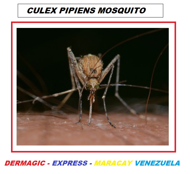
It is important to note that most of the studies were conducted in Europe, being perhaps the most relevant those made in the Czech Republic, where among them, in one study 5% of the mosquitoes studied were shown contaminated with spirochetes and one of them corresponded to the strain (BR-84) identified as Borrelia Afzelii.
 
Another detail to highlight is that the CDC does not mention these blood-sucking insects as a possible transmitter of Lyme borreliosis, which, although being low in the percentage shown in the studies, could be a factor in the spread of this disease by the world.
Here I leave the bibliographical references that prove these facts and in the attach one of the species of mosquitoes in which the Borrelia Burgdorferi was found.
“Under the sun there is nothing hidden, and sooner or later the evidence appears that shows that what you tried to hide, became the evidence that became a truth”
================================================================
BIBLIOGRPHICAL REFERENCES / REFERENCIAS BIBLOGRAFICAS
================================================================

1.) Doby JM , Chastel C , Couatarmanac’h A , Cousanca C , Chevrant-Breton J , Martin A , Legay B , Guiguen C .  [Etiologic and epidemiologic questions posed by erythema chronicum migrans and Lyme disease. Apropos of 4 cases at the Regional Hospital Center, Rennes]. Bull Soc Pathol Exot Filiales. 1985;78(4):512-25.b[Article in French]

2.) Magnarelli LA1, Anderson JF. Ticks and biting insects infected with the etiologic agent of Lyme disease, Borrelia burgdorferi. J Clin Microbiol. 1988 Aug;26(8):1482-6.

3.) J.H. (Han) van der Kolk. Borrelia burgdorferi seeks vectors. Page 119 | Taylor and Francsi online. Published online: 15 Dec 2014.
Source:https://www.tandfonline.com/doi/full/10.1080/01652176.2014.972609?scroll=top&needAccess=true

4.) Chang YF, Novosel V, Chang CF, Summers BA, Ma DP, Chiang YW, Acree WM, Chu HJ, Shin S, Lein DH. 2001 Jul. Experimental induction of chronic borreliosis in adult dogs exposed to Borrelia burgdorferi-infected ticks and treated with dexamethasone. Am J Vet Res. 62:1104–1112.[Crossref], [PubMed], [Web of Science ®], [Google Scholar]

5.) Chang YF, Novosol V, McDonough SP, Chang CF, Jacobson RH, Divers T, Quimby FW, Shin S, Lein DH. 2000. Experimental infection of ponies with Borrelia burgdorferi by exposure to Ixodid ticks. Vet Pathol. 37:68–76.[Crossref], [PubMed], [Web of Science ®], [Google Scholar]

6.) Fernando SS. 1983. The giant kidney worm (Dioctophyma renale) infection in man in Australia. Am J Surg Pathol. 7:281–284.[Crossref], [PubMed], [Web of Science ®], [Google Scholar]

7.) Halouzka J, Postic D, Hubálek Z. 1998. Isolation of the spirochaete Borrelia afzelii from the mosquito Aedes vexans in the Czech Republic. Med Vet Entomol. 12:103–105.[Crossref], [PubMed], [Web of Science ®], [Google Scholar]

8.) Halouzka J, Wilske B, Stünzner D, Sanogo YO, Hubálek Z. 1999. Isolation of Borrelia afzelii from overwintering Culex pipiens biotype molestus mosquitoes. Infection. 27(4-5):275–277.[Crossref], [PubMed], [Web of Science ®], [Google Scholar]

9.) Liu D. 2012. Dioctophyme. In: Liu D, editor. Molecular detection of human parasitic pathogens. Boca Raton (FL): Taylor & Francis; p. 535–538.[Crossref], [Google Scholar]

10.) Qiu WG, Schutzer SE, Bruno JF, Attie O, Xu Y, Dunn JJ, Fraser CM, Casjens SR, Luft BJ. 2004. Genetic exchange and plasmid transfers in Borrelia burgdorferi sensu stricto revealed by three-way genome comparisons and multilocus sequence typing. Proc Natl Acad Sci USA. 101:14150–14155.[Crossref], [PubMed], [Web of Science ®], [Google Scholar]

11.) Stricker RB, Lautin A, Burrascano JJ. 2005. Lyme disease: point/counterpoint. Expert Rev Anti Infect Ther. 3:155–165.[Crossref], [PubMed], [Google Scholar]

12.) Wagner B, Erb HN. Dogs and horses with antibodies to outer-surface protein C as on-time sentinels for ticks infected with Borrelia burgdorferi in New York State in 2011. 2012. Prev Vet Med. 107:275–279.[Crossref], [PubMed], [Web of Science ®], [Google Scholar]

13.) Zákovská A, Nejedla P, Holíková A, Dendis M. 2002. Positive findings of Borrelia burgdorferi in Culex (Culex) pipiens pipiens larvae in the surrounding of Brno city determined by the PCR method. Ann Agric Environ Med. 9:257–259.[PubMed], [Web of Science ®], [Google Scholar]

14.) J. A. ROBERTS. Experimental Transmission of Borrelia anserine (Sakharoff 1891) by Aedes aegypti. Letter | Published: 16 September 1961. Nature volume 191, page 1225 (16 September 1961)

15.) Hubálek Z1, Halouzka J, Juricová Z. Investigation of haematophagous arthropods for borreliae–summarized data, 1988-1996. Folia Parasitol (Praha). 1998;45(1):67-72.

16.) Petr Zeman. Borrelia-infection rates in tick and insect vectors accompanying human risk of acquiring Lyme borreliosis in a highly endemic region in Central Europe. Folia Parasitologica 45[4] 319-325 (1998). Regional Center of Hygiene, Dittrichova 17, 120 07 Prague 2, Czech Republic

17.) Zákovská A1, Nejedla P, Holíková A, Dendis M. Positive findings of Borrelia burgdorferi in Culex (Culex) pipiens pipiens larvae in the surrounding of Brno city determined by the PCR method. Ann Agric Environ Med. 2002;9(2):257-9.

18.) Kosik-Bogacka D1, Bukowska K, Kuźna-Grygiel W. Detection of Borrelia burgdorferi sensu lato in mosquitoes (Culicidae) in recreational areas of the city of Szczecin. Ann Agric Environ Med. 2002;9(1):55-7.

19.) Kosik-Bogacka D1, Kuźna-Grygiel W, Bukowska K. The prevalence of spirochete Borrelia burgdorferi sensu lato in ticks Ixodes ricinus and mosquitoes Aedes spp. within a selected recreational area in the city of Szczecin. Ann Agric Environ Med. 2004;11(1):105-8.

20.) Zákovská A1, Capková L, Serý O, Halouzka J, Dendis M. Isolation of Borrelia afzelii from overwintering Culex pipiens biotype molestus mosquitoes. Ann Agric Environ Med. 2006;13(2):345-8.

21.) Kosik-Bogacka DI1, Kuźna-Grygiel W, Górnik K. Borrelia burgdorferi sensu lato infection in mosquitoes from Szczecin area. Folia Biol (Krakow). 2006;54(1-2):55-9.

22.) Alexandre C.Atalibaa. José S.Resendeb. NatalinoYoshinaric. Marcelo B.Labrunaa. Isolation and molecular characterization of a Brazilian strain of Borrelia anserina, the agent of fowl spirochaetosis.Research in Veterinary Science. Volume 83, Issue 2, October 2007, Pages 145-149https://doi.org/10.1016/j.rvsc.2006.11.014

23.) Nejedla P1, Norek A, Vostal K, Zakovska A. What is the percentage of pathogenic borreliae in spirochaetal findings of mosquito larvae? Ann Agric Environ Med. 2009;16(2):273-6.

24.) Petra Nejedla 1,   Adam Norek 1,   Karel Vostal 1,   Alena Žákovská 1. What is the percentage of pathogenic borreliae in spirochaetal findings of mosquito larvae?. Ann Agric Environ Med. 2009;16(2):273–276

25.) Sikutová S1, Halouzka J, Mendel J, Knoz J, Rudolf I. Novel spirochetes isolated from mosquitoes and black flies in the Czech Republic. J Vector Ecol. 2010 Jun;35(1):50-5. doi: 10.1111/j.1948-7134.2010.00027.x.

26.) Melaun C1, Zotzmann S1, Santaella VG1, Werblow A1, Zumkowski-Xylander H2, Kraiczy P3, Klimpel S4. Occurrence of Borrelia burgdorferi s.l. in different genera of mosquitoes (Culicidae) in Central Europe. Ticks Tick Borne Dis. 2016 Mar;7(2):256-63. doi: 10.1016/j.ttbdis.2015.10.018. Epub 2015 Nov 12.

__________________

For more:  

https://madisonarealymesupportgroup.com/2016/07/23/german-study-finds-borrelia-in-mosquitos/

https://madisonarealymesupportgroup.com/2018/10/04/deer-fly-lyme-carrying-ectoparasite-on-the-move/