Archive for the ‘research’ Category

Matcha Tea Decreases Anxiety by Activating Dopamine & Seratonin Receptors

https://todayspractitioner.com/mind-body-medicine/matcha-tea-decreases-anxiety-by-activating-dopamine-and-seratonin-receptors

iu-30

Matcha Tea Decreases Anxiety by Activating Dopamine and Seratonin Receptors

See link to learn how Japanese researchers from Kumamoto University have shown that anxious behavior in mice is reduced after consuming Matcha powder or Matcha extract.

For more:  https://articles.mercola.com/teas/matcha-green-tea.aspx


Lyme Disease Common in Turkey. Patients With Neurological Symptoms Misdiagnosed With MS

https://www.ejbps.com/admin/assets/article_issue/volume_6_april_issue_4/1553939970.pdf?

Frequency of Borrelia Burgdorferi Western Blot & LTT Positivity Among Multiple Sclerosis Patients From Turkey

Barbaros Çetin*

Dokuz Eylul University, Faculty of Science, Department of Biology, Izmir, Turkey.

Article Received on 29/01/2019 Article Revised on 19/02/2019 Article Accepted on 12/03/2019

*Corresponding Author: Dr. Barbaros Çetin

ABSTRACT

In Turkey, Borrelia burgdorferi infections are not well known among physicians and almost completely overlooked. On the other hand, a small number of seropositivity studies (%3.3-%73) show that Borrelia burgdorferi is common in Turkey. There is no diagnostic biological marker in multiple sclerosis (MS). Only several clinical criteria used for diagnosis. These criteria are also compatible with other diseases. Lyme disease is currently among them.

In the chronic phase of Lyme, demyelination can form and this can be confused with MS. In this study 126 patients, between ages 17 and 66, with a definite diagnosis of multiple sclerosis was evaluated, and were found to be found positive Borrelia burgdorferi western blot and LTT test results 108 (%85.72). Only 18 (%14.28) patients have negative test results.

The results show that LYME disease is very common in Turkey and LYME patients with neurological symptoms are misdiagnosed with multiple sclerosis.

_________________

**Comment**

Important excerpt from study:

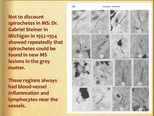
“Spirochetes in MS” (Buzzard, E.F.), published in the famous Lancet magazine in 1911, revealed the presence of Lyme spirochetes in the brains of MS patients. Over a period of more than a century, more than 50 international scientific papers proving the MS-Lyme relationship have been published in prestigious medical journals.[16-111]

Please look at the date for that…...1911, yet, how many doctors are considering Lyme/MSIDS in those patients thought to have MS?

I will answer that question – very, very few. 

68956343_10218363594908655_6861034436578770944_n

Courtesy, Tom Grier 

For an informative article on the subject of cell-wall deficient forms (CWD) and other neurological diseases: http://www.livinglyme.com/notes-and-observations-on-cell-wall-deficient-forms/

Dr. Lida Mattman, was a protégé of Gabriel Stiener who was the first to establish that MS was associated with a spirochete.

Grier states that this area of microbiology has long been neglected, and that we are now paying a price for that neglect. Mattman’s work suggests that cell wall deficient forms are prevalent, pathogenic, and may be behind sarcoidosis, Crohn’s disease, coronary thrombosis, Kaposi’s sarcoma, endocarditis, and MS.

Mattman explains her simple lab technique here:  https://madisonarealymesupportgroup.com/2019/04/02/transmission-of-lyme-disease-lida-mattman-phd/  Transcript included. 

According to Grier, Mattman said she frequently isolates L-forms from Lyme patients with aseptic meningitis and endocarditis, but that traditional culture media is virtually worthless, as are traditional heat fixed blood smears. Instead, she creates a live wet mount using the patients blood or bully coat. The blood sample is placed on a wet slide with acrodine orange dye to stain the nucleic acids. Then a monoclonal antibody fluorescent stain that is specific for Borrelia burgdorferi is added and is examined under a microscope. Grier states most labs are clueless about cell wall deficient forms and that we will not have answers until more labs agree to test for CWD forms.


Prevalence of Babesia in Canadian Blood Donors: June – October 2018

https://www.ncbi.nlm.nih.gov/pubmed/31385317

2019 Aug 5. doi: 10.1111/trf.15470. [Epub ahead of print]

Prevalence of Babesia in Canadian blood donors: June-October 2018.

Abstract

BACKGROUND:

The erythrocytic protozoan parasite Babesia microti, the cause of human babesiosis, is transmitted not only by tick bites but also via blood transfusion. B. microti is endemic in the northeastern/upper midwestern United States, where partial screening of blood donations has been implemented. In Canada, a 2013 study of approximately 14,000 donors found no B. microti antibody-positive samples, suggesting low risk at that time.

METHODS:

Between June and October 2018, 50,752 Canadian donations collected from sites near the US border were tested for Babesia nucleic acid by transcription-mediated amplification (TMA). Reactive donations were tested for B. microti by IgG immunofluorescence assay and polymerase chain reaction. A subset of 14,758 TMA nonreactive samples was also screened for B. microti antibody. Donors who tested reactive/positive were deferred, asked about risk factors, and were requested to provide a follow-up sample for supplemental testing.

RESULTS:

One sample from Winnipeg, Manitoba, was TMA and antibody reactive. Of the 14,758 TMA-nonreactive donations tested for antibody, four reactive donations were identified from southwestern Ontario near Lake Erie. None of the interviewed donors remembered any symptoms, likely tick exposure, or relevant travel within Canada or the United States.

CONCLUSIONS:

This is the largest B. microti prevalence study performed in Canada. The results indicate very low prevalence, with only one TMA-confirmed-positive donation of 50,752 tested. This donor was from the only region in Canada where autochthonous infection has been reported. Seropositive donations in southwestern Ontario suggest low prevalence; travel should not be ruled out given the proximity to the US border.

____________________

For more: I would caution authorities in believing there is a low prevalence of Babesa. I’ve heard it takes a trained eye to see it and is rarely detected using only 1 diagnostic test. I think the word is out on the seriousness of tick-borne disease. Let’s not go back in time by adopting a carefree approach. We should be looking hard and using accurate testing methods.

 https://madisonarealymesupportgroup.com/2016/06/02/study-showing-results-testing-babesia-microti/

https://madisonarealymesupportgroup.com/2019/07/28/tick-borne-infection-risk-in-blood-transfusion/

https://madisonarealymesupportgroup.com/2017/09/27/premature-infants-develop-babesia-via-blood-transfusion/

https://madisonarealymesupportgroup.com/2019/07/11/characteristics-of-transfusion-transmitted-babesia-microti-american-red-cross-2010-2017/  This clearly shows there were more than 200 Babesia transfusion-transmissions reported. It also shows you don’t have to reside in an endemic area or travel to an endemic area to get it. The article also clearly points out that the geographic range of ticks is expanding, which means the pathogens they carry will as well.

According to Dr. Ken Singleton, Babesia is rarely detected using one diagnostic test alone.  http://www.lymebook.com/chronic-lyme-testing-and-diagnosis

Excerpts:

Lyme-aware physicians generally screen for 2 strains—Babesia microti and WA-1 (Babesia duncani)—by testing for antibodies (by IFA or ELISA testing) made by the body against those organisms.

Another very useful test for Babesia is known as the FISH (fluorescent in situ hybridization) test. The FISH test is performed on thin blood smears (tests used to detect germs in white blood cells) and is able to detect the RNA (genetic material) of Babesia. If this test is positive, it is very strong evidence of the presence of active Babesia. The advantage of the FISH test is that it will detect other subspecies of Babesia in addition to B. microti and B. duncani. (A direct thick and thin blood smear using a staining technique called “Giemsa” can also be done by one’s local or commercial labs to look for Babesia organisms in red blood cells; however, it is an insensitive test except during acute Babesia, particularly when fever is present.)

A final potentially useful test is the Babesia PCR (polymerase chain reaction). Unfortunately, in my experience it is also not a sensitive test and is the least useful of the three tests mentioned.

All three of these tests—Babesia IFA, FISH, PCR—are available through IgeneX, a laboratory specializing in Lyme disease and other tick-borne organisms. Medical Diagnostics Laboratory (MDL) has two of the tests—Babesia ELISA and PCR. Both labs are excellent and I utilize both regularly. (See the resources section for more information.) However, as mentioned, Babesia can frequently escape detection by diagnostic tests. Therefore, many times babesiois must be a clinical diagnosis made by physicians who are experienced in its detection and treatment.

How many Canadian people slipped through the cracks?


Seropositivity to Tick Endosymbiont As A Marker To Determine Tick Bite Exposure

https://www.ncbi.nlm.nih.gov/pubmed/31397213/

Seropositivity to Midichloria mitochondrii (order Rickettsiales) as a marker to determine the exposure of humans to tick bite.

Abstract

Ixodes ricinus is the most common tick species parasitizing humans in Europe, and the main vector of Borrelia burgdorferi sensu lato, the causative agent of Lyme disease in the continent. This tick species also harbors the endosymbiont Midichloria mitochondrii, and there is strong evidence that this bacterium is inoculated into the vertebrate host during the blood meal. A high proportion of tick bites remains unnoticed due to rarity of immediate symptoms, implying the risk of occult tick-borne infections in turn a potential risk factor for the onset of chronic-degenerative diseases. Since suitable tools to determine the previous exposure to I. ricinus bites are needed, this work investigated whether seropositivity toward a protein of M. mitochondrii (rFliD) could represent a marker for diagnosis of I. ricinus bite.

We screened 274 sera collected from patients from several European countries, at different risk of tick bite, using an ELISA protocol. Our results show a clear trend indicating that positivity to rFliD is higher where the tick bite can be regarded as certain/almost certain, and lower where there is an uncertainty on the bite, with the highest positivity in Lyme patients (47.30%) and the lowest (2.00%) in negative controls.

According to the obtained results, M. mitochondrii can be regarded as a useful source of antigens, with the potential to be used to assess the exposure to ticks harboring this bacterium. In prospect, additional antigens from M. mitochondrii and tick salivary glands should be investigated and incorporated in a multi-antigen test for tick bite diagnosis.

___________________

**Comment**

This is an interesting study with future potential. Essentially they are saying since Lyme is so hard to detect – find its friends and you may find Lyme.

I’ve written about endosymbionts before, particularly Wolbachia:

https://madisonarealymesupportgroup.com/2017/07/10/wolbachia-the-next-frankenstein/

Briefly, endosymbionts are organisms living in the body or cells of another organism in a symbiotic relationship – which isn’t always of mutual benefit. An example of a mutualistic relationship is the protozoan endosymbionts inside a termite which help it to break down the wood it eats.
However, in the case of Wolbachia, while the benefit between itself and the worms it lives in may be mutualistic, it’s caused harm in dogs being treated for heart worm. Heart worm medication causes Wolbachia to be released into the blood and tissues causing severe Inflammation in pulmonary artery endothelium which may form thrombi and interstitial inflammation. Wolbachia also activates pro inflammatory cytokines. For human Lyme/MSIDS patients this could translate out to a similar result when they are treated for worms, which ticks also carry.
What I find interesting here is that both Midichloria and Wolbachia are in the same subclass of Rickettsidae and order Rickettsiales. Guess who else is in these groups?
  • Rickettsia
  • Ehrlichia
Both of which cause a variety of human and animal illness.

The question of course is, could these supposedly harmless endosymbionts be responsible for more than they are given credit for? Testing for them may not only reveal that Lyme is present but in fact that they are contributing to the problem. Sounds like an exciting field of discovery.

Endocarditis Caused by Bartonella Quintana, A Rare Case in the U.S.

https://www.ncbi.nlm.nih.gov/pmc/articles/PMC6667705/

. 2019; 17: e00533.
Published online 2019 Apr 6. doi: 10.1016/j.idcr.2019.e00533
PMCID: PMC6667705
PMID: 31384552

Endocarditis caused by Bartonella Quintana, a rare case in the United States

Abstract

Bartonella quintana is a relatively rare cause of endocarditis in the United States (USA). Historically it was linked with trench fever, but cardiac involvement seems to be more prevalent recently. There are some known risk factors associated with Bartonella quintana endocarditis such as human immunodeficiency virus (HIV) infection, alcoholism, homelessness and poor hygiene. We report a case of 37-year-old African man, with culture negative endocarditis, emboli and rising B. quintana and B. henselae IgG titers. B. quintana DNA was subsequently detected from the mitral valve sample with 16S rRNA gene and ribC primer sets. Eventually, blood culture for B. quintana was positive after 21 days. Patient was successfully treated with doxycycline and gentamicin. There have been a few cases of B. quintana endocarditis in the USA and most of them were associated with HIV infection, homelessness or alcoholism. The case reported here highlights the importance of high clinical suspicious for Bartonella species in blood culture negative endocarditis in the USA in appropriate setting and will help to increase awareness among physicians for early diagnosis and treatment.

___________________

**Comment**

A few points of interest:

  • Patient’s chief complaints:  progressive shortness of breath, chest pain, occasional non-drenching night sweats, fatigue, unintentional ten pound weight loss, and intermittent sharp chest pain radiating to the neck
  • Past medical history significant for latent tuberculosis infection and treatment completed 3 months prior to the presentation
  • While living in the Democratic Republic of Congo before migrating to Indiana, patient had a cow at his home and used to drink raw cow’s milk
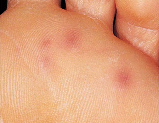
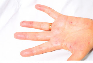
  • Had Janeway Lesions on hands & feet http://www.healthgala.info/2017/08/Janeway-lesion-Pictures-Definition-Symptoms-Causes-Treatment.html

Janeway lesion Pictures, Definition, Symptoms, Causes, Treatment

Janeway lesion Pictures, Definition, Symptoms, Causes, Treatment

  • Was considered immunocompetent 

For more:  https://madisonarealymesupportgroup.com/2017/01/04/endocarditis-consider-bartonella/

https://madisonarealymesupportgroup.com/2018/09/28/bartonella-infective-endocarditis-with-dissemination-a-case-report-literature-review/

https://madisonarealymesupportgroup.com/2019/08/12/tick-borne-pathogens-bartonella-spp-borrelia-burgdorferi-sensu-lato-coxiella-burnetii-rickettsia-spp-may-trigger-endocarditis/

https://madisonarealymesupportgroup.com/2019/04/25/case-of-endocarditis-caused-by-bartonella-after-mitral-valve-repair/

https://madisonarealymesupportgroup.com/2018/07/10/infective-endocarditis-associated-with-bartonella-henselae-a-case-series/

https://madisonarealymesupportgroup.com/2019/04/24/human-bartonellosis-an-underappreciated-public-health-problem/