Background: The incidence of Lyme disease (LD) in North America has increased substantially in the past two decades. Concomitant with the increased incidence of infection has been an enhancement in the recognition of LD complications. Here, we report a case of Lyme carditis complicated by heart block in a pediatric patient admitted to our children’s hospital. What is unique about this case is that the complaint of chest palpitations is an infrequent presentation of LD, and what it adds to the scientific literature is an improved understanding of LD in the pediatric population.
Case presentation: The patient was a 16-year-old male who presented with the main concerns of acute onset of palpitations and chest pain. An important clinical finding was Erythema migrans (EM) on physical exam. The primary diagnoses were LD with associated Lyme carditis, based on the finding of 1st degree atrioventricular heart block (AVB) and positive IgM and IgG antibodies to Borrelia burgdorferi. Interventions included echocardiography, electrocardiography (EKG), and intravenous antibiotics. The hospital course was further remarkable for transition to 2nd degree heart block and transient episodes of complete heart block. A normal sinus rhythm and PR interval were restored after antibiotic therapy and the primary outcome was that of an uneventful recovery.
Conclusions: Lyme carditis occurs in < 5% of LD cases, but the “take-away” lesson of this case is that carditis can be the presenting manifestation of B. burgdorferi infection in pediatric patients. Any patient with suspected Lyme carditis manifesting cardiac symptoms such as syncope, chest pain, or EKG changes should be admitted for parenteral antibiotic therapy and cardiac monitoring. The most common manifestation of Lyme carditis is AVB. AVB may manifest as first-degree block, or may present as high-grade second or third-degree block. Other manifestations of Lyme carditis may include myopericarditis, left ventricular dysfunction, and cardiomegaly.Resolution of carditis is typically achieved through antibiotic administration, although pacemaker placement should be considered if the PR interval fails to normalize or if higher degrees of heart block, with accompanying symptoms, are encountered. With the rising incidence of LD, providers must maintain a high level of suspicion in order to promptly diagnose and treat Lyme carditis.
___________________
**Comment**
Repeat: Researchers need to refrain from stating that something is “rare” with Lyme disease as:
testing misses more than 70% of cases (thousands go undiagnosed)
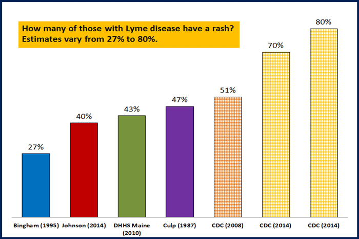
much fewer report the EM rash than is quoted
this is still a misunderstood illness that doctors are woefully uneducated on. If a patient doesn’t present with the EM rash, doctors unfamiliar with the wide symptom presentation are not going to catching this.
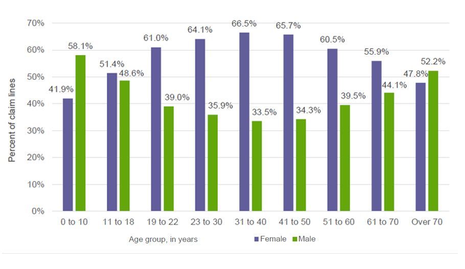
Click on image to see better. The graph is clear that there is a wide range of those with those finding the EM rash. While the EM rash is diagnostic for Lyme disease, many do not get it. In the first ever patient group in Lyme, Connecticut, only a quarter had the rash:
Again, mainstream medicine continues with abysmal testing and ancient dogma that is hurting patients.
More accurate scientific language would be: “Heart palpitations are rarely reported in the literature.”What happens in the real world is often quite different than what is reported in the literature – particularly with Lyme as research has been hijacked by The Cabal: https://madisonarealymesupportgroup.com/2017/01/13/lyme-science-owned-by-good-ol-boys/
BTW: I had chest palpitations, my husband had chest palpitations, and most of the patients I work with have them. I don’t think it’s nearly as rare as is being reported.
In 90% of cases, the most common consequence of Lyme carditis is heart block. The severity of the heart block can fluctuate rapidly and the progression to complete heart block can be fatal. Importantly, the heart block in Lyme carditis can be transient and usually resolves with antibiotic therapy. Additionally, Lyme carditis can affect other parts of the heart’s conduction system, as well as the heart’s muscle, valves, and outer layer of the heart wall.
The broad range of signs and symptoms of Lyme disease and the varying presentations from person to person make diagnosing the disease challenging. Furthermore, Borrelia burgdorferi spirochete are adept at evading the immune system. The bacterium can travel through the bloodstream, burrow into tissue and remain dormant for days, months, or even years before symptoms arise.
While many people associate Lyme disease with manifestations such as Bell’s palsy, the circular Bull’s-eye rash, and flu-like symptoms, Lyme disease can also cause sensory, cognitive, neurologic, and cardiac complications, even in its earliest stage. But, the signs and symptoms of Lyme disease are all too frequently attributed to another medical condition.
Objective signs of Lyme disease include Bell’s palsy, synovitis of the knee, and the presence of a Bull’s-eye or erythema migrans rash. However, most people exhibit a wide range of signs and symptoms of Lyme disease that may come and go and fluctuate in their intensity.
Furthermore, researchers in the UK remind clinicians to consider Lyme disease in children who present with Bell’s palsy. “In areas endemic with Lyme disease, Lyme disease should be considered as the likely cause of facial nerve palsy in children until proven otherwise.”
Wide range of signs and symptoms of Lyme disease
Studies indicate that at least 50% of patients with Lyme disease do not exhibit the classic Bull’s-eye rash. When a rash is present, it can appear anywhere on the body. It does not always appear at the site of the tick bite. The rash usually appears between 3 – 30 days after the tick bite.
A rash due to Lyme disease is typically not itchy or painful. It may fade and then reappear and it can be confused with a spider bite. Atypical rashes can also occur. And when multiple rashes appear on the body, it may be an indication that the Borrelia burgdorferi spirochete has disseminated beyond the tick bite and the disease is in a more advanced stage.
If left untreated, the infection can spread to other parts of the body, including the brain and central nervous system, cardiovascular system, peripheral and autonomic nervous system, along with the muscles and joints, and eyes.
Neurologic and cardiac manifestations
Lyme disease can cause neurological and cardiac symptoms such as meningitis, encephalitis, and carditis. But, more often symptoms include severe and unrelenting fatigue, joint pain (with or without swelling), sore muscles, neck and back pain, headaches, light, sound and temperature sensitivity, sleep disturbance, night sweats, irritability, anxiety, despair, sadness, lightheadedness, crying, poor memory and concentration, stiff neck, numbness, and tingling sensations.
Although rare, Lyme carditiscan cause sudden death. The authors of a case series warn, “These two cases highlight the importance of early recognition. And treatment, even if it’s empirical, may save lives.”
Another researcher reports, “The burden of Lyme disease and Lyme carditis in U.S. children’s hospitals has increased in recent years.” The authors identified 189 children diagnosed with Lyme carditis between 2007 and 2013.
Lyme-induced encephalopathy
Lyme disease can trigger encephalopathy in some patients, as well. A Tufts University School of Medicine study reports that 24 of 27 Lyme disease patients presented with a mild encephalopathy, which began 1 month to 14 years after the onset of the disease and was characterized by memory loss, mood changes, and sleep disturbances.
Fallon et al. described pain reported by patients with Lyme encephalopathy as being “similar to those of post-surgery patients,” and their fatigue “was similar to that of patients with multiple sclerosis.”
And another study found that 14% of 86 children with Lyme disease exhibited neurocognitive complications including behavioral changes, forgetfulness, declining school performance, headache or fatigue, and in 2 cases, a partial complex seizures disorder.
Long-term complications
Signs and symptoms of Lyme disease may be incorrectly attributed to another illness. Patients are often misdiagnosed with a psychiatric disorder, chronic fatigue syndrome, or fibromyalgia. An incorrect diagnosis and delay in treatment can lead to long-term consequences for Lyme disease patients.
Postural orthostatic tachycardia syndrome (POTS) is an autonomic dysfunction that can appear years after antibiotic treatment for Lyme disease, resulting in fatigue, cognitive impairment, and orthostatic intolerance such as palpitations, lightheadedness, chest discomfort, and shortness of breath, among others.
Other types of chronic manifestations include Lyme encephalopathy, neurocognitive complications, post-Lyme disease syndrome, neuropsychiatric Lyme disease, sensory neuropathy, and carditis.
Psychiatric symptoms common in Lyme patients
An estimated 1 in 3 patients with chronic neurologic Lyme disease suffer from depression, in addition to fatigue, headaches, poor concentration, lightheadedness, and joint pain, according to one study.
All too frequently patients with complex, multisystem illnesses are dismissed by clinicians, their symptoms often attributed to a psychiatric illness simply as “a diagnosis of default,” Bransfield writes.
This attitude can lead to tragic delays in identifying a correct diagnosis and appropriate treatment.
Apologies for the tardiness of this, but PLEASE watch it while you can & share it widely. The information contained within the documentary is not only eye-opening but imperative for women to understand for their health.
The documentary “Boobs: The War on Women’s Breasts” explores how mammography is causing more harm than good for many women
One of the harms of using mammography as a screening tool is that it can often lead to overdiagnosis and overtreatment, including false-positive tests and unnecessary biopsies
Due to the introduction of mammography, breast cancer was overdiagnosed — meaning that tumors were detected that would never lead to clinical symptoms — in 1.3 million women over a 30-year period
Mammograms and their resulting biopsies may increase the spread of cancer, and up to 1 in 4 breast tissue biopsies may be incorrectly diagnosed by pathologists, with unnecessary mastectomies being performed as a result
There’s also the issue that mammograms use ionizing radiation at a relatively high dose, which in and of itself can contribute to the development of breast cancer
Other potentially safer options to mammography such as thermography and ultrasound are not widely available because the billion-dollar mammagoraphy industry prevents the widespread use of these beneficial tests
When asked how much radiation women are exposed to from mammograms, Dr. Ben Johnson, author of “No Ma’am-ograms,” says, “At least the amount of 100 chest X-rays.”
It’s a statement that would surprise many women who are advised to receive a mammogram as the standard of care for breast cancer prevention in the U.S. — and it’s just one of the eye-opening facts presented in the documentary “Boobs: The War on Women’s Breasts.”
The film begins with a written statement informing viewers that all of the information you’re about to hear is supported by scientific literature, even though much of it opposes the assertions made by conventional medicine. Doctors from across the United States and around the world — including California, Georgia, Switzerland, Virginia and Arizona — are interviewed, providing expert testimony on why you may want to rethink this controversial procedure.
The ‘Early Detection’ Myth and Biopsy Risks
One myth perpetuated by conventional medicine is that mammography is the most important screening test for breast cancer because it can detect breast cancer “up to two years before the tumor can be felt by you or your doctor.”1
However, the film points out that by the time you can feel a cancerous lump in your breast, the cancer has already been growing for two to five years. “Mammograms are not early detection,” Johnson says.
The myth that mammograms don’t spread cancer is also addressed, via the story of one woman who was diagnosed with stage 4 cancer and given only 1 year to live. She made significant changes to her lifestyle and many of the tumors disappeared. The cancer was stable years later, until she received a biopsy and subsequent mammograms, which she says made the cancer spread — and a tumor appeared in the area where the biopsy was done.
During a biopsy, a piece of tissue from a tumor or organ is removed so that it can be examined under a microscope, often to determine if it is cancerous. Needle biopsies, for instance, are widely used as part of the traditional allopathic approach to diagnosing breast cancer. But they may accidentally cause malignant cells to break away from a tumor, resulting in its spreading to other areas of your body.
One of the harms of using mammography as a screening tool is that it can often lead to overdiagnosis and overtreatment, including false-positive tests and unnecessary biopsies.2
One study from the John Wayne Cancer Institute revealed that a needle biopsy may increase the spread of cancer compared to patients who receive excisional biopsies, also known as lumpectomies.3 They concluded, “Manipulation of an intact tumor by FNA [fine-needle aspiration] or large-gauge needle core biopsy is associated with an increase in the incidence of SN [sentinel node] metastases, perhaps due in part to the mechanical disruption of the tumor by the needle.”4
Johnson said he calls biopsies “the kiss of death,” describing how the needle gets driven through a billion cells, blows through the cancer and goes out the other side, dragging cells back through and spreading the cancer. Dr. Manfred Doepp, medical director of the Couros Center in Switzerland, agrees that biopsies can spread cancer, while the physical act of squeezing the breast during mammography may also trigger the cancer to spread.
The film cites a rapid response published in the BMJ, which states “robust scientific data, published in prestigious medical journals, have lent meaningful support to the concept” that the squeezing motion of mammography is in itself potentially harmful.5
Breast Cancer Overdiagnosed in 1.3 Million Women
The notion that mammography is an accurate screening tool is another myth busted in the film. Both false positives and false negatives occur, and even the skill of the radiologist in reading the mammogram is a factor in whether or not mammography is able to detect cancer.
Overall, when researchers examined trends in early-stage breast cancer and late-stage breast cancer from 1976 to 2008 among women 40 years and older, they found the introduction of mammography in the U.S. was linked to a doubling in the number of early-stage breast cancer cases detected each year.6
However, the rate at which women developed late-stage cancer decreased by 8% during that time, leading the researchers to suggest that only a small number of early-stage cancer diagnosed would progress into advanced disease:7
“Despite substantial increases in the number of cases of early-stage breast cancer detected, screening mammography has only marginally reduced the rate at which women present with advanced cancer.
Although it is not certain which women have been affected, the imbalance suggests that there is substantial overdiagnosis, accounting for nearly a third of all newly diagnosed breast cancers, and that screening is having, at best, only a small effect on the rate of death from breast cancer.”
They concluded that due to the introduction of mammography, breast cancer was overdiagnosed — meaning that tumors were detected that would never lead to clinical symptoms — in 1.3 million women over a 30-year period.
Up to 80% of Breast Biopsies Are Benign
The overdiagnosis of breast cancer comes with serious risks, including needless treatments and unnecessary stress and anxiety. In another study cited in the film, it’s stated that more than 1 million breast biopsies are performed annually in the U.S., 75% to 80% of which turn out to be benign.8 Sometimes, the benign result isn’t uncovered until an unnecessary mastectomy has already occurred.
Up to 1 in 4 breast tissue biopsies may be incorrectly diagnosed by pathologists,9 and unnecessary mastectomies have been performed as a result. In a case that went to trial in 2020 in Israel, a woman was diagnosed with breast cancer after a biopsy tested at two medical centers came back positive.
She underwent six months of chemotherapy as a result, as well as a partial mastectomy to remove the growth. After the growth was removed, the lawsuit alleges, it turned out to be benign.10 In 2015, researchers with Boston Children’s Hospital in Massachusetts revealed that false-positive mammograms and breast cancer overdiagnoses among women ages 40 to 59 cost the U.S. $4 billion each year.11
Mammograms May Not Work for Women With Dense Breasts
The story of Nancy Cappello is also covered in the film. Cappello was diagnosed with breast cancer after receiving two normal mammograms. The cancer was missed because she has dense breast tissue, and was only revealed when her doctor felt a ridge in her breast and prescribed an ultrasound test in addition to a mammogram.
Cappello was a pioneer in the movement to teach women about dense breast tissue and how using a mammogram to detect cancer in such tissue is “like finding a polar bear in a snowstorm.” She said:12
“So I went on a quest — for research — and I discovered for nearly a decade BEFORE my diagnosis, six major studies with over 42,000 women concluded that by supplementing a mammogram with an ultrasound increases detection from 48% to 97% for women with dense tissue.
I also learned that women with extremely dense tissue are 5x more likely to have breast cancer when compared with women with fatty breasts and that research on dense breast tissue as an independent risk factor for breast cancer has been studied since the mid 70s.
… I endured a mastectomy, reconstruction, 8 chemotherapy treatments and 24 radiation treatments. The pathology report confirmed — stage 3c cancer because the cancer had traveled outside of the breast to my lymph nodes. Eighteen lymph nodes were removed and thirteen contained cancer — AND REMEMBER — a “normal” mammogram just weeks before. Is that early detection?”
As a result of Cappello’s movement, 38 states have passed mandatory breast density reporting laws. The film states that up to 90% of women may have some degree of dense breast tissue that may affect a mammogram’s outcome and could benefit from whole breast ultrasound — a procedure that’s not widely available. Women are also interviewed that were told they had dense breast tissue, but not what that meant for the effectiveness of mammography.
‘Precancer’: Ductal Carcinoma in Situ
Ductal carcinoma in situ (DCIS) refers to the abnormal growth of cells within the milk ducts of the breast forming a lesion commonly between 1 to 1.5 centimeters (cm) in diameter. While the cells appear malignant under a microscope, they have not invaded surrounding tissue and are considered “precancer,” noninvasive or “stage zero breast cancer.” Some experts have also argued that DCIS should be considered noncancerous.
Despite this, the film tells the story of one woman who had both breasts removed due to a DCIS diagnosis. Such diagnoses now represent 20% to 25% of all “breast cancers,”13 and the standard of care is to treat all DCIS with mastectomy or breast-conserving surgery with radiotherapy. According to a review in the British Journal of Cancer:14
“In effect, women with DCIS are labelled as ‘cancer patients’, with concomitant anxiety and negative impact on their lives, despite the fact that most DCIS lesions will probably never progress to invasive breast cancer.
Owing to the uncertainty regarding which lesions run the risk of progression to invasive cancer, current risk perceptions are misleading and consequently bias the dialogue between clinicians and women diagnosed with DCIS, resulting in overtreatment for some, and potentially many, women.”
Is Radiation From Mammograms Dangerous?
There’s also the issue that mammograms use ionizing radiation at a relatively high dose, which in and of itself can contribute to the development of breast cancer. In a 2016 study, it’s stated, “ionizing radiation as used in low-dose X-ray mammography may be associated with a risk of radiation-induced carcinogenesis.”15
They point out that people who carry certain genetic variations or have an inherited disposition of breast cancer, in particular, should avoid ionizing radiation as much as possible — the latter group being one that conventional medicine often recommends for routine or even extra mammography screening. Again, they refer individuals to a different, safer, form of testing: ultrasound or magnetic resonance imaging (MRI).16
A “new and improved” type of mammogram called 3D tomosynthesis, which is basically a CT scan for your breasts, is particularly problematic, as your radiation exposure is even greater than from standard mammograms — and by a significant margin.
According to one study,17 annual screening using digital or screen-film mammography on women aged 40 to 80 years is associated with an induced cancer incidence and fatal breast cancer rate of 20 to 25 cases per 100,000 mammograms. Meaning, annual mammograms cause 20 to 25 cases of fatal cancer for every 100,000 women getting the test.
The 3D mammogram requires multiple views in order to achieve three-dimensionality, so it stands to reason your total radiation exposure would be considerably higher than from a standard 2D mammogram.
Thermography and Ultrasound Use No Radiation
In order to make informed decisions, women should be provided with all of their screening options, along with an explanation of their strengths and weaknesses, and be allowed to utilize the option of their choice.
Mammography is only one option, with considerable drawbacks. Other potentially safer options include self and clinical breast exams, thermography, ultrasound and MRI. Thermography and ultrasound use no radiation, and may detect abnormalities that mammograms miss, especially in women with dense breast tissue.
These are explained in detail toward the end of the film, but they can be difficult to access in the U.S., as the billion-dollar mammography industry prevents the widespread use of these beneficial tests.
It’s also important to understand that mammography doesn’t prevent breast cancer in any way. Breast cancer prevention involves a healthy lifestyle, avoidance of toxins and attention to certain nutritional factors, such as vitamin D. It is absolutely vital to know and optimize your vitamin D level as it can radically reduce your risk of breast cancer.
Ignoring these factors and simply getting an annual mammogram and believing you’re protected is the end result being foisted upon many women trusting the advice of conventional medicine. By leading a healthy lifestyle, and seeking to get informed about all of your breast cancer screening options, you can avoid this potentially deadly pitfall.
About the Director
I believe in bringing quality to my readers, which is why I would like to share some information about the filmmaker, Megan S. Smith, M.S., and what went in to making “bOObs: The War on Women’s Breasts.” Thank you, Megan, for sharing with us.
What was your inspiration for making this film?
My inspiration for making this film, as well as in making my next film on alternative cancer therapies, was the death of my husband 11 years ago from lung cancer. Specifically, for “bOObs,” my inspiration was emboldened by the death of my three dear friends in as many years after their breast cancer returned following 10-plus years in remission.
What was your favorite part of making the film?
My favorite part of making this film was interviewing the patients and doctors. I learned during my interviews of Drs. Ben Johnson and Galina Migalko about the detriments of mammograms and that we might instead be using thermography in conjunction with ultrasound. This nonradiation protocol made sense to me.
Also, my interview with The American Cancer Society’s Dr. Otis Brawley was an eye opener, as he disclosed, “Mammography is a suboptimal test” and “We need a better test.” All these doctors were spot on. More information on the film can be found here.
A September 2020 meta-analysis concludes there is a significant relationship between autism and concentrations of lead and mercury in the body
According to the researchers, mercury concentration is a pathogenic cause for autism, meaning it’s a causative factor
According to a 2014 review, there is evidence of malfeasance and conflicts of interest in studies claiming that thimerosal in vaccines is safe
Serious flaws and errors also plague studies that claim aluminum in vaccines is safe. A mathematical error found in a key FDA study has reignited concerns about the safety of aluminum in vaccines
Glutathione is the dominant agent that binds to and helps move mercury and other heavy metals out of your tissues. Part of effective detox involves upregulating your biochemistry to facilitate the mobilization and elimination of metals
The controversy over whether mercury overexposure can trigger autism is a long-standing one. A new meta-analysis of previous studies sheds much needed light on the matter, concluding there’s a “significant relationship” between the two.
The review,1,2 published in the September 2020 issue of Pediatric Health, Medicine and Therapeutics, looked at 18 studies conducted between 1982 and 2019 that examined the relationship between concentrations of copper, lead or mercury in blood, plasma, hair or nails and the prevalence of autism. While no relationship was found between autism and copper concentrations, a high degree of correlation was found for mercury and lead.
According to the authors,3 the relationship between mercury and autism is so strong that “the concentration of mercury can be listed as a pathogenic cause (disease-causing) for autism.” This held true even when outlier studies that might unduly influence the results were removed.
Mercury Is a Causative Factor
In the introduction, the authors point out that studies carried out in this area suggest mercury and other toxins are involved in the cause of autism, which include abnormal brain development that affects social interaction and communication skills.
“Metals’ biological effects are associated with their chemical properties, suggesting that excessive metal exposure can cause brain abnormalities around the world,” the researchers state.4
“Mercury is considered as a risk factor for autism since, according to previous studies, it has been recognized as a neurotrophic toxin. Reduction in mercury content in hair and teeth of the children with autism aroused the low disposal of mercury hypothesis.
Blaurock-Bush et al found that heavy metals are effective in the development of autism disorder. The role of mercury in the pathogenesis of autism has also been proven in other studies …
According to points raised in the present study … it would be quite reasonable to advise prevention of exposure to mercury and lead in children and provision of suitable conditions during the sensitive period of mothers’ pregnancy as vital measures to prevent the disease …”
A 2017 review paper,5 “The Toxicology of Mercury: Current Research and Emerging Trends,” details the “kinetics of this metal,” including “its metabolism, interaction with other metals, distribution, internal doses and targets and reservoir organs.” The paper cites several studies linking mercury and autism among its references, noting that:6
“Autism spectrum disorder (ASD) has been demonstrated to be accompanied by distorted metal homeostasis. The degree to which people are affected by the metals seems to be largely influenced by the individual genetic makeup.
Especially Hg [mercury] exposure has become a suspected causative factor for many pathological conditions, and several sources of exposure to Hg compounds can be listed, including dental amalgam fillings, seafood, vaccines and increasingly from energy saving light bulbs as well.”
Malfeasance in Research Showing Thimerosal Safety
In the video above, the University of Calgary faculty of medicine illustrate how mercury causes neuronal degeneration in your brain. While there are many environmental sources of mercury exposure, some of the most prominent ones include high-mercury fish,dental amalgam and thimerosal-containing vaccines.
Thimerosal is a mercury-based preservative used in certain vaccines. While it has been removed from most childhood vaccines, it is still used in some multidose vials, meaning vials that contain more than a single dose of the vaccine.
Remarkably, while the fact that mercury is neurotoxic is noncontroversial, health authorities still insist injected thimerosal is perfectly safe and has never been linked to neurological dysfunction. How could that be?
In 2014, a review article7 in the BioMed Research International journal titled, “Methodological Issues and Evidence of Malfeasance in Research Purporting to Show Thimerosal in Vaccines Is Safe,” noted that:
“The studies upon which the CDC relies and over which it exerted some level of control report that there is no increased risk of autism from exposure to organic Hg in vaccines, and some of these studies even reported that exposure to Thimerosal appeared to decrease the risk of autism.
These six studies are in sharp contrast to research conducted by independent researchers over the past 75+ years that have consistently found Thimerosal to be harmful … Many studies conducted by independent investigators have found Thimerosal to be associated with neurodevelopmental disorders.
Several studies, for example, including three of the six studies covered in this review, have found Thimerosal to be a risk factor for tics. In addition, Thimerosal has been found to be a risk factor in speech delay, language delay, attention deficit disorder, and autism.
Considering that there are many studies conducted by independent researchers which show a relationship between Thimerosal and neurodevelopmental disorders, the results of the six studies examined in this review, particularly those showing the protective effects of Thimerosal, should bring into question the validity of the methodology used in the studies …
Importantly … five of the publications examined in this review were directly commissioned by the CDC, raising the possible issue of conflict of interests or research bias, since vaccine promotion is a central mission of the CDC.
Conceivably, if serious neurological disorders are found to be related to Thimerosal in vaccines, such findings could possibly be viewed as damaging to the vaccine program.”
Aluminum Is Another Neurotoxic Poison
Today, the most commonly used vaccine preservative is aluminum, not thimerosal.It’s unfortunate that the Pediatric Health, Medicine and Therapeutics review did not include it, because it’s likely that aluminum has a similar impact on autism as mercury.
According to a 2018 study,8people with autism were found to have high amounts of aluminum in their brains.
“The mean (standard deviation) aluminium content across all 5 individuals for each lobe were 3.82(5.42), 2.30(2.00), 2.79(4.05) and 3.82(5.17) μg/g dry wt. for the occipital, frontal, temporal and parietal lobes respectively,” the researchers noted.9
The lead author on this paper was Dr. Christopher Exley, a leading expert in aluminum toxicology. He and a team of international scientists have also published a paper10 in the (preprint) December 2020 issue of the Journal of Trace Elements in Medicine and Biology.
In it, they provide evidence for their position that “the safety of aluminium-based vaccine adjuvants … must be seriously evaluated without further delay, particularly at a time when the CDC is announcing a still increasing prevalence of autism spectrum disorders, of 1 child in 54 in the USA.”
As with thimerosal above, serious flaws and errors plague studies that claim aluminum in vaccines is safe. As reported in “Major Error Found in Vaccine Aluminum Safety Calculation,” a mathematical error found in a key U.S. Food and Drug Administration study has reignited concerns about its safety.
The FDA study,11 published in 2011, compared aluminum exposure from vaccines in infants to the Agency for Toxic Substances and Disease Registry’s (ATSDR) safety limit of oral aluminum, concluding that:12
“… the body burden of aluminum from vaccines and diet throughout an infant’s first year of life is significantly less than the corresponding safe body burden of aluminum modeled using the regulatory MRL.
We conclude that episodic exposures to vaccines that contain aluminum adjuvant continue to be extremely low risk to infants and that the benefits of using vaccines containing aluminum adjuvant outweigh any theoretical concerns.”
The problem, found by Physicians for Informed Consent, is that the FDA based its calculations on 0.78% of oral aluminum being absorbed into the bloodstream instead of the value of 0.1% used by the ATSDR.
“As a result,” Physicians for Informed Consent noted,13 “the FDA paper assumed that nearly 8 (0.78%/0.1%) times more aluminum can safely enter the bloodstream, and this led the authors to incorrectly conclude that aluminum exposure from vaccines was well below the safety limit.” Christopher Shaw, a professor at the University of British Columbia who has studied the effects of injected aluminum, explained in a news release:14
“We knew that the [2011] Mitkus et al. paper modeling aluminum clearance had to be inaccurate since it was assuming that injected aluminum kinetics were the same as the kinetics of aluminum acquired through diet.
Now, in addition, we see that they did their modeling based on using the incorrect level of aluminum absorption. What is particularly striking is that despite all these errors, since 2011, Mitkus et al. is used by CDC and other entities as the basis for claiming that aluminum adjuvants are safe.”
The Dangers of Lead
Lead is a naturally occurring metal that was once commonly used in gasoline, paint and children’s toys, and is still a part of batteries, pipes, pottery, roofing materials and cosmetics. Due to environmental pollution, food and water has also become a source of this dangerous toxin.
If you live in an urban area or near a busy road, it’s probably best to assume that your soil is contaminated with lead to some extent. This is also an issue if you plan to plant a vegetable garden, as vegetables can take up lead from the soil very efficiently.
Lead damages your brain and nervous system, and has been shown to lower IQ. Even small amounts can be dangerous, as lead builds up in your body over time. Children under 6 are especially at risk, as they absorb lead more easily than adults.
As detailed in “The Heroes Who Sunk Lead,” Herbert Needleman performed much of the foundational research showing even low levels of lead were dangerous. Another crucial crusader against lead was geochemist Clair Cameron Patterson, Ph.D.
It’s thanks to Patterson’s tireless work that lead was finally removed from gasoline, thereby saving untold billions of people from serious harm.15 He’s an unsung public health hero of the 20th century that most people have never heard of.
The video below is a short summary of the evolution of leaded gas, and ultimately, its removal, which was no small feat. Unfortunately, there are many other sources of toxic metals, and unless we address them all, we’re unlikely to get a handle on the autism epidemic.
We’re Getting Mercury Out of Dentistry
As mentioned, dental mercury is one pernicious source of mercury. Here, there is good news. After years of pressure from Consumers for Dental Choice and its allies, the FDA has finally released a long-overdue safety communication on dental amalgam.16 September 24, 2020, the FDA issued a warning that mercury fillings may adversely affect:
Pregnant women and their developing fetuses
Women who are planning to become pregnant
Nursing women and their newborns and infants
Children, especially those younger than 6
People with pre-existing neurological disease such as multiple sclerosis, Alzheimer’s disease or Parkinson’s disease
People with impaired kidney function
People with known heightened sensitivity (allergy) to mercury or other components of dental amalgam
While the FDA downplays the importance of its changed recommendation by stressing that the benefits of dental amalgam likely “outweigh their risks for most patients,” this update is nothing short of monumental, and opens the door, finally, for the elimination of dental mercury for all patients in the U.S., as has been done in many other countries already.
Detoxifying Heavy Metals
Heavy metal detoxification is no simple matter. As explained in “The Three Pillars of Heavy Metal Detoxification,” glutathione is the dominant agent that binds to and helps move mercury and other heavy metals out of your tissues. Part of effective detox involves upregulating your biochemistry to facilitate the mobilization and elimination of metals. In summary, the three pillars of heavy metal detox are:
Cleanse and clear your GI tract of metals and toxins
Optimize glutathione
Upregulate detox genes
My mercury detox protocol is detailed in “Revised Protocol for Detoxifying Your Body from Mercury Exposure.” One way to help improve your glutathione is by taking N-acetylcysteine (NAC), which is a precursor to and rate-limiting nutrient for the formation of glutathione.
In addition to upregulating your biochemistry to mobilize and eliminate heavy metals, sauna bathing can go a long way toward eliminating mercury and other toxins from your body. You can learn more about this in “How to Achieve Superior Detoxification With Near-Infrared Light.”
In January 2020, I also interviewed Boyd Haley, Ph.D., is a chemist specializing in the development of chemicals to chelate toxic metals. Haley has developed a nontoxic chelating compound called emeramide or NBMI (brand name Irminix), which tightly binds to mercury and free iron (which is also highly toxic), and acts as a potent antioxidant, as it has two glutathione arms.
Emeramid is still under drug development but can be obtained via expanded access, named patient use, compassionate use or special use, depending on the country you’re in. An early access application and prescription, required by the EMA, is available on the company’s website, EmeraMed.com.17
In closing, the evidence strongly suggests exposure to mercury, lead and aluminum are significant risk factors for autism and other neuropathologies. The simplest answer to the autism epidemic is therefore to prevent children from these kinds of exposures. That includes banning dental amalgam and getting thimerosal and aluminum out of all vaccines.
Antibiotics for Lyme Disease – Useful For Treating Chronic Lyme Patients?
By Lorraine Johnson
10/9/20
One of the thorniest issues in Lyme disease has to do with the use of antibiotics for Lyme disease. According to the Infectious Diseases Society of America (IDSA) and Centers for Disease Control and Prevention, 2-4 weeks of antibiotic treatment is more than enough to knock out the illness.
Here’s the problem with that. Every year, tens of thousands of people who take that prescribed short course of treatment remain ill.
This happens to some of those diagnosed early, and it happens more to those whose diagnosis is delayed. But the IDSA and CDC recommend no further treatment for them. They just say, “Don’t take any more antibiotics.”
Patients who still hope to recover their health often find their way to doctors willing to prescribe longer courses of antibiotics for Lyme disease. And as a result, some people get much better. Unfortunately, however, not everybody.
This is one of the questions we have sought to answer through our MyLymeData patient registry: Why do some people improve substantially with antibiotic treatment, while others do not?
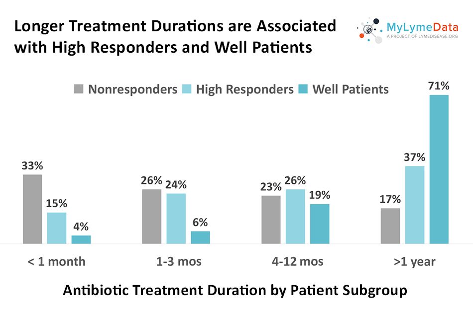
Longer antibiotic treatment durations were associated with better treatment response — with most high responders and well patients reporting treatment durations of four or more months and many reported durations exceeding a year.
Our recently published peer-reviewed study of over 2,000 participants from MyLymeData begins to answer those questions.
How do patients respond to treatment?
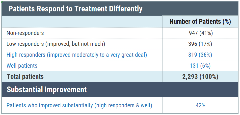
We looked at patients with chronic Lyme disease—those who remained ill for six or more months following treatment with antibiotics for Lyme disease (Shor 2019). The first thing we did was identify different patients as well, high responders, low responders, or non-responders. Well patients responded positively to a survey question asking if they were well or remained ill. Those who remained ill were asked whether their condition had changed as a result of treatment. Those who said they were unchanged or worse (41%) were categorized as non-responders. Patients who said that they were better or worse following treatment, were asked how much better or worse. Those who had improved substantially (moderate to a very great deal of improvement) were deemed high responders.
You can see the break-down of patients in the table below:
59% of patients had improved with treatment and 42% were either well or high responders. The focus of our study was on this latter group. You might wonder whether 42% response is considered good compared to other drugs. (I know I did.) Here’s what the prior head of GlaxoSmithKline said about treatment effectiveness rates of drugs in general (Connor, 2003).
“The vast majority of drugs – more than 90 per cent – only work in 30 or 50 per cent of the people. Drugs out there on the market work, but they don’t work in everybody.” — Dr. Allen Roses, GlaxoSmithKline
So you can see that a 42% rate of substantial improvement is within the range of most drugs on the market.
Compared to the general population and patients with other chronic diseases, chronic Lyme patients report a substantially lower quality of life and a significant symptom disease burden. In this study, high responders and well patients reported substantially better quality of life, a greater percentage of improvement, and reduction in symptom severity. Patients who identified as well reported their quality of life comparable to the US general population.
What factors determine how patients respond to treatment?
To find out what made some patient substantially improve or become well, we turned to our academic partners at the University of California at Los Angeles who specialize in artificial intelligence and machine learning. Their team looked at 215 features related to diagnostic factors, treatment approach, duration of individual antibiotics, alternative treatments, symptoms, type of clinician, and functional impairment to identify the 30 top predictive features for treatment response (Vendrow 2020). Most of the top 30 features identified in their study related to chronic Lyme disease treatment (20), symptom severity (9), and type of clinician treating Lyme disease. We analyzed three of these factors associated with treatment response:
Treatment approach (antibiotics, alternative, both or no treatment)
Treatment durations
Type of clinician overseeing the patients care
What treatment approach did well patients and high responders use?
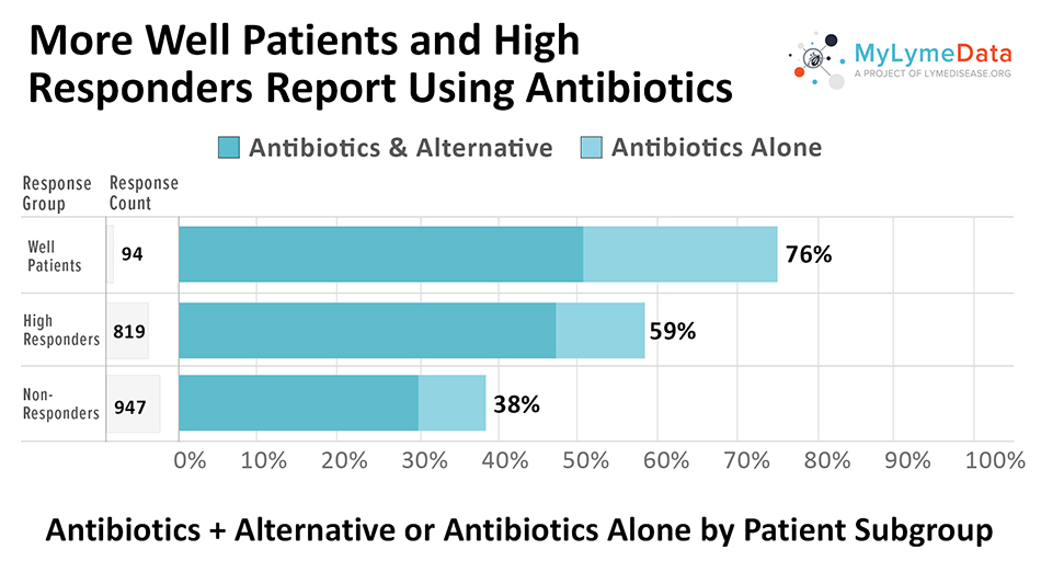
We asked patients what their treatment approach was and listed four options a) antibiotics, b) alternative treatments, c) both antibiotics and alternative treatments, and d) no treatment at all. High treatment response was most closely associated with the use of antibiotics compared to patients who were using alternative treatments alone or forgoing treatment altogether. Treatment with antibiotics for Lyme disease was far higher among well patients (76%) and high responders (59%) compared to non-responders (38%).
As you can see in the chart below, many patients who were taking antibiotics were also taking herbal supplements, which can be antimicrobial (kill bacteria and viruses). So it is possible that there was a synergistic effect between antibiotics and herbal supplements.
Why this is important
The use and duration of antibiotics for chronic Lyme disease treatment is controversial because there is no biomarker that can determine whether the Lyme bacteria has been eradicated in CLD patients. Patients are often told that either chronic Lyme disease does not exist or that it is “incurable.”If this were true, we would not expect more well and substantially improved patients to be taking antibiotics. Instead, we might have expected the percentage of people using antibiotics to be roughly the same among the patient subgroups.
Treatment duration
The optimal chronic Lyme disease treatment is unknown. The four retreatment trials by the National Institute of Health on small samples of patients have produced conflicting results—and most importantly — were all limited to 90 days of treatment (Krupp 2003, Fallon 2008, Klempner 2001, Delong 2012). The Infectious Diseases Society of America (IDSA) and the CDC generally do not recommend treatment for longer than a month (Wormser 2006).
However, a study by the CDC found that patients with Lyme disease generally reported longer treatment durations – with 60% of patients treated for five or more weeks and 36% treated for more than eight weeks (Hook 2015). Unfortunately, that study did not ask patients how long they had been ill. And, the CDC assumed these patients had acute disease – an assumption that may have been wrong. Without looking further, the CDC simply concluded that physicians needed to be better trained to shorten their treatment duration to less than a month.
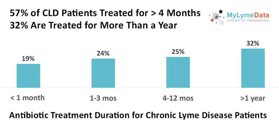
But, as the chart below illustrates, in the study longer chronic Lyme disease treatment durations were common. Most were treated for four or more months (57%), and 32% were treated for more than a year.
More importantly, longer treatment durations were associated with better treatment response—with most high responders and well patients reporting treatment durations of four or more months and many reported durations exceeding a year. As the chart below reveals, those treated for less than a month were unlikely to report improvement.
Why this is important
Insurance companies often deny payment for extended antibiotic care for Lyme disease. Furthermore, many physicians who prescribe long-term antibiotics have been targeted by medical boards. Yet, longer treatments are seen across the board here, with 57% exceeding four months and 32% exceeding one year. This means that these lengths of treatment are common for patients with chronic Lyme disease and reflect clinical practice in the community.
As many as 3 million people have chronic Lyme disease in the US, and nobody knows the best way to treat them,” said Lorraine Johnson, CEO of LymeDisease.org. “The key finding here is that patients who are now well or who report substantial improvement have taken longer courses of antibiotics.”
The fact that more patients who are well or have substantial improvement report longer treatment durations tells us that many patients improve with longer treatment durations. This suggests that the CDC’s conclusion that better clinician training is needed to curb treatment durations is off-base. Instead, it appears that doctors are using their clinical judgement and doing what works for their individual patients. This is the approach recommended by the International Lyme and Associated Diseases Society (ILADS) (Cameron 2014).
The implications of this are profound because they suggest that the legal “standard of care” for patients with chronic Lyme disease is for longer treatment durations. When patients with the most improvement in their health report the longest treatment durations, how can you argue that it should be denied?
Type of clinician overseeing care
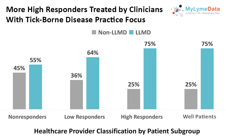
We asked patients to tell us the type of clinician overseeing their care. Choices included: family physicians, internists, rheumatologists, infectious disease specialists, and clinicians whose practice focused on tick-borne diseases (often referred to as Lyme Literate MDs or LLMDs). Very few patients (11%) selected an infectious disease specialist. Seventy-five percent of high responders and well patients report having their care overseen by an LLMD.
Physicians who treat Lyme disease as their primary focus might be expected to have better results than physicians who don’t simply because volume of cases handled means a greater experience level. It is commonly recognized in medicine that volume of cases is associated with better treatment outcomes (Joynt 2013). Just as patients with cancer commonly seek out physicians who specialize in that area, perhaps patients with chronic Lyme disease should also.
Why this is important
Medical boards, insurance companies, and the government often rely on the treatment guidelines of the IDSA on the basis that IDSA clinicians have the most expertise in infectious diseases and, accordingly, know what is best for patients with Lyme disease. However, patients with chronic Lyme disease are generally not treated by infectious disease experts. When short term treatment protocols fail, they find another physician to provide more individualized and longer term care. Many of these clinicians specialize in treating chronic Lyme disease and have developed greater experience and knowledge about what works. It is important that high responders and well patients more often report using LLMDs. This shows that LLMDs are associated with better patient outcomes and their expertise in this area should be more highly valued.
Main take-aways
Let me leave you with these three take-aways from the study. Chronic Lyme disease patients who improve substantially more often:
Use antibiotics as part of their treatment approach
Take antibiotics for more than 4 months and often for more than a year
Rely on LLMDs to oversee their care
A note of caution: None of this should be taken to suggest that patients should blindly take antibiotics for longer durations to get well. These findings are preliminary – observational studies like this do not have control groups and cannot prove cause and effect. What they can do is suggest approaches that may improve patient outcomes. Patients vary considerably in their treatment response. In chronic Lyme disease treatment, one size does not fit all. In fact, when patients who were not taking antibiotics were ask why, about 25% responded that antibiotics had never worked for them. And some responded that antibiotics “no longer” worked for them. But for now, it seems that doctors should be using individualized care based on their diagnosis and the response of this particular patient to treatments.
The other thing this study suggests is that we have a long way to go to determine optimal care for chronic Lyme patients. For example, why do some patients not respond to treatment and which antibiotics are the most effective?
Personal Reflections
Back in 2004, when I was just beginning to get well after being ill for about five years, I co-authored a commentary with Dr. Raphael Stricker about Lyme disease having “two standards of care.” Before becoming ill, I had a robust career as a lawyer. Thus, it was natural that I would address a legal topic as my first opportunity to give back to a community that had made it possible for me to recover. But at that time, I was thinking that the two standards of care related to the divergent guidelines of the IDSA and the International Lyme and Associated Diseases Society.
With this study, it now seems that the two standards of care still exist, but that they reflect how physicians are treating different stages of the disease. People treated under the IDSA guidelines are mainly those with acute disease—the rash, Bell’s palsy, or a flu-like illness after a recent tick bite. When patients remain ill after the short courses of antibiotics offered by the IDSA, they look for clinicians who are more experienced with treating chronic Lyme disease—typically LLMDs.
A few years ago, someone asked me whether it was important to be treated by an LLMD or if they should see someone who did not focus on Lyme disease. And, although I told the person “you know, I think you might do better with an LLMD,” I had no data to back up that suggestion. Now we do.
If you are a patient who is not enrolled in MyLymeData, please enroll today. If you are a researcher who wants to collaborate with us, please contact me directly.
The MyLymeData Viz Blog is written by Lorraine Johnson, JD, MBA, who is the Chief Executive Officer of LymeDisease.org. You can contact her at lbjohnson@lymedisease.org. On Twitter, follow her @lymepolicywonk. If you have not signed up for our patient-centered big data project, MyLymeData, please register now.
About MyLymeData Lyme Disease Research
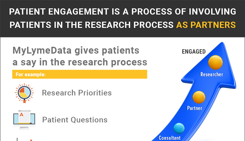
MyLymeData is one of the largest patient-driven registries in the nation, with over 13,500 patients enrolled. It was created by patients, is run by patients and will address the issues that Lyme disease patients care about. MyLymeData Viz provides the community with results from MyLymeData. If you are enrolled in MyLymeData, we thank you for providing the data that will accelerate the pace of research in Lyme disease. If you are not enrolled, please enroll today.
You might be interested in more MyLymeData results:
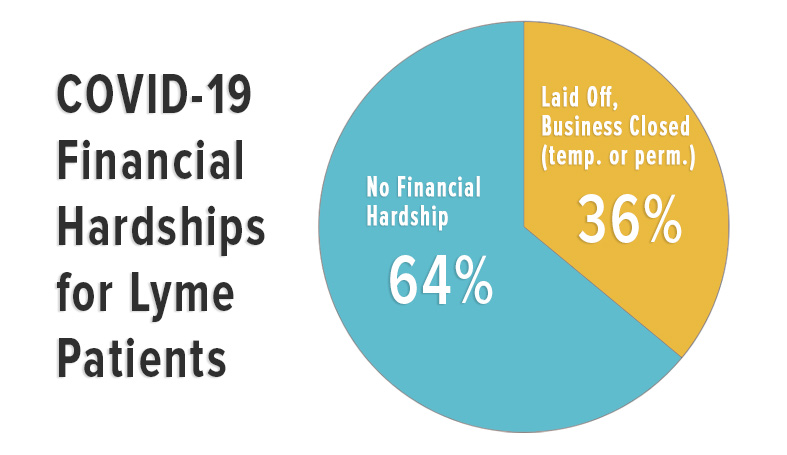
COVID-19 IMPACT ON LYME PATIENTS: SURVEY RESULTS THROUGH SEPTEMBER 10, 2020
LYME DISEASE PREVALENCE: DOES SEX MATTER?
MYLYMEDATA: MY DATA, MY TRUST
______________________
**Comment**
The results of this study are identical to my experience both as a patient and as an advocate working with many patients. When people solicit my advice, I immediately tell them mainstream medicine will not treat this appropriately. Period. That’s not to say it can’t be used for other side-issues that arise, but for any hope of recovering from tick-borne illness, they should get to an ILADS trained doctor with experience.
It’s truly saddening that polarization continues unabated with this complex illness, but that is the true state of things. I’ve watched similar issues arise with the COVID-19 debacle. Due to severe conflicts of interest our public ‘authorities’ are allowed to have, they completely malign any test or treatment they don’t control and obtain profits from:







MyLymeData is one of the largest patient-driven registries in the nation, with over 13,500 patients enrolled. It was created by patients, is run by patients and will address the issues that Lyme disease patients care about. MyLymeData Viz provides the community with results from MyLymeData. If you are enrolled in MyLymeData, we thank you for providing the data that will accelerate the pace of research in Lyme disease. If you are not enrolled, please enroll today.