Archive for the ‘Ticks’ Category

Connection Between ALS & Lyme?

http://www.digitaljournal.com/life/health/is-there-a-connection-between-als-and-lyme-disease/article/527874#ixzz5MNIwrXs6

Is there a connection between ALS and Lyme Disease?

Listen (Audio here)
By Tim Sandle     Jul 25, 2018 in Health
In the U.S. cases of Lyme Disease appear to be rising. Some researchers have drawn a link between the tick-transmitted bacterial infection and the neurodegenerative condition ALS. Jo Ann Simon explains more.
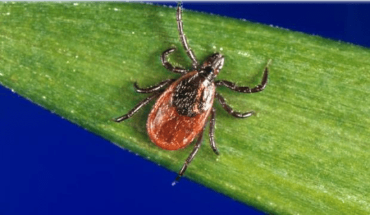
Screenshot_2018-09-21 Deer tick - Image - Digital Journal
Black legged Deer Tick – Jim Gathnany/CDC
According to the Center for Disease Control and Prevention, 376,000 cases of reported Lyme disease occur in the U.S. Lyme disease is a bacterial illness that can cause serious neurological problems. First discovered in the 1970s, Lyme disease draws its name from the Connecticut area, including the towns of Lyme and Old Lyme (see the Digital Journal article “Discoverer of Lyme disease dies”).

Lyme disease is difficult to detect until the symptoms, which arise in a person following the transfer of the pathogenic bacteria into the human blood stream as the result of a tick bite, appear.

The condition of amyotrophic lateral sclerosis (ALS), also known as motor neurone disease, received considerable attention a couple of years ago through the awareness campaign ‘Ice Bucket Challenge’. There are 20,000 people living with ALS at a given time, with 6,000 more being diagnosed in the U.S. every year.

Jo Ann Simon has been examining the connection between ALS and Lyme Disease based on her medical experiences and relevant statistics.

According to Simon,

“ALS, or amyotrophic lateral sclerosis, is a progressive neurodegenerative disease that affects nerve cells in the brain and the spinal cord. A-myo-trophic comes from the Greek language. “A” means no. “Myo” refers to muscle, and “Trophic” means nourishment – “No muscle nourishment.”

She explains further:
“When a muscle has no nourishment, it “atrophies” or wastes away. “Lateral” identifies the areas in a person’s spinal cord where portions of the nerve cells that signal and control the muscles are located. As this area degenerates, it leads to scarring or hardening (“sclerosis”) in the region.”
Digital Journal: Is there a connection between Lyme Disease and ALS?

Jo Ann Simon: The culprit can be the spirochete borrelia burgodorferi bacteria of Lyme Disease or the unnamed foreign invader of ALS in the brain that triggers motor neuron disease, or are they the same?”

Our research, testing and discovery brought us through a maze of doctors, hospitals, treatments and various results. We celebrated when we thought it might be MMN Multi Focal Motor Neurothopy or Guilliane Barre, both treatable motor neuron diseases, but further testing eliminated that glimmer of hope.

DJ: What are the research highlights?

Simon: There are five little known facts about ALS and Lyme Disease, which research draws out. First, ALS and Lyme Disease have common ground with the auto immune and the central nervous systems. Second, common symptoms range from fatigue, numbness, muscle weakness and twitches, speech impairment, and cramping.

Third, recent studies that show that a significant percentage of ALS diagnosed patients test positive for Lyme Disease. Fourth, in some cases, patients diagnosed with ALS actually had Lyme Disease instead. And fifth, Lou Gehrig, the namesake of ALS lived very close to Lyme, Connecticut, where the disease was born.

DJ: What can people do to reduce the chance of infection?

Simon: The most important take away from this experience is that everyone needs to protect themselves, their family, friends and pets from ticks. Prevention of a tick bite could save your life! Prevention is the best medicine.

Also, use bug spray that has DEET (Off or Repel products) or Picaridin (Sawyer, Fisherman, Skin So Soft products). These are effective to deter ticks and can be found online, or at your local pharmacy, or department store.

If you are walking in grassy wooded areas, tuck in your pants to socks and wear long sleeve shirts so that your skin is not exposed. They might still jump on you for a ride, but you can eliminate them by running your clothes in a hot dryer for 10 minutes so they turn into harmless toast.

DJ: Is there anything else?

Simon: Yes, you can treat your shoes and clothes with Permithrin, a synthetic pesticide that repels ticks from 5 to 70 washes, depending on the product. Insect Shield in North Carolina will treat your clothes for up to 70 washes, or you can treat yourself with different products such as Sawyer insect repellent which can be purchased on line or at your local pharmacy, grocery or department store. L.L. Bean and Cabela’s sell pretreated clothes and camping gear.

Protect your pet. If you stopped your pet’s tick preventive over the winter, get it started again now. Outdoor dogs and cats will likely be the first family members to find a tick and bring it home to you. There are two types of products to use. Products that kill ticks on contact – quick tick gone or kill ticks after their lunch – bite to die. Talk to your vet to decide the best product for your pet.

Also, get professional treatment for your property to eliminate the threat of ticks in your outside living areas. This does not stop the threat elsewhere, but at least you can sleep at night not worrying about the ticks on your doorstep. Do a tick check every day. This is especially important for your children and pets since they normally spend the most time outside.

___________________

For More:  https://madisonarealymesupportgroup.com/2017/10/14/lost-link-als-lyme/    She found that Lyme was likely the cause of her deterioration in health, got treated for it and stopped the progression of the ‘ALS’. She’s still alive now, although hardly after the damage the original documentary had done to her care plan.

http://www.caregivershome.com/news/article.cfm?UID=1151after years of medical observations that some people with amyotrophic lateral sclerosis — better known as Lou Gehrig’s disease, or ALS — also suffer from a form of dementia. Conversely, some patients with dementia also have been observed developing crippling symptoms similar to those in ALS, where patients gradually lose control of their muscles.  This latest common protein discovery in a way adds another link in the chain of research into the major neurodegenerative diseases. Now, a faulty protein has been uncovered in Alzheimer’s, Parkinson’s and Huntington’s diseases, among others. Each of these diseases remains incurable, but scientists believe discoveries such as this represent a major step forward in finding a cure.

A relationship between ALS / MND (motor neuron disease) and Lyme makes sense, looking at the findings of the 1990 research that was published in the article ‘Immunological Reactivity against in Borrelia burgdorferi in Patients with Motor Neuron Disease’ by Halperin et al.  This study showed that in almost 50% of the 19 people diagnosed with ALS, Lyme was the cause. Once treated, several of these patients improved. In that same year, 1990, the CDC published its first definition about Lyme and described the complex, systemic, multi-symptom and sometimes devastating chronic disease experienced by many Lyme patients – then and still today.

Did anyone ever do a follow-up on this promising research? No. It was simply hidden away and Halperin chose to become a co-author of the 2006 IDSA Lyme Guidelines instead, which maintain that ‘Lyme is a mild disease that is hard to get, easy to treat and hardly ever becomes a chronic condition’. Any possible connection with ALS or any other of the serious and previously acknowledged debilitating or even deadly conditions was no longer mentioned. Any long-term health issues are reasoned away, using semantics rather than ‘evidence based’ science.

https://madisonarealymesupportgroup.com/2017/05/11/dr-al-miller-lyme-disease-series/  Watch parts 1 & 4 to see how Lyme can masquerade as other neurodegenerative illnesses.

And lastly, if you want to see first-hand a doctor (Dr. Martz) diagnosed with ALS who got his life back after Lyme was discovered & treated, watch the excellent documentary, “Under Our Skin.”  Story here:  https://www.lymedisease.org/372/

You can also read about Dr. Martz in “Cure Unknown: Inside the Lyme Epidemic,” by Pamela Weintraub.

Tick Infestations of Wildlife & Companion Animals in Ontario, Canada, With Detection of Human Pathogens in Ixodes Scapularis Ticks

https://www.ncbi.nlm.nih.gov/m/pubmed/30206012/

Tick infestations of wildlife and companion animals in Ontario, Canada, with detection of human pathogens in Ixodes scapularis ticks.

Smith KA, et al. Ticks Tick Borne Dis. 2018.

Abstract

The growing risk of transmission of tick-borne zoonotic pathogens to humans in Ontario, Canada, warrants investigations into regional tick distribution, tick burdens of local peridomestic animals, and prevalence of tick-borne pathogens. The objectives of this study were to investigate the geographic distribution and magnitude of tick infestations in opportunistically sampled mammalian wildlife and companion animals (i.e., dogs) in southern Ontario and to test these ticks for evidence of zoonotic tick-borne pathogens. Ticks collected from wildlife carcasses, live-trapped wildlife and companion animals (2015-2016), as well as wildlife diagnostic cases (2011-2013), were identified to species and life stage.

Ixodes scapularis ticks were tested by real-time PCR for Anaplasma phagocytophilum, Babesia microti, Borrelia miyamotoi and Borrelia burgdorferi sensu stricto (s.s.). Amblyomma americanum ticks were tested for Ehrlichia chaffeensis. A total of 1687 ticks of six species were collected from 334 animals, including 224 raccoons (n = 1381 ticks) and 50 dogs (n = 67 ticks).

The most common tick species collected from parasitized raccoons were Ixodes texanus (n = 666 ticks) and Dermacentor variabilis (n = 600 ticks), which were removed from 58.5% (median: 2 ticks; range: 1-36) and 49.1% (median: 2 ticks; range: 1-64) of raccoons, respectively. Of I. scapularis tested, 9.3% (4/43) were positive for Bo. burgdorferi s.s. and 2.3% (1/43) for A. phagocytophilum. These results reveal that numerous tick species parasitize common, peridomestic wildlife and that at least two zoonotic, tick-borne pathogens circulate in southern Ontario. Host-tick vector-pathogen dynamics should continue to be monitored in the face of global climate change, landscape alterations and expanding human populations.

________________

**Comment**

This researcher obviously hasn’t read his own countryman’s work:  https://madisonarealymesupportgroup.com/2018/08/13/study-shows-lyme-not-propelled-by-climate-change/   Another problem with the climate change models is they overlook the fact that deer ticks were established in northwestern Ontario, southern Manitoba and were already in central Canada prior to 1970. What they predict to happen in the future has already happened in Canada. Their oversight caused a skewed rate of tick expansion and a miscalculation of northward projected movement.

“For blacklegged ticks, climate change is an apocryphal issue.” -John Scott

apocryphal:
adj. Of questionable authorship or authenticity
adj. Erroneous; fictitious

Human Bartonellosis: Perspectives of a Veterinary Internist

https://dta0yqvfnusiq.cloudfront.net/galax57722929/2018/03/Human-Bartonellosis-5ab03952a5057.pdf

HUMAN BARTONELLOSIS:  PERSPECTIVES OF A VETERINARY INTERNIST

Edward B. Breitschwerdt, DVM, DACVIM
Chief Scientific Officer, Galaxy Diagnostics, Inc.
Professor, Internal Medicine, NCSU, Raleigh, NC
Adjunct Professor of Medicine, Duke University Medical Center
INTRODUCTION
Bartonella species are fastidious gram-negative bacteria that are highly adapted to a mammalian reservoir host and within which the bacteria usually cause a long-lasting intraerythrocytic bacteremia.  1-3  These facts are of particular importance to veterinarians and physicians, as an increasing number of animal reservoir hosts have been identified for various Bartonella species. Among numerous other examples, Bartonella henselae has co-evolved with cats, Bartonella vinsonii subsp. berkhoffii has co-evolved with dogs and wild canines, and Bartonella bovid has co-evolved with cattle.  1-2  Importantly, the list of reservoir-adapted Bartonella species, including a large number of rodent species that might serve as “pocket pets,” continues to grow exponentially, as new Bartonella spp. are discovered. Prior to 1990, there were only two named Bartonella species, whereas there are now at least 24 named and numerous unnamed or candidatus species, based upon deposited Gen Bank sequences or preliminary reports, respectively, seventeen Bartonella spp. including B.alsatica, B. bacilliformis, B. clarridgeiae, B. doshiae, B. elizabethae, B. grahamii, B. henselae (Houston 1 and San Antonio 2 strains), B. koehlerae, B. melophagi, B. quintana, B. rochalimaea, B. tamiae, B. vinsonii subsp. berkhoffii (Genotypes I, II and III), and B. washoensis have been associated with an expanding spectrum of human diseases.
Epidemiological evidence and experimental flea transmission studies support an important role for fleas in the transmission of B. henselae, B. clarridgeae and most likely B. koehlerae among cats. 1  Three other Bartonella species, B. bovid, B. quintana and B. vinsonii subsp. berkhoffii have been isolated from cat blood, but the modes of transmission and the reservoir potential of these species in felids has not been definitively established. Recently, we isolated Bartonella vinsonii subsp. berkhoffii from a cat with recurrent osteomyelitis spanning an eighteen month time period. 2  Thus, cats can maintain a chronic bacteremia with at least six Bartonella spp., of which five are known zoonotic pathogens.  1-3 In addition to fleas, an increasing number of arthropod vectors, including biting flies, keds, lice, sandflys and ticks have been implicated in the transmission of Bartonella species. Although there is clinical and epidemiological evidence to support tick transmission of B. vinsonii subspecies berkhoffii to dogs and coyotes, the mode of transmission of this Bartonella subsp. to cats and dogs has not been determined. Recent evidence supports tick transmission of B. henselae by Ixodes scapularis and Ixodes ricinus. Considering the diversity of Bartonella species and subspecies, the large number of reservoir hosts and the spectrum of arthropod vectors, the clinical and diagnostic challenges posed by Bartonella transmission in nature may be much more complex than is currently appreciated in human and veterinary medicine.
In the natural reservoir host, such as a cat or rodent, chronic bacteremia with a Bartonella species can frequently be detected by blood cultre or PCR in outwardly healthy individuals.  1-3  In contrast, the diagnostic detection of a Bartonella spp. in a non-reservoir adapted host, such as a dog, horse or human patient, can be extremely difficult. Most, although not all diseases caused by Bartonella spp. occur in accidental hosts and these organisms are being increasingly implicated as a cause of zoonotic infections.  4-8  It is important to recognize that strains of a Bartonella sp. vary in their virulence. Therefore, highly pathogenic strains of B. henselae, for which the cat is the primary reservoir, can induce granulomatous myocarditis in cats, presumably following flea transmission.  Until recently, mechanisms that facilitate persistent Bartonella bacteremia in mammals were not well understood. Recent reports have identified an intra-endothelial, as well as intra-erythrocytic localization for these bacteria, which represents a unique strategy for bacterial persistence. Non-hemolytic intracellular colonization of erythrocytes in conjunction with the ability to invade and replicate within endothelial cells would preserve the organisms for efficient vector transmission, protect Bartonella from the host immune response, and potentially contribute to decreased antimicrobial efficacy. Although the clinical implications are not understood, other in vitro studies indicate that Bartonella spp. can infect dendritic cells, microglial cells, monocytes and CD34+ bone marrow progenitor cells.
CAT SCRATCH DISEASE
For over a century regional lymphadenopathy has been associated with animal contact, particularly cat scratches. Over the years, numerous microorganisms were implicated as the cause of CSD. In 1992, Regnery and colleagues at the Centers for Disease Control, identified seroreactivity to B. henselae antigens in 88% of 41 human patients with suspected CSD compared to 3% of controls.  Subsequently, additional support for B. henselae as the predominant cause of CSD was provided when Bartonella DNA was amplified from lymph node samples of 21 of 25 (84%) patients with suspected
CSD, using a polymerase chain reaction assay. A similar study from Sweden identified B. henselae DNA, but failed to identify A. felis DNA, in a large number of patients with suspected CSD. Prior to the
recognition of B. henselae as the cause of CSD, Afipia felis, named for the Armed Forces Institute of Pathology, was considered the sole cause of CSD. Subsequently, we blood cultured B. henselae or B. clarridgeae
from 17 of 19 cats owned by 14 patients with CSD, which indicated that bacteremia is a frequent occurrence in cats that transmit B. henselae
to a human being. 1-2
Historically, atypical manifestations of CSD have included tonsillitis, encephalitis, cerebral arteritis, transverse myelitis, granulomatous hepatitis and/or splenitis, osteolysis, pneumonia, pleural effusion, and thrombocytopenic purpura. With the advent of specific diagnostic techniques, (culture, serology, and PCR), there has been a dramatic increase in reports describing human patients with “atypical” manifestations of CSD. Osteomyelitis, granulomatous hepatitis and granulomatous splenitis have been increasingly recognized in children infected with B. henselae, who frequently lack the classical lymphadenopathy of CSD. Previously, Bartonella infection would not have been considered a likely differential diagnosis by the physician in patients lacking a history of lymphadenopathy or animal contact. As evidenced by reports in the past four years, the spectrum of human disease associated with the genus Bartonella continues to expand, requiring periodic reassessment as new information becomes available. On a comparative medical (“One Health”) basis, our research group has documented many of the same CSD atypical manifestations in cats or dogs, including encephalitis, transverse myelitis, granulomatous hepatitis, osteolysis, pleural effusion, and thrombocytopenic purpura. In this context, a highly prevalent, naturally-occurring human disease (CSD) can be used as a “model” to determine the potential behavior of these bacteria in companion animal patients.
Because cat scratch disease generally denotes a self-limiting illness characterized by fever and lymphadenopathy and because the recognized spectrum of human disease manifestations associated with Bartonella infections (which may not include fever or lymphadenopathy) has expanded considerably in recent years, it is becoming obvious that the designation CSD lacks clinical, microbiologic and zoonotic utility. Although cats are a major reservoir for B. henselae, B. clarridgeiae, and B. koehlerae, some patients deny the possibility of a cat scratch or bite wound, or indicate no contact with cats. Transmission from environmental sources, various arthropod vectors, perinatally or by other animal hosts is probable and the more inclusive term bartonellosis may facilitate enhanced future understanding of diseases caused by members of the genus Bartonella. As physicians have been taught that CSD is self-limiting, there is an ongoing lack of appreciation that B. henselae can cause chronic, asymptomatic or intermittently symptomatic illness, accompanied by persistent bacteremia in people. In this context, the documentation of chronic, relapsing bacteremia in cats, dogs and other animal species provides a “model” for better understanding human bartonellosis.
BARTONELLA ENDOCARDITIS
Endocarditis can be induced by a spectrum of Bartonella species in dogs and human patients and is the best example of documented disease causation for this genus. Historically, Bartonella species have been a cause of culture-negative endocarditis in people and dogs because the diagnostic methods used by microbiology laboratories were not adequate to isolate these bacteria. Now, by using
specialized techniques, a spectrum of Bartonella species have been identified in research and diagnostic laboratories in different parts of the world—in heart valves or in blood cultures from dogs
and people with endocarditis. 3  It is important for physicians and veterinarians to recognize that some of these Bartonella species are found in the blood of cats, dogs, rats, ground squirrels, and rabbits.
ISOLATION AND MOLECULAR DETECTION OF BARTONELLA SPECIES
Because conventional microbiological techniques lack sensitivity, bartonellosis is usually diagnosed by PCR amplification of organism specific DNA sequences and/or through serological testing. Recently, the development of a more sensitive isolation approach, using BAPGM (Bartonella alpha Proteobacteria growth medium) followed by PCR has greatly facilitated the molecular detection or isolation of Bartonella species from the blood of sick or healthy animals, including cats, dogs, horses and human beings. Most importantly, the use of this enrichment growth medium prior to PCR testing has allowed our research group to confirm that immunocompetent human patients, in particular veterinarians and veterinary technicians, can have chronic intravascular infections with Bartonella spp. 4-5 Information relative to this EnrichmentPCRTM testing platform for animal and human patients is available at www.galaxydx.com.
It is increasingly clear that no single diagnostic strategy will confirm infection with a Bartonella sp. in the immunocompetent patient population.  As described in studies from our NCSU laboratory, B. henselae, B. koehlerae and B. vinsonii subsp berkhoffii seroreactivity was found in only 58.6% of the patients in which Bartonella spp. infection was confirmed by EnrichmentPCR TM and sequencing. Therefore, Bartonella serology lacks sensitivity and can only be used to implicate prior exposure to a Bartonella sp. Even when serum from cat scratch disease patients, which is caused by B. henselae, is used in various diagnostic laboratories for IFA testing, test sensitivities have ranged from 14 to 100%.
EVOLVING IMPLICATIONS OF CHRONIC BARTONELLA SPP. BACTEREMIA IN IMMUNOCOMPETENT PEOPLE
Previously, we described B. quintana bacteremia in a woman who was tested following the development of an infected cat bite lesion involving the hand. 6  Two months later, the feral cat that had
induced the bite wound was captured and was also shown to be B. quintana bacteremic. In a cumulative study involving 392 patients with occupational animal contact or extensive arthropod exposure 31.9% were bacteremic with one or more Bartonella spp., when blood, serum and BAPGM enrichment culture PCR results were combined. Although this high prevalence of bacteremia is biased by testing at risk, sick individuals, it clearly demonstrates that intravascular infection with Bartonella sp. is much more common in immunocompetent patients, than was previously suspected. By IFA testing, only 75 out of 128 (58.6%) PCR positive patients were seroreactive to a panel consisting of five Bartonella sp. test antigens.
In a recent study, Bartonella vinsonii subsp. berkhoffii, Bartonella henselae or DNA of both organisms were amplified and sequenced from blood, BAPGM enrichment blood cultures or autopsy tissues from four family members. 7  Historical and microbiological results derived from this family support human perinatal transmission of Bartonella species. To date, there have been a limited number of studies that address the potential impact of intravascular infection with a Bartonella sp. on reproductive performance, however, studies involving experimentally-infected cats, rodents and naturally-infected cows with various Bartonella sp. have identified decreased reproductive performance involving both males and females. The parents of these children had attempted to conceive children for several years prior to resorting to in vitro fertilization.
We have also described a veterinarian, who experienced a needle stick while obtaining a fine needle aspiration sample from a cutaneous histiocytic neoplasm. 8  Subsequently symptoms, including headaches, fatigue and intermittent paresthesias (numbness) developed. This patient seroconverted to B. vinsonii subsp. berkhoffii genotypes I and III and B. vinsonii subsp. berkhoffii genotype I DNA was amplified and sequenced from sequentially obtained blood samples, whereas genotype III DNA was amplified from the cytological specimen. All symptoms resolved following antibiotic treatment.
It is increasingly evident that dogs can serve as a source for human infection with B. vinsonii subsp. berkhoffii. Bartonella vinsonii subsp. berkhoffii genotype II was amplified and sequenced from
a liver biopsy from a patient with epithelioid hemangioendothelioma (soft tissue tumor considered a vascular cancer), after which the organism was isolated by BAPGM blood culture. 9  The unique capability of Bartonella to invade and induce long lasting intraerythrocytic and intraendothelial infections, in conjunction with the ability of at least three Bartonella spp. (Bh, Bq, and B. bacilliformi) to induce VEGF-mediated vasoproliferative disease in immunocompromised or immunocompetent individuals suggests that these novel emerging bacterial pathogens might contribute to the development of vascular tumors.
Bartonella koehlerae bacteremia was documented in eight immunocompetent patients by PCR amplification and DNA sequencing, either prior to or after BAPGM enrichment blood culture.10  Presenting symptoms most often included fatigue, insomnia, joint pain, headache, memory loss, and muscle pain. Four patients were also infected with Bartonella vinsonii subsp. berkhoffii genotype II. Bartonella koehlerae antibodies were not detected (titers<1:16) in 30 healthy human control sera, whereas five of eight patient samples had B. koehlerae antibody titers of 1:64 or greater. Studies are needed to determine if B. koehlerae is a cause or cofactor in the development of arthritis, peripheral neuropathies or tachyarrhythmias in human patients. Co-infection with B. henselae and two hemotropic Mycoplasma variants resembling Mycoplasma obis were also found in the blood of a veterinarian with a historical diagnosis of multiple sclerosis. 11
PUBLIC AND OCCUPATIONAL HEALTH CONSIDERATIONS
Due to extensive contact with a spectrum of animal species, veterinary professionals appear to have an occupational risk of infection because of frequent exposure to Bartonella spp., therefore these individuals should exercise increased precautions to avoid arthropod bites, arthropod feces (i.e. fleas and lice), animal bites or scratches and direct contact with bodily fluids from sick animals. As Bartonella spp. have been isolated from cat, dog or human blood, cerebrospinal fluid, joint fluid,aqueous fluid, seroma fluid and from pleural, pericardial and abdominal effusions, a substantial number of diagnostic biological samples collected on a daily basis in veterinary practices could contain viable bacteria.
The increasing number of defined Bartonella spp., in conjunction with the high level of bacteremia found in reservoir adapted hosts, which represent the veterinary patient population, ensures that all veterinary professionals will experience frequent and repeated exposure to animals harboring these bacteria. Therefore, personal protective equipment, frequent hand washing and avoiding cuts and needle sticks have become more important as our knowledge of this genus has improved and various modes of transmission have been defined.
Physicians should be educated as to the large number of Bartonella spp. in nature, the extensive spectrum of animal reservoir hosts, the diversity of confirmed and potential arthropod vectors, current limitations associated with diagnosis and treatment efficacy, and the ecological and evolving medical complexity of these highly evolved intravascular, endotheliotropic bacteria.
REFERENCES
1  Chomel BB, et al. Vet Res 2009;40:29.
2  Breitschwerdt EB, et al. J Vet Emerg Crit Care 2010; 20:8.
3  Chomel BB, et al. Ann N Y Acad Sci 2009;1166:120.
4  Breitschwerdt EB, et al. J Clin Microbiol 2008;46:2856.
5  Breitschwerdt EB, et al. Parasit Vectors 2010;3:29.
6  Breitschwerdt EB, et al. J Clin Microbiol 2007;45:270.
7  Breitschwerdt EB, et al. J Clin Microbiol 2010;48:2289.
8  Oliveira AM et al. J Vet Intern Med 2010;24:1229.
9  Breitschwerdt EB, et al. J Clin Microbio 2009;47:1957.
10 Breitschwerdt EB, et al. Parasit Vectors 2010;3:76.
11 Sykes JE, et al. J Clin Microbiol 2010;48:3782.
____________________

Glandular Tularemia

https://www.nejm.org/doi/full/10.1056/NEJMicm1801531

Glandular Tularemia

  • Laura Marks, M.D., Ph.D.,
  • and Andrej Spec, M.D.
nejmicm1801531_f1

A 68-year-old man from Missouri presented to the primary care clinic with a history of 1 week of fever followed by 2 months of progressive, painful swelling on the right side of his neck. Approximately 2 days before the onset of the patient’s symptoms, his outdoor cat died from a subacute illness; a veterinarian had diagnosed feline leukemia without laboratory testing, and the cat had been treated with prednisone, which the patient administered. The patient’s physical examination revealed three erythematous, tender lymph nodes. The remainder of the physical examination was normal. Serologic testing with IgM antibody was positive for Francisella tularensis (titer, 1:1280). A diagnosis of glandular tularemia was made. Glandular tularemia is the second most common manifestation of tularemia after the ulceroglandular form. Because culture requires biosafety level 3 conditions, diagnosis is often confirmed serologically. Domestic cats can become infected through the consumption of infected prey and can transmit the bacteria to humans. The patient was treated with doxycycline for 4 weeks; the lesions improved within 5 days and resolved within 3 weeks.


Laura Marks, M.D., Ph.D.
Barnes–Jewish Hospital, St. Louis, MO

Andrej Spec, M.D.
Washington University in St. Louis, St. Louis, MO

___________________


**Comment**

I remember hearing Timothy Lepore, MD, FACS, surgeon at Nantucket Cottage Hospital, at a Lyme conference.  He explained that Tularemia is also a disease of those who work with the land such as landscapers and farmers, as well as those who get bit by a tick. There are cases reported in every state but Hawaii, and many other wild and domestic animals can be infected. The highest rates of infection are in Arkansas.  Please see this link for more details but know that this is a bioweaponized pathogen:  https://madisonarealymesupportgroup.com/2016/10/25/of-rabbits-and-men/  The WHO estimates that an aerosol dispersal of 50 kg of F. tularensis over an area with 5 million people would result in 25,000 incapacitating casualties including 19,000 deaths.

For hunters:  https://madisonarealymesupportgroup.com/2018/08/07/tularemia-hunting-dogs-as-possible-vectors/  “The frequency of about seven percent shows that hunting dogs can also become infected regularly. As vectors of the disease, even without symptoms, the animals must also be considered unexpected carriers,” Posautz adds.

Study Shows Tick Infection & Transmission Potential for Both DTV & WNV

https://www.liebertpub.com/doi/abs/10.1089/vbz.2017.2224#utm_source=ETOC&utm_medium=email&utm_campaign=vbz

Generation of a Lineage II Powassan Virus (Deer Tick Virus) cDNA Clone: Assessment of Flaviviral Genetic Determinants of Tick and Mosquito Vector Competence

Kenney Joan L. , Anishchenko Michael , Hermance Meghan , Romo Hannah , Chen Ching-I , Thangamani Saravanan , and Brault Aaron C.
Published Online:1 Jul 2018https://doi.org/10.1089/vbz.2017.2224

Abstract

The Flavivirus genus comprises a diverse group of viruses that utilize a wide range of vertebrate hosts and arthropod vectors. The genus includes viruses that are transmitted solely by mosquitoes or vertebrate hosts as well as viruses that alternate transmission between mosquitoes or ticks and vertebrates. Nevertheless, the viral genetic determinants that dictate these unique flaviviral host and vector specificities have been poorly characterized. In this report, a cDNA clone of a flavivirus that is transmitted between ticks and vertebrates (Powassan lineage II, deer tick virus [DTV]) was generated and chimeric viruses between the mosquito/vertebrate flavivirus, West Nile virus (WNV), were constructed. These chimeric viruses expressed the prM and E genes of either WNV or DTV in the heterologous (from one species to another) nonstructural (NS) backbone. Recombinant chimeric viruses rescued from cDNAs were characterized for their capacity to grow in vertebrate and arthropod (mosquito and tick) cells as well as for in vivo vector competence in mosquitoes and ticks.

Results demonstrated that the NS elements were insufficient to impart the complete mosquito or tick growth phenotypes of parental viruses; however, these NS genetic elements did contribute to a 100- and 100,000-fold increase in viral growth in vitro in tick and mosquito cells, respectively. Mosquito competence was observed only with parental WNV, while infection and transmission potential by ticks were observed with both DTV and WNV-prME/DTV chimeric viruses. These data indicate that NS genetic elements play a significant, but not exclusive, role for vector usage of mosquito- and tick-borne flaviviruses.

________________

**Comment**

I’m no microbiologist and without the full article and better understanding of what this NS backbone is, 

The study shows 4 things:

  1.  The NS elements gave a 100 fold “test tube” increase in viral growth in tick cells.  These organisms are extremely fastidious and difficult to study in a lab.  It’s even tougher to figure out how this plays out in the human body.
  2. INFECTION & TRANSMISSION potential by ticks was observed with both DTV and WNV.  Read that sentence again.
  3. Why didn’t this make the news?
  4. Mosquitoes are nasty but ticks are a whole other monster.  Mosquito research gets all the money.  Why?

http://www.dutchessny.gov/CountyGov/Departments/Legislature/2017Auerbach.pdf  This pdf by Lyme Advocate Jill Auerbach shows that while there were only 5,700 cases of WNV in 2012, research dollars were $29 million, whereas, Lyme cases in 2012 were 312,000 but received only $25 million.  While the number of the infected continue to soar the research dollars for Lyme are radically reduced in successive years:

Disease New Cases (annual) NIH Funding
 

Hepatitis C 2012


1,300


$112 million

West Nile Virus 2012

5,700

$29 million

HIV/AIDS 2012

56,000

$3 billion (11% total NIH budget)

Influenza 2012

73,000

$251 million

Lyme disease 2012

312,000

$25 million

Lyme disease 2013

363,070

$20 million

*Lyme disease 2004             198,040                                  $34.4 million

Disease

      New Cases 2015

CDC funding 2016

Lyme Disease

           380,690  (10 x 38,069)
2016 numbers not yet available

 $10 million

This does NOT include other Tick-borne diseases

Houston, we have a problem.