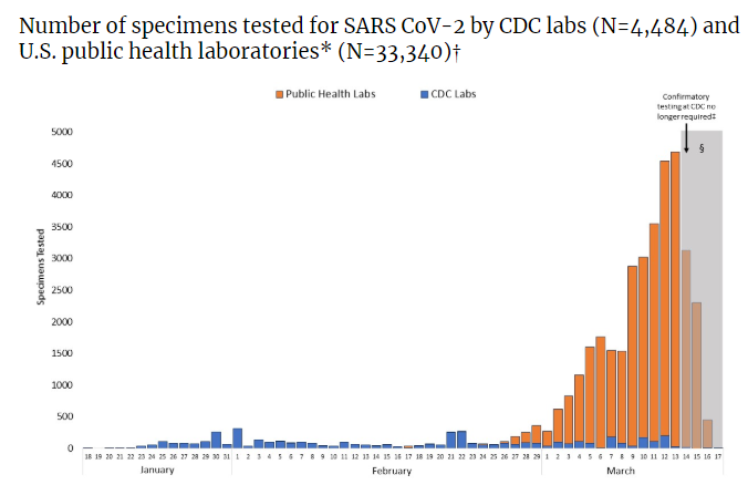
CDC’s Deadly Testing Fiasco: Centralization of Public Health Authority a Threat to National Security
The following article is a “Must Read” as it points out what the Lyme community knows all too well: The CDC can not be trusted. History shows it has severe conflicts of interests by owning licenses and patents that directly interfere with testing and diagnosis of numerous diseases – including COVID-19 & Lyme.
https://www.linkedin.com/pulse/cdcs-deadly-testing-fiasco-centralization-public-james-lyons-weiler/
CDC’s Deadly Testing Fiasco: Centralization of Public Health Authority a Threat to National Security
This is a companian article to augment the first of six Unbreaking Science episodes dedicated to insuring that those who are responsible for the failures of a rapid and effective response to the COVID-19 outbreak are held responsible. The article and the episode are complimentary – that is they overlap in content, but each has some unique information not found in the other. I recommend the video first, followed by a read of the article.
By the end of this first article and episode, the public will understand that
- WHO Offered the US a Perfectly Good, Validated Test
- CDC Refused, Wanting “Their Own”
- This is Not the First Time CDC Wanted to Corner the Testing Market
- HHS Whistleblower Chris Meekin Says CDC Lied to President Trump About the CDC’s Testing Abilities
- Due to Capacity Alone, CDC is Incapable of Mounting an Effective Response to Outbreaks (Obvious National Security Issue)
- Delay in Accurate Testing Could Help CDC – But It May Have Cost Us Everything
WHEN THE WORLD HEALTH ORGANIZATION offered the CDC a test – made by Germany – that had already been tested, one has to wonder who held the conversation within CDC leading to the disastrous conclusion that CDC should make “their own” test. One also has to wonder what factors went into play and which were considered. Most importantly, one has to wonder how much more time the US and its medical infrastructure would have had if patients were not released from the Princess Cruise ship and unknown numbers of other places if they had not tested negative due to CDC’s faulty test.
Whoever made those fateful decisions made them according to the status quo of the CDC – they prefer that which leads to massive amounts of positive revenue, regardless of the effect of that decision on safety.
The prevention paradigm at CDC displaced the rest of allopathy completely in the 1980s. David Lewis recalls this switch from controlling disease via contact tracing and use of treatments that prevent new infection (prevent tranmission) to the vaccination model – in which the vaccines were represented as preventing “new cases” via the prevention of symptoms following transmission. Most people think that vaccines prevent infection. They only do so if they reduce viremia – the growth of viruses – or bacterial growth – in someone who already has an infection. Vaccines can pass as “effective” if they suppress symptoms but fail to prevent infection.
Drugs that treat virus infections prevent the next infection by reducing viremia to the those where new infections are prevented. “Effective” when evaluating drugs is determined by testing for the presence of the virus – an assessment of the person who has been treated. “Effective” for a vaccine is defined as the presence of antibodies – and thus the assumption is that the next person will also be vaccinated, so if transmission still occurs, as is known in mumps, rubella, pertussis and most likely measles – the next person will not develop symptoms, even though the virus or bacterium might reach them from another vaccinated individual.
World Health Organization Offered CDC An Accurate Test: CDC Refused
Germany is not suffering from a raging infection outbreak because they have been conducting accurate testing since the onset of their first infection. The World Health Organization (WHO) offered the US CDC the test from Germany – and yet CDC made the deadly decision to develop their own test.
The German test was ready and available on January 17th 2020 – including published details of the protocol.
For those who know how, it is trivially easy to design primer probe sets that are both sensitive enough to amplify tiny amounts of nucleotide material in biological samples. It is also also trivially simple to make sure the primers selected are specific for the target sequences, and will not amplify off-target sequences, such as other viruses and human sequences.
The test had also already been validated for specificity to SARS-2-CoV – using human samples – meaning it would not accidentally give false positives for other respiratory viruses – and also meaning that it would not give a false positive from human genes in the human samples.
The CDC’s penchant for having “their own test” has been their MO in my experience for some time. During the 2014 Ebola Outbreak, as Director of the Ebola Rapid Assay Development Conortium (ERADC), I called CDC to inquire if they would test a protoype of a novel test technology that would wick saliva into a glass bottle – and the wick would change color in the presence of Ebola virus particles.
Sadly, I was turned down cold. Upon asking why, the reply was “We have our own.” (All of this is chronicled in detail in “Ebola:An Evolving Story”. I then had to suffer witness to lies from CDC on the genetics of the strain circulating in three countries in west Africa – lies from CDC’s then director Thomas Frieden, who testified to Congress that there were no mutations in “this virus” (meaning Ebola isolated from patients in Africa). In fact there were at least 396 mutations (difference) between Ebola 2014 and Ebola from Zaire in 2005. I also had suffer witness to lies from Dr. Stuart Nichol who, when I asked about Frieden’s stunningly incorrect claim, told a rather large group of scientists on a secret (Obama-era) White House phone call that my question was unfounded because Ebola 2015 was “99.9999%” identical to the Zaire 1995 Ebola strain. That lie is also chronicled in “Ebola: An Evolving Story”. The mutations – all 396 of them – amount to a significant difference between Ebola 2014 and Ebola 2005.
Why would he refuse? Surely, he knew about the published variants. Why would they lie? Because they had a PCR test they wanted to bring to market for airport screening. “Their” test, I was told by Nichols – who was the individual who refused to test our prototype – was designed to be used on blood drawn from patients. I pointed out to him how unsafe drawing blood in an airport during an Ebola outbreak might be.
This time, with SARS-CoV-2, there was no need for a new test. The Germans had one. CDC cost us precious time by insisting that they develop “their own”.
How many clinical tests does CDC hold patents on? (UBS Info request)
The value of accurate tests at the beginning of an outbreak cannot be underestimated. It aids in identifying cases- then clusters. After the “boom”, however, the utility of testing is severely diminished to triage for quarantine, and when that becomes pointless, triage for interventions – access to increasingly rare medical care, including access to hospital beds.
The data from China tell us that about 80% of cases are mild – meaning 20% are serious. 14% of patients will have serious ilness, and 6% will require intensive care. In the US, we have about 920,000 hospital beds check, of which we have only 740,000 community beds, and around 68,000 intensive care beds (American Hospital Association). A Johns Hopkins report has projected that we could need as many at least 1,000,000 general hospital beds, perhaps as many as 9.6 million if the outbreak is severe – and at least 200,000 intensive care beds, as many as 2.9 million if the outbreak is severe. They projected that the US may need at least 740,000 ventilators.
With the caveat that projections are dependent on what we do now, we clearly also don’t have enough ventilators. As SARS-CoV-2 infections spread across the US, predictions of a severe shortfall of ventilators for intensive care units throughout the country have caught every state off guard. According to the Johns Hopkins University, U.S. hospitals have a total of 160,000 ventilators — 62,000 full-featured ventilators, and 98,000 more basic ones that can be used in an emergency. The national stockpile is estimated to be an additional 20,000. Projections of the number of needed ventilators range up to 740,000. At $5,000 per ventilator, the estimated cost of 740,000 ventilators is $3.7 Billion.
PCR test kits like CDC’s use “primer probe sets” – segment of nucleotide sequences that match the pathogen’s sequence in carefully selected regions that make the probes specific to the pathogen. They are used in a reaction to make millions of copies of one or more target sequences.
I analyzed CDC’s primer probe sets to see if I could help identify the problem – or problems with their test. On 2/27/2020, I sent CDC the details of my analysis. Of the twelve pairs of primer probe sets, 1/4 of them had matches to human protein-coding genes. This means they could fail to amplify the viral target sequences. I also sent this information to the FDA. The problem is that reagents could be used up amplifying human genes – causing FALSE NEGATIVES.
My review took an afternoon. The FDA gave the CDC an Emergency Use Authorization to start distributing the test. The problem was that someone told the world that ONLY the CDC’s test would be considered valid- and that any competing tests would also have to receive an EUA. On Feb 29, FDA relented and allowed local labs to develop their own tests.
CDC had let it out that they wanted their test to also test for SARS and MERS – a far more complex test than the WHO test from Germany. This reveals a move to get a corner on the market and beat out competitors and making the test unnecessarily complex. Those considerations do not amount to much until we remember that if CDC has used Germany’s more accurate and readily available test, we would have stretched the time to the beginning of the curve – allowing all parts of society to figure out how to help bring an end to the pandemic. Instead, we are in the exponential part of the curve with cases rising steeply – an experience that was utterly unnecessary.
The CDC wanted to re-invent the wheel. We will see in this series that there is a common theme – US scientists pretending that we “need to better understand this virus” before we know how to react. In what could otherwise be a shining moment, US public health infastructure and the US medical enterprise is providing example after example of being unable – or unwilling – to perform lateral learning. We know everything we need to know to understand how we should react from China – now verified by the experience by Italy – and by comparison the outstanding successes in Singapore and Hong Kong, and to a lesser degree South Korea, who have experienced linear increases in the number of cases due to extensive testing and quarantine of people who have been in contact with those who test positive.
Anthony Fauci Admits Testing is “Not Working”
Dr. Anthony Fauci of the NIAID told a Congressional Committee: “Let’s admit it: the test is not working”.
Fauci told CBS News on Face the Nation:
“What we can say is that now that we have the private sector involved, we’re going to see an entirely different scene than we’ve seen the weeks previously, for sure.”
The US turning to the private sector sends a major signal: no trust in the CDC regarding testing accuracy and capacity.
On 3/17/2020, he told Hugh Hewitt that, in his opinion, it’s no one’s fault – it just “happened”. Just happened? That’s what a kid says when he knocks over and breaks the cookie jar trying to steal a cookie.
Here’s the exchange from the Hugh Hewitt interview:
•HH: Now my last technical question has to do with the CDC and the testing breakdown. Was that because of regulatory capture? Was that because the CDC always prefers its own work? Was that simply because we were not, we’ve never, I think what the President was saying yesterday is while we’ve confronted Ebola and H1N1, we’ve never confronted as fast a moving virus as this one before that is so elusive to diagnose. What happened on the testing development?
•AF: You know, it was a complicated series of multiple things that conflated that just, you know, went the wrong way. One of them was a technical glitch that slowed things down in the beginning. Nobody’s fault. There wasn’t any bad guys there. It just happened. And then when we realized, when the CDC realized, and the FDA said both the system itself as it was set up, which serves certain circumstances very well, was not well-suited to the kind of broad testing that we needed the private sector to get involved in. The regulatory constraints, which under certain circumstances are helpful and protective of the American people were not suited to the emergence of this particular outbreak. So there was a confluence of a bunch of things. I believe now that the CDC and the FDA and the Department, that we’ve got it right now, because we’re handing much of it over to the private sector to heavy hitter companies that do this for a living. And I think what you’re going to be seeing looking forward is a major, major improvement in the availability of testing.
• HH: Was the glitch or anything about the production of the test President Trump’s fault? Or actually, let me put it more broadly, would every president have run into the same problem?
•AF: Oh, absolutely. This has nothing to do with anybody’s fault, certainly not the President’s fault.
In testimony to a Congressional Committee, Fauci told the US that the system was never designed to handle something like this – to which one Congressional representative replied “Well, that’s disturbing”.
On 3/19/2020, Fauci told Mark Zuckerberg in a rare interview on Facebook that the CDC had failed. He also said we should involve the private sector – the ones who can mass-produce the tests for clinical use. Showing an unbelievable lack of remorse or regret, Fauci told Zuckerberg that it (involving the private sector) was a
“good lesson to learn for ‘next time’”
Some time a go, I called for locally developed testing ahead of this fiasco. It is my considered opinion that CDC should not be involved “next time” – or at least those involved in the initial consideration of the offer from WHO for an accurate test should not be involved the “next time”.
The fact that the large medical facilities are making their own tests means the CDC’s test will be largely if not completely irrelevant THIS TIME – and if they are allowed to enforce their “all testing is to be validated by CDC”, well, we’re just a nation of dupes. The medical community has now developed nearly zero faith in CDC’s clinical testing abilities, and this is a reputation that is truly well-earned.
CDC’s deadly mistake is directly responsible for the death and permnent lung damage to come across the US. It is also a major factor in the economic downturn in which the stock market lost 20% of its value – a whollop that make be one of many to come.
Rosen Interview of HHS Whistleblower Chris Meekin
CDC not only completely botched the testing, which is absolutely essential in anyone’s opinion for resource allocation, contact tracing, and dispensing medicines: CDC also lied to the President.
From an interview with James Rosen (Sinclair Investigative Reporting):
“[A] former senior federal health official nominated to his post by President Trump, alleges that the delays in testing occurred because leaders at the Centers for Disease Control “lied” to the president, and to Health and Human Services Secretary Alex Azar, about the center’s ability to produce the kits.”
From my conversations with members of the task force, both inside and outside the administration,” Meekins told Sinclair in an exclusive interview, “The U.S. government, from Secretary Azar to the president relied on the Centers for Disease Control to produce a test; they failed….CDC said they would handle it….What we have found out is that these leaders at the CDC lied to both the HHS secretary and, by extension, the president. And as a result the nation got weeks behind.”
The CDC originally attributed the failed test to specimen labeling.
[CDC Telebriefing: CDC Update on Novel Coronavirus, 2/12/2020]
Dr. Nancy Messonnier: Thank you for joining us. Today, I would like to provide a few updates on important developments over the last few days. First, I want to extend my condolences to the family of the American who died in China over the weekend. As far as we know, this is the first American to die from this new coronavirus. Though more than a thousand people in China have died. My sympathy and my thanks go to the people of China, for those who have lost loved ones and those who are on the front lines battling this virus. In China, they are taking aggressive measures just as we are in the United States. Since we briefed you last, there has been one new confirmed novel coronavirus infection detected in the United States. The new confirmed infection is an individual who returned from Wuhan and was quarantined at Marine Corps Air Station Miramar. This individual was on one of the last Department of State flights out of Wuhan, the epicenter of the outbreak in China.
Given the spread of the virus in Wuhan, it is not surprising to see a positive case among people who recently returned from there. That is in fact the reason they are being quarantined. Currently the person has mild illness but is hospitalized. This brings the total number of confirmed positives in the United States to 13. I want to clarify some of the reports that have been circulating about this case. Last Thursday when one of the planes from Wuhan landed at Miramar, a few people were sick and transported to local hospitals for further evaluation. These people were placed in isolation and samples were taken for testing. When running laboratory diagnostics for any disease, anywhere in the world, the ability to match the individual to the specimen is key, and is part of the normal procedures put in place to ensure that that matching is done correctly. But in this situation with this patient, it didn’t work correctly, and the patient was misidentified initially as negative. The issue was identified within 24 hours. The CDC tested the sample, the positive result was conveyed quickly to the local public health and CDC teams. The mishap was unfortunate, but we have corrected this from happening again in the future by adding additional quality control. And it’s really important to emphasize that during this time appropriate infection control precautions were taken around everyone, including around this patient who, again, is doing well.
Now I’d also like to update you on our diagnostic test kits. As you know, this is a dynamic, rapidly evolving situation, and our response continues to be based on the latest science. We continue to be flexible to meet the public health challenges that the virus presents, and clearly a success is the CDC rapid development of a diagnostic and rapid deployments to the states, which was clearly important to try to bring the testing closer to patients to avoid delays that have been inherent in sending samples to CDC. When the state receives these test kits, their procedure is to do quality control themselves in their own laboratories. Again, that is part of the normal procedures, but in doing it, some of the states identified some inconclusive laboratory results. We are working closely with them to correct the issues and as we’ve said all along, speed is important, but equally or more important in this situation is making sure that the laboratory results are correct. During a response like this, we know things may not always go as smoothly as we would like. We have multiple levels of quality control to detect issues just like this one. We’re looking into all of these issues to understand what went wrong, and to prevent these same things from happening in the future.
Before I take questions, I want to give you a couple more updates. Since the airport screening began in mid-January, CDC and its partners have screened more than 30,000 passengers from China. With the temporary restrictions on travel, we are seeing fewer and fewer travelers from China, especially from Hubei province. Passengers are being funneled through 11 airports, most of these people are coming from parts of mainland China outside of Hubei, show no symptoms and have not been assessed as high risk. Those who passed the screening continue on to their final destination where they self-monitor their health for 14 days in cooperation with their state and local health departments. We’re asking these people to limit their activities and stay home during that 14-day period. Our goal is to be as least restrictive as possible while ensuring the safety and health of all Americans. Since starting our travel restrictions and funneling through airports, we have not detected any cases among returning travelers from China.
Most of the U.S. cases were found before the travel restrictions were put in place among travelers who returned from Wuhan and later sought medical care for their illnesses. These cases were picked up by astute clinicians and reported to CDC. We are continually reassessing our recommendations around quarantine and self-monitoring and will continue to work with state and local public health departments to refine and improve this process. Most of the diseases in China, however, we can and should be prepared for this new virus to gain a foothold in the U.S. The goal of the measures we have taken to date are to slow the introduction and impact of this disease in the United States but at some point, we are likely to see community spread in the U.S. Or other countries and this will trigger a change in our response strategy. This will require the effort of all levels of Government, the public health system and our communities as we face these challenges together. We are focusing now on preparing in other areas, including development of guidance for our health care practitioners, and planning for increased demand on our health care system. One important aspect of this is taking steps to make sure there are enough supplies and appropriate guidance to prevent the spread of the disease, especially among health care personnel caring for patients.
Only later did CDC attribute failure of the test to a flaw in “one of its components”. We still don’t know what component, or the nature of the flaw, or, for that matter, that it is correct. As of today, 3/23/2020, the CDC website reports that it is still testing its kit.
The forward-looking statements by Messonier on the importance of CDC in fighting outbreaks is overblown and not substantiated by evidence of their performance. The CDC’s webpage on the SARS outbreak boasts that they were essential during the outbreak and provided extensive testing. They did the same in the H1N1 outbreak.
They insisted that they be the only source of a test for Ebola- and tried also to corner the market on testing for Lyme disease.
Due to CDC’s self-centeredness and its gross incompetence, the US lags far – FAR behind other countries in testing.
Testing reduces the rate of spread as well as mortality because people who test positive can receive better care. Testing done at home would allow people to return to work. Our entire infrastructure for testing – with CDC at the center, validating results – wastes precious time and resources. And CDC’s test itself is not validated. PCR is the wrong tool – it takes too much time and requires validation by an agency incapable of producing a valid test.
There is even a test that can be conducted on-site for a COVID-19 diagnosis in 15 minutes by Biomedomics; it is thoroughly described in the Unbreaking Science companion video. (Not a Paid Endorsement). It could readily be used – it was used by China and other countries who have better control of COVID-19 rates.
The test requires a drop of blood – like a sugar test – and give results in 15 minutes.
It is simple to interpret, and people who test negative 1.5 weeks with three tests could send photos of their test to their doctors.
In spite of how trivially easy it is to create an accurate PCR test, it appears that CDC has forgotten how to do so. It is ludicrous to send CDC specimens so they can validate tests now that they have proven incompetent at developing a useful COVID-19 test. Local research hospitals have far more talent and creativity in diagnostic testing – as do commerical entities. They tend to test and validate their assays with science, like the Germans did. They had their test ready – and validated – January 16, 2020.
Many more details on the fiasco and CDC’s illegitimate and stalling responses are available in this powerpoint presentation (See link at the beginning of article for powerpoint)
Other questions remain: Does raw incompetence compute in this space? There can be no other plausible explanation. Or can there? Is there another reason? Would an errant test benefit CDC? Did someone put another flaw into the test intentionally? What possible reason – or reasons- could exist that CDC would want SARS-CoV-2 to take a foothold in the US? Either way, centralizing public health responsibility has proven to be a seroius threat to public health and to national security.
Watch “Ending the Pandemic – CDC’s Deadly Testing Fiasco” – 3/24/2020
All of the discussion on the flaws in CDC’s test – which dominates the coverage by the press – eclipses the real flaw: that CDC had, in it hands, the information on primers and protocols for a test that was validated.
The culture of CDC primacy – making itself appear more relevant than it really is – and placing itself at the center of society for all issues on Public Health – have already cost us far too much. These delays were utterly unnecessary and preventable.
Those at the CDC who decided, for the rest of us, that the validated test from Germany was useless should be held accountable and forced to resign. There simply is no excuse for – and now clearly no need for centralization of public health “authority”. CDC does not know everything; it does not represent cutting edge understanding of key public health information. They have put the US population’s health at risk, at great cost (Read: IPAK Statement on CDC Diagnostics).
The CDC does not even produce reliable and useful information on influenza infection and death rates – an infection we have been diagnosing and tracking for over 100 years – by overestimating death by two orders of magnitude to to convince the public to accept the basically useless influenza vaccines.
Which brings us to another topic worth exploring in the near future: What possible reasons could lead allopathy to so suddenly become anti-antiviral? During the H1N1 outbreak, CDC repositioned 25% of the nation’s antivirals to fight the virus.
CDC is a regulatory agency, part of HHS, which is part of the Executive Branch of the US Government. The US Government should not – but does – have technology that it licenses (see technology available from CDC here) – including many in diagnostics for pathogens. That arrangement places it in competition with the private sector – and agency scientists should not be allowed use US taxpayer’s funds to develop “their own” test. CDC, and HHS, in fact, should not own anything: regardless, they do obviously own and license diagnostic kits, competing (unfairly) wih the private sector.
So what reason could there be for CDC to intentionally fail at testing? Fail at swift adoption of antivirals and therapeutics that are working for other nations? Who benefits from an unchecked outbreak? There is one reason that causes many I know to reevaluate the entire CDC testing fiasco: the COVID-19 vaccine – and, more importantly, its mandate. We’ll review that issue in Episode 2 of “Ending the Pandemic”.
Sources and Links
- EXCLUSIVE: Former HHS official claims CDC leaders “lied” to Trump over coronavirus testing https://wwmt.com/news/coronavirus/exclusive-former-hhs-official-claims-cdc-leaders-lied-to-trump-over-coronavirus-testing
- Politicohttps://www.politico.com/news/2020/03/06/coronavirus-testing-failure-123166
- Meekinhttps://wwmt.com/news/coronavirus/exclusive-former-hhs-official-claims-cdc-leaders-lied-to-trump-over-coronavirus-testing
- Medication Junctionhttps://www.medicationjunction.com/are-coronavirus-tests-accurate/
- CDC https://www.cdc.gov/coronavirus/2019-ncov/about/testing.html
- https://www.cdc.gov/vhf/ebola/laboratory-personnel/specimens.html
- https://www.upmc.com/media/news/031420-covid19-testing
- https://www.cdc.gov/vhf/ebola/laboratory-personnel/specimens.html
- https://www.politico.com/news/2020/03/06/coronavirus-testing-failure-123166
- https://www.cdc.gov/mmwr/preview/mmwrhtml/mm6315a4.htm?s_cid=mm6315a4_w
- https://www.aha.org/statistics/fast-facts-us-hospitals
- https://www.hughhewitt.com/dr-anthony-fauci-and-senator-tom-cotton-on-coronavirus-and-day-two-of-the-great-american-shutdown
- https://www.forbes.com/sites/rachelsandler/2020/03/02/how-the-cdc-botched-its-initial-coronavirus-response-with-faulty-tests/#1f052590670e
- https://www.cdc.gov/media/releases/2020/t0212-cdc-telebriefing-transcript.html
- https://act.nationalnursesunited.org/page/-/files/graphics/NU-Quarantine-RN-press-conf-statement.pdfs
- https://www.investmentwatchblog.com/did-the-diamond-princess-cruise-ship-quarantine-just-infect-more-people-with-covid-19-and-spread-it-further/